


Introduction
Head and neck cancers represent a significant and growing global health burden, with incidence and mortality rates continuing to rise in England and internationally.1, Reference Zhou, Huang, Wang, Zhang, Zhou and Tu2 Radiotherapy is a central component in the treatment of head and neck cancers; however, it is associated with adverse effects such as xerostomia, mucositis and osteonecrosis.Reference Anderson, Ebadi, Vo, Novak, Govindarajan and Amini3 Xerostomia, a reduction of salivary flow from the salivary glands due to damage to glandular tissue, is the most common side effect of head and neck radiotherapy and has a significant negative impact on patients’ speech, swallowing and taste sensation, along with increasing the risk of oral infections, substantially impairing quality of life.Reference Langendijk, Doornaert, Verdonck-de Leeuw, Leemans, Aaronson and Slotman4, Reference Alsahafi5 Once a patient is experiencing xerostomia, this can take up to two years to resolve, despite cessation of radiotherapy treatment,Reference Deasy, Moiseenko, Marks, Chao, Nam and Eisbruch6 so early identification of at-risk patients allows for balancing of tumour suppression and minimisation of side effects.Reference Takada, Tambas, Clementel, Leeuwenberg, Sharabiani and Damen7 The risk of a patient experiencing xerostomia has traditionally been managed through constraining the radiation dose delivered to surrounding tissues, known as organs at risk; however, this approach is imprecise and does not account for variability in tissue response, both within and between individuals.Reference Van den Bosch, van der Schaaf, van der Laan, Steenbakkers, Schuit and Langendijk8
Mean radiation dose to organs at risk is commonly used to determine the risk of xerostomia after radiotherapy. It is widely accepted that limiting the radiation dose to the parotid glands, particularly the contralateral gland, is key to reducing this risk. Keeping the mean dose to one parotid below 20 Gy, or to both parotids below this threshold, is generally associated with a low risk of severe xerostomia.Reference Deasy, Moiseenko, Marks, Chao, Nam and Eisbruch6 Baseline xerostomia prior to treatment has also been identified as an important predictor of post-treatment xerostomia and is routinely used in predictive models alongside dose metrics.Reference Van den Bosch, van der Schaaf, van der Laan, Steenbakkers, Schuit and Langendijk8 For example, baseline xerostomia is incorporated into normal tissue complication probability models; these are widely used statistical tools that describe the relationship between the radiation dose delivered to the normal tissues and the probability of toxicity. These models provide an estimate of treatment-related risk and help guide clinical decision-making.Reference Takada, Tambas, Clementel, Leeuwenberg, Sharabiani and Damen7 Despite widespread use, these approaches have important limitations. Dose-based metrics assume uniform function across the parotid glands; however, emerging evidence suggests that radiation dose to functional subregions, particularly ductal areas containing salivary stem or progenitor cells, may be more strongly associated with long-term salivary function.Reference Van Luijk, Pringle, Deasy, Moiseenko, Faber and Hovan9 In addition, these methods focus primarily on the parotid glands and are informed by pre-treatment conditions, often overlooking the contribution of the submandibular glands and minor salivary glands to saliva production and the changes that radiotherapy makes to gland structure and function.Reference Van Dijk, Langendijk, Zhai, Vedelaar, Noordzij and Steenbakkers10 Whilst mean parotid dose predicts mild to moderate xerostomia, it appears less accurate for predicting severe toxicity.Reference Gabryś, Buettner, Sterzing, Hauswald and Bangert11 Furthermore, many models lack external validation, limiting confidence in their generalisability across patient populations.Reference Sharabiani, Clementel, Andratschke and Hurkmans12
Radiomics is an image analysis approach which extracts quantitative data from radiological images to characterise tissue beyond what is visible to the human eye. These features, such as shape, intensity and heterogeneity, can be combined with clinical and treatment data to improve prediction of treatment response and toxicity. Unlike biopsy-based approaches, radiomics enables non-invasive assessment of entire organs or tumours.Reference Liu, Wang, Dong, Wei, Fang and Zhou13, Reference Mayerhoefer, Materka, Langs, Häggström, Szczypiński and Gibbs14 Machine learning refers to a set of methods that identify patterns within data and use these to make predictions on new datasets: whereas radiomics can supply the input data, machine learning provides a predictive modelling approach. Deep learning is a subset of machine learning that uses neural network architectures to automatically learn complex features from data without pre-defined inputs. Whilst powerful, deep learning models are particularly susceptible to overfitting, especially in small datasets, where models may capture noise rather than true biological signal.Reference Zaharchuk, Gong, Wintermark, Rubin and Langlotz15, Reference Wagner, Namdar, Biswas, Monah, Khalvati and Ertl-Wagner16
A Radiomics Quality Score has been developed and later refined to assess the quality of radiomics studies. It assigns a score out of 36 based on key methodological features, including how clearly the imaging protocol is described, the robustness of the statistical analysis, and whether the findings are reproducible and validated.Reference Lambin, Leijenaar, Deist, Peerlings, de Jong and van Timmeren17, Reference Lambin, Woodruff, Mali, Zhong, Kuang and Lavrova18 Methodological assessment is important because highly complex models may achieve impressive internal performance while remaining unsuitable for routine clinical use.
The literature evaluating radiomics and machine learning for predicting radiation-induced xerostomia has expanded rapidly in recent years. Whilst many studies report encouraging predictive performance, uncertainty remains regarding methodological quality, reproducibility, external validation, and whether these increasingly complex models provide clinically meaningful improvements over established dosimetric and clinical prediction models or sufficient incremental value to justify routine clinical implementation. This critical review therefore evaluates the current evidence for radiomics, machine learning and deep learning in predicting xerostomia following radiotherapy for head and neck cancer. In addition to summarising reported predictive performance, it critically appraises methodological quality using the Radiomics Quality Score and considers the current readiness of these approaches for clinical implementation.
Materials and methods
A structured narrative review was undertaken to evaluate the evidence for radiomics, machine learning and deep learning approaches in predicting xerostomia following radiotherapy for head and neck cancer. PubMed and Embase databases were searched in October 2025 using combinations of terms relating to radiomics, xerostomia, radiotherapy and head and neck cancer. Search terms included: ‘radiomics and xerostomia’, ‘radiomics and parotid’, ‘machine learning and xerostomia’, ‘deep learning and xerostomia’ and ‘radiomics and radiotherapy and head and neck’. No date parameters were used.
Duplicate records were removed and remaining studies screened by title and abstract. English-language full-text studies evaluating radiomics, machine learning or deep learning approaches for the prediction of xerostomia following radiotherapy for head and neck cancer were included. Studies using radiomics for unrelated outcomes or not involving xerostomia prediction were excluded. Additional relevant studies identified during screening were also included. Studies using voxel-based dosiomics and spatial dose modelling were included because they utilise quantitative image-derived features analogous to radiomic approaches. Two reviewers independently screened the articles for inclusion.
Given substantial heterogeneity in imaging modality, modelling approach, outcome definition and validation methods, findings were synthesised narratively rather than by meta-analysis. Methodological quality of the included studies was assessed using the Radiomics Quality Score.Reference Lambin, Leijenaar, Deist, Peerlings, de Jong and van Timmeren17
Results and analysis
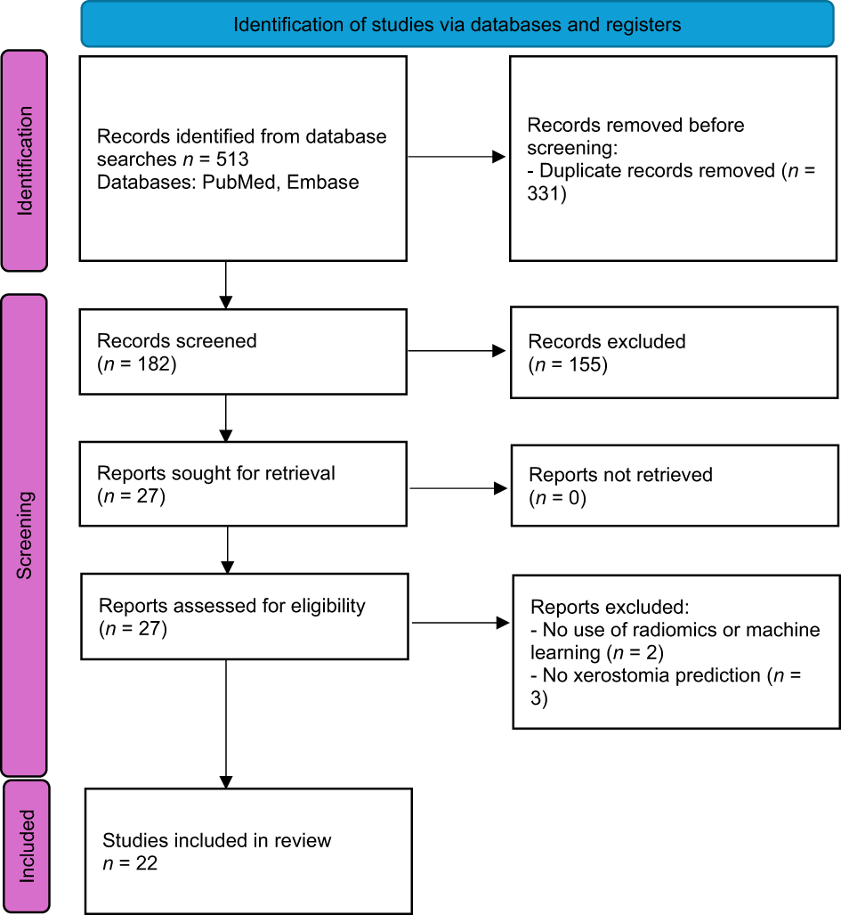
Twenty-two studies met the inclusion criteria (Figure 1). Study characteristics, including cohort size, imaging modality, modelling approach, predictors, outcome definitions and performance metrics, are summarised in Table 1.Reference Van Dijk, Langendijk, Zhai, Vedelaar, Noordzij and Steenbakkers10, Reference Van Dijk, Brouwer, van der Schaaf, Burgerhof, Beukinga and Langendijk19–Reference Lin, Ren, Jian, Yang, Zhang and Zhu39
Preferred Reporting Items for Systematic Reviews and Meta-Analyses (‘PRISMA’) flow chart.

Summary of literature on the application of radiomics in prediction of xerostomia following radiotherapy for head and neck cancer

Table 1 Long description
Table I summarises the 22 studies included in this review evaluating radiomics, machine learning and deep learning approaches for predicting radiation-induced xerostomia following head and neck radiotherapy. For each study, the table reports the imaging modality, sample size, xerostomia endpoint, xerostomia assessment method, model type, software, validation approach, area under the receiver operating characteristic curve (AUC), principal findings and key limitations. Most studies used CT-based radiomics, with additional studies using MRI, megavoltage CT and PET-CT. Sample sizes ranged from 19 to 1208 participants. Delta-radiomics and deep learning generally showed the highest predictive performance, although external validation was uncommon and most studies were limited by small cohorts and methodological heterogeneity.
AUC = area under the receiver operating characteristic curve; CT = computed tomography; EORTC QLQ-H&N35 = European Organisation for Research and Treatment of Cancer Quality of Life Questionnaire Head and Neck Module, 35-item version; NTCP = normal tissue complication probability; LASSO = least absolute shrinkage and selection operator; XGBoost = extreme gradient boosting; SHAP = Shapley additive explanation; DVH = dose–volume histogram; SVM = support vector machine; MRI = magnetic resonance imaging; MR = magnetic resonance; CTCAE = Common Terminology Criteria for Adverse Events; DWI = diffusion-weighted imaging; DCE-PWI = dynamic contrast-enhanced perfusion-weighted imaging; IMC1 = informational measure of correlation 1; GLM = generalised linear model; PET = positron emission tomography; 3D = three-dimensional; CNN = convolutional neural network; MDADI = MD Anderson Dysphagia Inventory; MONAI = Medical Open Network for AI; GTVp = gross tumour volume of the primary tumour
Study characteristics
Twenty-two studies with cohort numbers ranging from 19 to 1208 patients met the inclusion criteria. Computed tomography (CT) was the most prevalent imaging modality (n = 14 studies), while magnetic resonance imaging (MRI), megavoltage CT and positron emission tomography (PET)-CT were less frequently evaluated. Most studies combined radiomic features with dosimetric and/or clinical variables, with several deep learning studies utilising raw imaging and dose data.
All the studies used imaging of the parotid glands; six studies included the submandibular glands.
Outcome definitions
Outcome definitions for xerostomia varied across studies. Many studies assessed xerostomia using validated patient-reported quality of life measurement tools such as the European Organisation for Research and Treatment of Cancer Quality of Life Questionnaire Head and Neck Module,40 or Common Terminology Criteria for Adverse Events.41 Other studies used objective measures such as stimulated saliva output or surrogate markers including parotid gland volume change. The timing of outcome assessment also varied, ranging from during radiotherapy to more than 24 months following treatment. Several studies reported both early and late xerostomia outcomes.
Model performance
Area under the receiver operating characteristic curve represents the ability of a model to correctly assign a higher probability score to an actual positive instance than to a negative one, with 1 representing the best possible score and 0.5 signifying a result obtained by chance.Reference Li42 This was used as a measure of the effectiveness of models in xerostomia prediction.
Of the six studies that incorporated imaging of the submandibular glands, there was a lack of consensus around the benefit of their inclusion. Three studiesReference Li, van Rijn-Dekker, de Vette, van der Schaaf, Van den Bosch and Langendijk32, Reference Men, Geng, Zhong, Fan, Lin and Xiao37, Reference Chu, de Vette, Neh, Sijtsema, Steenbakkers and Moreno38 found improved predictive power when submandibular features were studied; however, two studiesReference Jiang, Lakshminarayanan, Hui, Han, Cheng and Bowers33, Reference Guo, Jiang, Lakshminarayanan, Han, Cheng and Bowers34 found that use of parotid imaging was superior to submandibular features, and one found no improvement over a conventional normal tissue complication probability model with models utilising either parotid or submandibular features.Reference Berger, Noble, Shelley, McMullan, Bates and Thomas30
Model performance varied widely across studies, with area under the receiver operating characteristic curve values ranging from 0.64 to 0.93. Computed tomography radiomics models generally achieved area under the receiver operating characteristic curve values between 0.64 and 0.93. The strongest CT performance was observed in delta-radiomics studies,Reference Van Dijk, Langendijk, Zhai, Vedelaar, Noordzij and Steenbakkers10, Reference Berger, Noble, Shelley, McMullan, Bates and Thomas30, Reference Berger, Noble, Yang, Shelley, McMullan and Bates31 with reported area under the receiver operating characteristic curve values of 0.86–0.93 and consistently stronger predictive performance than models based on pre-treatment imaging alone, suggesting that treatment-induced temporal changes may provide additional predictive information.
Whilst most studies focused on CT-based radiomics, several MRI-based studiesReference Van Dijk, Thor, Steenbakkers, Apte, Zhai and Borra25–Reference Calamandrei, Mariotti, Bicci, Calistri, Barcali and Orlandi27 reported encouraging model performance. One study reported improved performance with the inclusion of MRI radiomic features,Reference Van Dijk, Thor, Steenbakkers, Apte, Zhai and Borra25 with area under the receiver operating characteristic curve value increasing from 0.81 to 0.88 and remaining high during external validation (area under the receiver operating characteristic curve = 0.83); however, other studies demonstrated lower predictive performance, particularly for late xerostomia outcomes.
Deep learning models consistently demonstrated high internal predictive performance. However, this performance was not consistently maintained during external validation. One studyReference Chu, de Vette, Neh, Sijtsema, Steenbakkers and Moreno38 reported an improvement in area under the receiver operating characteristic curve from 0.75 to 0.79 during model development, but performance fell to 0.62 in the validation cohort, highlighting the challenges of model generalisability.
Multimodal studies combining CT and MRI features with dosimetric variablesReference Sheikh, Lee, Cheng, Lakshminarayanan, Peng and Han28, Reference Khajetash, Hajianfar, Talebi, Ghavidel, Mahdavi and Lei29 reported their most promising model area under the receiver operating characteristic curve values of 0.79 and 0.81, whilst the single study using PET-CT–based modellingReference Li, van Rijn-Dekker, de Vette, van der Schaaf, Van den Bosch and Langendijk32 demonstrated a more modest area under the receiver operating characteristic curve improvement from 0.69 to 0.75. Whilst many studies report encouraging results, the majority combined radiomic features with variables such as radiation dose, baseline xerostomia and patient characteristics, with the addition of radiomic features often contributing improvements in area under the receiver operating characteristic curve of around 0.05–0.1 over reference models.
Methodological quality
Radiomics Quality Score assessment demonstrated substantial methodological variability across studies (Figure 2).Reference Van Dijk, Langendijk, Zhai, Vedelaar, Noordzij and Steenbakkers10, Reference Van Dijk, Brouwer, van der Schaaf, Burgerhof, Beukinga and Langendijk19–Reference Lin, Ren, Jian, Yang, Zhang and Zhu39 Mean Radiomics Quality Score was 14 out of 36 (range = 4–20, median = 16). The most consistently met domains were feature reduction and multivariable modelling; however, common deficiencies included the absence of prospective studies, lack of cost-effectiveness analyses, limited open-science practices and inadequate external validation. None of the studies performed cost-benefit analysis. One studyReference Chu, de Vette, Neh, Sijtsema, Steenbakkers and Moreno38 provided publicly available source code and none made imaging data publicly available.
Summary of Radiomics Quality Score (RQS) points per scoring domain, for each study. Note: green indicates fully scored items, orange reflects not fully scored items and red indicates zero points. *Numbers in brackets reflect the maximum possible score for each domain.

Figure 2 Long description
Figure II presents the Radiomics Quality Score (RQS) assessment for each of the 22 included studies. Sixteen methodological domains are assessed: imaging protocol quality, segmentation, imaging at multiple timepoints, use of phantom scanners, feature selection, model validation, prospective study design, cut-off analyses, biological correlates, description and calibration statistics, clinical utility, comparison to gold standard, multi variable analyses, cost-effectiveness and open science. Each study is assigned an overall score out of a maximum of 36. Overall methodological quality was moderate, with a mean RQS of 14. Most studies performed well in feature reduction and reporting of model discrimination, but prospective study designs, external validation, cost-effectiveness analyses and open-science practices such as publicly available datasets or source code were uncommon.
Whilst most studies clearly described imaging acquisition and feature extraction methods, reproducibility and validation remained the most frequent methodological weaknesses. Most studies relied solely on internal validation, with external validation performed in only 6 of 22 studies.Reference Berger, Noble, Yang, Shelley, McMullan and Bates20, Reference Pota, Scalco, Sanguineti, Farneti, Cattaneo and Rizzo22, Reference Van Dijk, Thor, Steenbakkers, Apte, Zhai and Borra25, Reference Chao, El Naqa, Bakst, Lo and Peñagarícano35, Reference Chao, Tomalin, Wei, Liu, Zhang and Liu36, Reference Chu, de Vette, Neh, Sijtsema, Steenbakkers and Moreno38 Where external validation was undertaken, predictive performance was generally reduced compared with internal analyses, suggesting limited model generalisability across institutions and imaging datasets.
Discussion
This review demonstrates that radiomics and machine learning have considerable potential to improve the prediction of radiation-induced xerostomia following treatment for head and neck cancer. However, whilst many studies reported promising predictive performance, particularly those employing delta-radiomics and deep learning approaches, the overall evidence suggests that radiomics has not yet demonstrated sufficient reproducibility or generalisability to justify routine clinical use.
Radiomic features consistently appeared to capture important information relating to salivary gland structure and response to radiotherapy. Features reflecting gland heterogeneity and fat content were associated with xerostomia risk, which aligns with known relationships between tissue composition and radiation sensitivity.Reference Redman43 Delta-radiomics studies evaluated changes in gland morphology and texture over time during radiotherapy, potentially capturing evolving biological responses to radiation-induced injury rather than relying only on baseline tissue characteristics. This is biologically credible given that salivary gland toxicity develops dynamically throughout treatment.Reference Blitzer, Paz, McCoy and Kimple44 However, serial imaging also increases the risk of variability in imaging protocols, image processing and gland segmentation, which may alter radiomic feature values and reduce reproducibility across centres and scanners.Reference Marcu, Grava and Marcu45 These observations indicate that radiomics is capturing meaningful biological information. However, this signal has not yet translated into consistently reliable predictive models.
Across studies, improvements in predictive performance following the addition of radiomic features were generally modest, with many studies demonstrating incremental area under the receiver operating characteristic curve increases of approximately 0.05–0.1 over established clinical or dosimetric models. Furthermore, dose-based variables remained strong predictors of xerostomia across most modelling approaches. This suggests that radiomics may capture complementary biological information, but conventional predictors such as radiation dose continue to account for the majority of predictive performance. Consequently, the current evidence supports radiomics and machine learning models as adjuncts to established normal tissue complication probability models, which use the radiation dose delivered to normal tissues to predict radiation toxicity,Reference Takada, Tambas, Clementel, Leeuwenberg, Sharabiani and Damen7 rather than replacements.
Recent work has highlighted other potential approaches to xerostomia prediction. A systematic review of molecular biomarkers associated with radiotherapy-related toxicities identified a small number of promising candidates, particularly within DNA repair pathways, but concluded that poor data quality, limited external validation and inconsistent reporting currently limit clinical translation.Reference Koch, Reinhardt, Elicin, Aebersold and Schanne46 These findings closely mirror those of the present review and suggest that the principal barriers to implementation are methodological rather than modality-specific. Rather than viewing radiomics and molecular biomarkers as competing approaches, they could be considered complementary. Radiomic features are capable of characterising spatial and temporal tissue changes,Reference Mayerhoefer, Materka, Langs, Häggström, Szczypiński and Gibbs14 whereas molecular biomarkers may capture intrinsic biological radiosensitivity.Reference Koch, Reinhardt, Elicin, Aebersold and Schanne46 Their integration may offer a plausible route towards more accurate and biologically informed prediction models than either approach alone.
Deep learning studies generally reported the strongest predictive results, with area under the receiver operating characteristic curve scores often over 0.80. Deep learning models are able to learn complex spatial relationships directly from radiological images, without the need for pre-defined feature extraction, but can be vulnerable to overfitting, whereby the model ‘overlearns and memorises’ details from its training dataset, relying on them for prediction and performing poorly when faced with previously unseen data.Reference Wagner, Namdar, Biswas, Monah, Khalvati and Ertl-Wagner16 The largest externally validated deep learning study in this reviewReference Chu, de Vette, Neh, Sijtsema, Steenbakkers and Moreno38 showed a substantial reduction in performance on external validation, which raises concerns about the generalisability of the findings. In addition to reproducibility concerns, many deep learning approaches function as ‘black box’ systems, whereby it is not possible to work back through processes to identify the source of any errors or bias and limiting transparency regarding which imaging features drive predictions.Reference Buhrmester, Münch and Arens47 The decline in performance observed during external validation suggests that deep learning models may be particularly susceptible to dataset-specific learning. This may reduce clinician trust and create additional barriers to routine clinical adoption.
Radiomics Quality Score assessment (Figure 2) gives further indication that, despite encouraging predictive performance, clinical translation of radiomics and machine learning models remains problematic. Common methodological deficiencies highlighted include a lack of prospective study designs, an absence of cost-effectiveness analysis, a lack of adherence to open-science principles and in some cases a lack of biological correlation. Small variations in voxel size, reconstruction algorithms, image preprocessing or contour delineation may substantially alter radiomic feature values, reducing model stability and limiting external generalisability;Reference Teng, Wang, Nicol, Ching, Wong and Lam48 however, public availability of imaging datasets, segmentation maps and source code was not routine, with only one study having its code open-source. As a result, independent validation and reproduction of studies is restricted, reducing confidence in their findings. Importantly, the studies that reported the highest predictive performance were not consistently those achieving the highest Radiomics Quality Score, suggesting that further studies are needed to address methodological gaps before the findings can be relied upon to inform clinical practice.
Another source of variability among studies related to outcome definitions. Studies differed in their xerostomia scoring systems, timing of assessment and use of patient-reported versus objective measures. There was also heterogeneity in the glands used for the imaging models, with some studies examining one or both parotid glands, and some also including the submandibular glands. The parotid glands are responsible for the majority of stimulated salivary production, with the rest coming from other salivary glands; however, the submandibular and sublingual glands, and other small salivary glands, are primarily responsible for resting salivary production.Reference Deasy, Moiseenko, Marks, Chao, Nam and Eisbruch6 Therefore, this is an important inconsistency which limits comparability across studies.
The current evidence does not support routine clinical implementation of radiomics-based xerostomia prediction. Whilst many studies show encouraging and biologically grounded results, the benefits over established models are limited and inconsistent. Future research should prioritise large, prospective, multi-centre studies, with standardised imaging protocols and outcome definitions, and open-source science principles. Emphasis should be placed on reproducibility, external validation, and integration with established normal tissue complication probability frameworks. Without addressing these challenges, further increases in model complexity are unlikely to result in any meaningful clinical benefit. If such robust prediction models become available, they could influence treatment planning by identifying patients most likely to benefit from interventions aimed at risk reduction – for example, proton therapy – which is less widely available than conventional radiotherapy but is much more targeted, reducing the risk of radiation toxicities to surrounding structures.Reference Li, Lee, Cohen, Sherman and Lee49 Conversely, patients predicted to be at lower risk may tolerate more aggressive target coverage when clinically appropriate.
Limitations
This review has the following limitations. As a narrative review, no formal meta-analysis was undertaken. Quantitative synthesis was not possible because of substantial heterogeneity in imaging modalities, outcome definitions, modelling approaches and validation strategies. Furthermore, although the Radiomics Quality Score provides a structured framework for assessing methodological quality, scoring of domains such as biological correlates and clinical utility involves a degree of subjective judgement, whereby a different reviewer may generate slightly different scoring for the included studies. Nevertheless, use of a standardised quality assessment framework allowed a comprehensive evaluation of methodological strengths and weaknesses across the included studies.
Since the completion of our literature search, Jazi et al.Reference Jazi, Khademi, Adebi, Shams, Mehnati and Khezerloo50 have published a systematic review of radiomics for radiation-induced xerostomia. Their review provides a comprehensive overview of imaging modalities, modelling strategies and predictive performance. The present review complements this work by focusing specifically on methodological quality using the Radiomics Quality Score, the incremental predictive value of radiomics beyond established dosimetric models, and the current readiness of these approaches for clinical implementation.
Conclusion
Radiomics and machine learning approaches offer a biologically plausible method for enhancing prediction of radiotherapy-induced xerostomia. However, current evidence demonstrates modest, inconsistent improvements over established dose-based models, with limited reproducibility and poor generalisability. At present, radiomics and machine learning approaches should be viewed as promising adjuncts to existing xerostomia prediction models rather than being ready for routine clinical implementation. Their future value may lie in enhancing established dosimetric and clinical predictors, enabling more personalised xerostomia risk assessment, while retaining the interpretability and robustness of existing normal tissue complication probability-based approaches.
Competing interests
None declared.


 Open access

