Introduction
Cerebral WMH are prevalent incident findings on brain MRI scans among the elderly people (Vernooij et al., Reference Vernooij2007). The prevalence and severity of these white matter (WM) lesions are reported to be greater among individuals with AD and those at risk of AD (Yoshita et al., Reference Yoshita2006). Underlying pathogenesis and the clinical relevance of WMH have yet to be fully understood. However, substantial amount of evidence supports the role of WMH in cognitive and functional decline in the aged population (Poggesi et al., Reference Poggesi2011). Results from a number of longitudinal studies have also revealed that WMH confer an increased risk for dementia independently of or in conjunction with hippocampal atrophy or amyloid pathology (Prins et al., Reference Prins2004; Brickman et al., Reference Brickman2012; Provenzano et al., Reference Provenzano2013).
However, differential roles of WMH by region and their associations with specific subtypes of dementia have not been clarified. WMH are generally categorized into deep subcortical WMH (DWMH) and periventricular WMH (PWMH) according to the location they are observed on MRI scans and different WMH are postulated to have distinct etiological basis (Kim et al., Reference Kim, MacFall and Payne2008). Regional WMH have been reported to differentially affect mood and cognition, yet with conflicting results. Studies reported the significance of DWMH especially in patients with late onset depression and vascular dementia (Verdelho et al., Reference Verdelho2010; Poggesi et al., Reference Poggesi2011). PWMH have been implicated in association with cognitive decline and the risk of AD (Prins et al., Reference Prins2004; Yoshita et al., Reference Yoshita2006).
The data from a large, multi-center cohort of Korean elderly adults who participated in the CREDOS study were utilized. In this study, we have investigated among participants with MCI, how WMH by region and severity were associated with baseline characteristics cross-sectionally, and how they affected cognition longitudinally. A recent CREDOS study by Lee et al. (Reference Lee, Lee, Park, Lee, Moon and Chung2014) reported on the independent prognostic significance of PWMH for dementia. The present study extends onto clarifying differential roles of PWMH and DWMH on the risk of specific subtypes of dementia: all-cause dementia, AD, and SVD in participants with MCI at baseline. We hypothesized that severity of each WMH would predict conversion to distinct subtypes of dementia: severity of either type of WMH would predict conversion to all-cause dementia, PWMH severity would predict conversion to AD, and DWMH severity to SVD after controlling for other predictors of dementia.
Methods
Participants Participants were recruited from the CREDOS study, a prospective, nationwide multi-center cohort study designed to assess the occurrence and risk factors of cognitive disorders. This report pertains to a cohort recruited from 2005 through 2010 and followed for up to three years (median 14.3; interquartile range (IQR) 12.1–24.3; range 6.0–36.5 months). More description about the CREDOS is detailed elsewhere (Park et al., Reference Park2011). Participants met the MCI criteria adopted by CREDOS at baseline and had at least one longitudinal clinical review after six months post-baseline. The criteria for MCI in the CREDOS study are as follows: (1) presence of memory complaints; (2) intact function in Activities of Daily Living (ADL); (3) objective cognitive impairment (at least 1.0 standard deviation (SD) below age-and education-adjusted norms) in more than one cognitive domain on neuropsychological testing (Busse et al., Reference Busse, Hensel, Guhne, Angermeyer and Riedel-Heller2006; Ahn et al., Reference Ahn2010); (4) Clinical Dementia Rating (CDR) (Morris, Reference Morris1993) of 0.5; and (5) without dementia according to Diagnostic and Statistical Manual of Mental Disorders (DSM)-IV criteria (American Psychiatric Association, 1994). None of the participants presented any of the following exclusion criteria: (1) significant hearing or visual impairment; (2) neurological disorders (e.g. territorial infarction, intracranial hemorrhage, brain tumor, hydrocephalus, multiple sclerosis); (3) major psychiatric disorders (e.g. schizophrenia, intellectual disability, mania); (4) significant physical illnesses (e.g. severe cardiac, respiratory diseases, uncontrolled diabetes, malignancy). There were 667 MCI participants drawn from the CREDOS cohort and screened for eligibility. Of these, three participants aged younger than 50 years at enrollment, and 7 participants who had insufficient follow-up intervals (< 6 months) were excluded from the analysis. Additional 35 participants were excluded due to missing data on one or more clinical variables. Thus, a total of 622 participants with MCI diagnoses fulfilling these criteria were included. We chose to use relatively liberal criteria (1.0 SD) for objective cognitive impairment in an attempt to identify cognitive changes at the earliest possible point as well as based on longitudinal studies that have produced better predictive power for dementia in those classified as MCI using 1.0 SD threshold (Busse et al., Reference Busse, Hensel, Guhne, Angermeyer and Riedel-Heller2006). This study was approved by the institutional review boards of the participating centers and informed consent was obtained from all participants.
Clinical evaluation
All participants underwent comprehensive clinical evaluations including neuropsychological tests, brain MRI scans, and laboratory tests. Each participant had an informant as a collateral source of information regarding the participants’ cognition, abnormal behaviors, ADL, demographic characteristics, vascular risk factors, and other comorbidities. The following questionnaires were additionally administered; the Korean Mini-Mental State Examination (K-MMSE) (Kang et al., Reference Kang, Na and Hahn1997), the CDR scale (Morris, Reference Morris1993), the Korean version of the Geriatric Depression Scale short form (SGDS-K) (Bae and Cho, Reference Bae and Cho2004) and the Hachinski Ischemic Score (HIS) (Hachinski et al., Reference Hachinski1975). A standardized neuropsychological battery, the Seoul Neuropsychological Screening Battery-Dementia Version (SNSB-D) was used to assess all participants at baseline and at follow up visits. The SNSB-D includes quantitative measures of attention, language, visuospatial function, memory, and frontal executive function domains. More information on the battery is detailed elsewhere (Ahn et al., Reference Ahn2010).
Diagnosis of incident dementia
Diagnosis of incident all-cause dementia was made in accordance with DSM-IV criteria (American Psychiatric Association, 1994) and required objective deficits on neuropsychological testing as well as impairment in ADL. Specific diagnostic criteria were used for designation of dementia classification. AD was diagnosed for those who met the National Institute of Neurological and Communicative Diseases and Stroke-AD and Related Disorders Association (NINCDS-ADRDA) research criteria for possible or probable AD (McKhann et al., Reference McKhann, Drachman, Folstein, Katzman, Price and Stadlan1984). SVD was diagnosed in accordance with National Institute of Neurological Disorders and Stroke-Association Internationale pour la Recherche et l’Enseignement en Neurosciences (NINDS-AIREN) criteria (Roman et al., Reference Roman1993) and imaging criteria proposed by Erkinjuntti (Reference Erkinjuntti2002). Other types of dementia were diagnosed using McKeith criteria for dementia with Lewy bodies (DLB) (McKeith et al., Reference McKeith1996) and the Lund and Manchester criteria for frontotemporal dementia (FTD) (The Lund and Manchester Groups, 1994). Onset of dementia was marked as the date on which the clinical symptoms and neuropsychological findings first allowed the diagnosis of dementia to be made. Each participant is entitled to only one diagnosis of dementia in CREDOS study.
Assessment of white matter hyperintensity severity
MRI scans were acquired at each center at baseline. All MRI scans were acquired according to the imaging protocol developed for the CREDOS with 1.5-T MRI scanners (Noh et al., Reference Noh2014). The MRI scans included trans-axial T2, T1-weighted, gradient-echo, fluid attenuated inversion recovery (FLAIR), and coronal T1-weighted images. Scans were rated for WMH on a visual rating scale developed for the CREDOS using the T2 axial or FLAIR images (Figure 1). The CREDOS WMH visual rating scale examines WMH separately in the deep subcortical and periventricular region. DWMH were rated as D1 (< 10 mm), D2 (10 mm–25 mm), or D3 (≥ 25 mm) based on the longest dimension of the lesions. PWMH were rated as P1 (cap and band < 5 mm), P2 (5 mm–10 mm), or P3 (cap or band ≥ 10 mm) based on the maximum length measured perpendicular and horizontal to the ventricle in caps and bands respectively. In CREDOS studies, researchers were trained to delineate between PWMH and DWMH by rating the DWMH in the axial images at least two slices above the lateral ventricle view slice and/or confirming that normal looking WM intervene between PWMH and DWMH. Ratings of each WMH were combined to provide minimal (D1P1, D1P2), moderate (D1P3, D2P1, D2P2, D2P3, D3P1, D3P2), and severe (D3P3) overall WMH severity measures (Figure 1). Either DWMH ≥ 10 mm or PWMH ≥ 10 mm is necessary to be categorized as moderate overall WMH severity. In a recent study by Noh et al. (Reference Noh2014), the CREDOS WMH visual rating scale showed a good correlation with the volumetric measurements of WMH (Spearman ρ 0.70–0.81). Visual ratings of both PWMH ≥ 10 mm and DWMH 10 mm–25 mm were equivalent to WMH volume of approximately 10 cm3 (Noh et al., Reference Noh2014). Thus, we adopted the cut-off of WMH = 10 mm to distinguish between minimal from more severe form of WMH for this study. The inter-rater reliability for ratings of PWMH, DWMH, and overall WMH in the CREDOS study was previously found to be good (Cohen κ 0.73–0.91) (Moon et al., Reference Moon2011).
Classification of cerebral white matter hyperintensities in CREDOS study.

Statistical analysis
Differences in baseline characteristics between individuals with different severity of WMH were assessed for DWMH and PWMH separately using the Mann–Whitney test for continuous variables and the χ2 test for categorical variables. The primary outcome of interest of this study was progression to all-cause dementia during a follow-up period of up to three years. The secondary outcome of interest was progression to different subtypes of dementia; AD and SVD. Those that had not converted to dementia during the study period were considered censored at their last follow-up assessments. The effect of baseline PWMH and DWMH severity on the risk of progression to each type of dementia was evaluated using the Cox proportional hazards model controlling for all other covariates. Covariates were selected based on their relevance and significance in cognitive function in the elderly population: age, sex, education, K-MMSE, CDR sum of boxes (CDR-SB), SGDS-K, HIS, and vascular risk factors including hypertension, diabetes, and hyperlipidemia. PWMH and DWMH severity were dichotomized at the cut-off of 10 mm to be included in the model as categorical variables. To verify the differential role of PWMH controlling the effect of DWMH and vice versa, we have entered both variables simultaneously in the multivariate model. The assumptions of proportional hazards were checked by goodness of fit test using Schoenfeld residual and Kolmogorov-type supremum test (Lin et al., Reference Lin, Wei and Ying1993) and found to be met. Results were considered statistically significant with p value < 0.05. IBM SPSS Statistics Version 22 (SPSS Inc, Chicago, Illinois) and SAS version 9.3 (SAS Institute, Cary, NC) were used for all statistical analyses.
Results
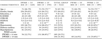
Baseline characteristics of the participants are summarized in Table 1. Distributions of each WMH by severity for the study participants are shown in Figure 1. Differences in characteristics of participants with different severity of DWMH and PWMH are shown in Table 1. Patients with more severe WMH were older, had higher ischemic scores (HIS), and were more frequently hypertensive. There were significantly more males among patients with PWMH ≥ 10 mm compared to those with PWMH < 10 mm, and they scored lower on SGDS-K. Patients with DWMH ≥ 10 mm were less educated than those with DWMH < 10 mm.
Patient characteristics

Values represent median (interquartile range) or number (percentage) unless otherwise indicated.
*p < 0.05; **p < 0.01; ***p < 0.001.
P values for comparison between DWMH < 10 mm and DWMH ≥ 10 mm, PWMH < 10 mm and PWMH ≥ 10 mm.
The p values are derived from Mann-Whitney tests and χ2 tests depending on the type of the variables.
PWMH = periventricular white matter hyperintensity; DWMH = deep subcortical white matter hyperintensity; K-MMSE = Korean Mini-Mental State Examination; CDR-SB = Clinical Dementia Rating scale sum of boxes; SGDS-K = Korean version of the Geriatric Depression Scale short form; HIS = Hachinski Ischemic Score.
Of the 622 MCI participants, a total of 139 patients (22.3%) converted to all-cause dementia during follow-up. Among them, 111 (79.9%) were classified as AD and 25 (18.0%) as SVD. A total of 94/426 (22.1%) patients with DWMH < 10 mm, 45/196 (23.0%) patients with DWMH ≥ 10 mm at baseline converted to all-cause dementia. A total of 80/437 (18.3%) patients with PWMH < 10 mm, 59/185 (32.0%) patients with PWMH ≥ 10 mm at baseline converted to all-cause dementia. For each subtype of dementia, a total of 89/426 (20.9%) patients with DWMH < 10 mm, 22/196 (11.2%) patients with DWMH ≥ 10 mm at baseline converted to AD. A total of 76/437 (17.4%) patients with PWMH < 10 mm, 35/185 (18.9%) patients with PWMH ≥ 10 mm at baseline converted to AD. Among patients with DWMH < 10 mm or PWMH < 10 mm at baseline, 2/426 (0.5%) and 1/437 (0.2%) converted to SVD respectively. Among patients with DWMH ≥ 10 mm or PWMH ≥ 10 mm at baseline, 23/196 (11.7%) and 24/185 (13.0%) converted to SVD respectively. There were three patients diagnosed with other types of dementia; FTD, DLB, and dementia due to strategic infarct respectively. Four hundred forty two (71.1%) participants had stable MCI while 41 (6.6%) have reverted to normal by the final follow up visits.
Differential roles of PWMH and DWMH in predicting incident dementia
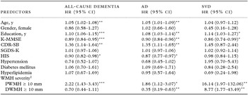
First, we sought to determine whether severity of each type of WMH predicted incident all-cause dementia. Severe baseline PWMH, but not DWMH significantly predicted progression to all-cause dementia (HR 2.22; 95% CI 1.43–3.43; p < 0.001) controlling other predictors of dementia (Table 2). Advanced age, poor performance on K-MMSE, and higher CDR-SB scores at baseline also significantly predicted all-cause dementia. Higher educational achievement and lower scores on HIS at baseline also predicted all-cause dementia (Table 2).
Predictors of progression to dementia (n = 622)

* p < 0.05; ** p < 0.01; *** p < 0.001.
AD = Alzheimer's disease; SVD = subcortical vascular dementia; PWMH = periventricular white matter hyperintensity; DWMH = deep subcortical white matter hyperintensity; K-MMSE = Korean Mini-Mental State Examination; CDR-SB = Clinical Dementia Rating scale sum of boxes; SGDS-K = Korean version of the Geriatric Depression Scale short form; HIS = Hachinski Ischemic Score; HR = hazard ratio; CI = confidence interval.
Hazard ratios and p values are derived from Cox proportional hazards model for multivariable analysis adjusting for age, sex, education, K-MMSE, CDR-SB, SGDS-K, HIS, vascular risk factors, and WMH severity.
†Reference category, WMH < 10 mm.
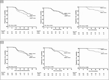
Second, we evaluated the role of each type of WMH in predicting progression to specific subtypes of dementia; AD and SVD. Table 2 summarizes the adjusted significance and HR of each variable for the risk of AD and SVD separately. PWMH ≥ 10 mm at baseline significantly predicted AD (HR 1.86; 95% CI 1.12–3.07; p = 0.016), whereas DWMH ≥ 10 mm at baseline was associated with decreased risk for AD (HR 0.35; 95% CI 0.19–0.63; p = 0.001) (Table 2). On the other hand, PWMH ≥ 10 mm (HR 16.14; 95% CI 1.97–132.06; p = 0.009) and DWMH ≥ 10 mm (HR 8.77; 95% CI 1.77–43.49; p = 0.008) at baseline were both significant predictors of progression to SVD (Table 2). The Kaplan–Meier progression free curves are shown in Figures 2A and 2B for each type of dementia according to the PWMH and DWMH severity. The results from multivariable analysis showed that PWMH ≥ 10 mm is significantly associated with the risk of AD (HR 1.86; 95% CI 1.12–3.07) controlling for all covariates including DWMH severity (Table 2). Other predictors of AD were advanced age, higher educational achievement, worse performance on K-MMSE, and higher CDR-SB scores at baseline (Table 2). Unlike in the case of AD, age or CDR-SB scores did not predict future incidence of SVD. Only the severity of both DWMH and PWMH, along with education and K-MMSE scores significantly predicted SVD (Table 2).
The Kaplan–Meier progression free curves for each type of dementia according to severity of white matter hyperintensities. (A) Kaplan–Meier progression free curves for all-cause dementia, AD, and SVD by PWMH severity groups. Curves were estimated using Kaplan–Meier method. Hazard ratios and p values were derived from Cox proportional hazard model for multivariable analysis (reference category: PWMH < 10 mm). (B) Kaplan–Meier progression free curves for all-cause dementia, AD, and SVD by DWMH severity groups. Curves were estimated using Kaplan–Meier method. Hazard ratios and p values were derived from Cox proportional hazard model for multivariable analysis (reference category: DWMH < 10 mm). AD = Alzheimer's disease; SVD = subcortical vascular dementia; PWMH = periventricular white matter hyperintensity; DWMH = deep subcortical white matter hyperintensity; HR = hazard ratio.

Discussion
In the present study, both PWMH ≥ 10 mm and DWMH < 10 mm at baseline independently predicted conversion to AD during follow-up whereas both PWMH ≥ 10 mm and DWMH ≥ 10 mm predicted conversion to SVD. Only PWMH ≥ 10 mm significantly predicted incident all-cause dementia. Our results are in line with the previous studies suggesting longitudinal relationship between baseline PWMH severity and future dementia (Prins et al., Reference Prins2004; van Straaten et al., Reference van Straaten2008). Although, there was no cross-sectional association between global cognitive function and severity of either WMH at baseline, we found that PWMH ≥ 10 mm significantly predicted incident AD and SVD during follow-up and these associations were both independent of DWMH severity. DWMH ≥ 10 mm at baseline was associated with increased risk of SVD but also with decreased risk of AD (HR, 0.35; 95% CI 0.19–0.63). Ischemic scores (HIS) also showed similar inverse relation with decreased risk for AD (HR, 0.87; 95% CI 0.77–0.97). DWMH are clinically implicated in association with the risk of SVD, and other vascular risk factors and the distinct etiological basis of DWMH is reported to be ischemic in origin (Verdelho et al., Reference Verdelho2010; Schmidt et al., Reference Schmidt2011). Causal relation cannot be established from this study. However, PWMH seem to play a role in the pathogenesis of dementia via a mechanism independent from DWMH and other vascular risk factors, especially in AD, evidenced from the opposite directions of the association in this study. The role of PWMH in mediating clinical manifestation of dementia still remains unclear and is, in part, attributed to the high density of association fibers and cholinergic fibers critical to cognition in the region. Recent diffusion tensor imaging (DTI) studies have demonstrated that WM pathologies in patients with AD selectively involve fiber tracts connecting cortical association areas, such as the cingulate bundles and the corpus callosum. These WM alterations are also detected in participants with MCI suggesting that WM pathology may serve as a potential imaging marker for AD (Zhang et al., Reference Zhang2007).
The periventricular WM is distinct from the deep subcortical WM in several aspects. Histopathological studies have revealed distinct pathological changes for each type of WMH; while DWMH are primarily ischemic foci, PWMH characteristically reflect disrupted ependymal lining, varying degrees of gliosis, and rarefaction of myelin (Schmidt et al., Reference Schmidt2011). Distinct vasculatures perfuse each WMH: non-collateralizing ventriculofugal vessels arising from subependymal arteries perfuse the periventricular WM whereas medullary arteries arising from middle cerebral arteries perfuse the deep WM, accounting in part, for the vulnerability of periventricular WM to hemodynamic compromise (Kim et al., Reference Kim, MacFall and Payne2008). Regenerative property is another distinctive feature of the periventricular WM region. Subventricular zone (SVZ), one of the two brain regions (along with subgranular zone of the hippocampus) enriched with multipotent neural stem cells, is situated in the periventricular WM (Gage, Reference Gage2000). Failure of endogenous neural stem cells to respond appropriately to neuronal loss are thought to be the common mechanism that contributes to functional compromise in neurodegenerative diseases (Armstrong and Barker, Reference Armstrong and Barker2001). Defect in neurogenesis in the SVZ has been detected in AD patients and abnormal accumulation of β amyloid protein toxic to SVZ progenitor cells seem to impair the neuroregenerative property of the region (Curtis et al., Reference Curtis, Faull and Eriksson2007). Rarefaction of myelin is one of the distinct features of the WMH, especially in the periventricular region. Remyelination occurs in response to axonal myelin loss. However, the attempts at remyelination are observed primarily in the periventricular WM cells (Simpson et al., Reference Simpson2007). Abnormalities in brain myelination patterns prior to the appearance of amyloid and tau pathology are demonstrated in mouse models of AD (Desai et al., Reference Desai, Sudol, Janelsins, Mastrangelo, Frazer and Bowers2009). When the brain loses its regenerative ability, it is likely to result in functional compromise.
In our study, no particular vascular risk factor was independently associated with greater hazard for any type of dementia. However, participants with PWMH ≥ 10 mm or DWMH ≥ 10 mm were more frequently hypertensive and had higher HIS scores at baseline. These findings are in line with the previous studies on vascular risk factors as etiological basis of WMH (Vuorinen et al., Reference Vuorinen2011). In our study, WMH severity rather than each vascular risk factor significantly predicted progression from MCI to dementia. It is plausible that vascular risk factors such as hypertension are not direct causations of dementia but are the risk factors of WMH, which in turn mediate the clinical manifestation of dementia.
Education is widely known for its protective effect in cognition. Higher education achievement in our sample however was associated with greater risk of both AD and SVD. One recent CREDOS study (Ye et al., Reference Ye2013) that evaluated the effects of education on progression to AD in early versus late-stage MCI indicated that protective effects of education against cognitive decline remain in the early-stage amnestic MCI but disappear into the late-stage. This explanation is in line with the previous studies that advocated cognitive reserve theory and might account for the paradoxical association of education and dementia risk in our study which included all stages of MCI.
Strengths of this study are the relatively large sample size and a prospective design. We note several limitations to our study. We calculated the power of this study to detect the effect of severity of PWMH (< 10 mm, ≥ 10 mm), and DWMH (< 10 mm, ≥ 10 mm) on conversion to dementia. The conversion rates to dementia within three years were 18.1%, and 21.8% in patients with PWMH < 10 mm, and DWMH < 10 mm respectively. With the period of 4 years and 11 months for accrual, and follow-up of three years, the powers associated with PWMH, and DWMH are 66.5%, and 61% respectively, expecting at least 10% increase of conversion rate (HR = 1.65 for PWMH, HR = 1.56 for DWMH). However, although the low power, the results of the significance for PWMH, and the non-significance for DWMH might be due to much higher HR = 2.22, and much lower HR = 0.70 than expected. The CREDOS study is a still on-going, prospective study from which we have utilized data of participants recruited from 2005 to 2010. Utilization of all data collected after completion of the study will yield more reliable results with appropriate power. The CREDOS study recruited participants from memory clinics at university hospitals, thus the generalizability of the study results is limited. We used MRI visual rating scales that distinguished PWMH and DWMH which allowed us to explore the differential roles of each type of WM pathology. However, ceiling effects with the use of visual rating scales and lack of medial temporal lobe atrophy measures as covariates remain limitations of this study.
Conclusions
Our findings suggest that severe PWMH at baseline significantly predict progression from MCI to all-cause dementia, AD, and SVD. PWMH seem to play a role in clinical manifestation of dementia via mechanism independent from DWMH, especially in AD and can therefore serve as a better surrogate marker for AD in this population of MCI participants. Risk factors associated with the formation and progression of WMH should be thoroughly investigated and prevented whenever possible.
Conflict of interest
None.
Description of authors’ roles
SH Kim, SH Choi, and DK Kim designed the study, analyzed the data, and wrote the paper. SH Kim and SH Choi contributed equally. SH Choi, YM Lee, HR Na, HJ Han, YS Shim, JH Kim, SJ Yoon, SY Kim, and DK Kim participated in implementation of the study, acquisition of the data, and supervision of data collection. SH Kim and MJ Kim carried out the statistical analysis. MJ Kim, YM Lee, WJ Myung, YD Kim, JY Kim, and JH Park assisted with data analysis and writing the paper.
Acknowledgments
This study was supported by the grants of the Korea Healthcare Technology R&D Project, Ministry of Health and Welfare, Republic of Korea (HI10C2020) and Samsung Biomedical Research Institute (GL1B20812). The funders had no role in study design, data collection and analysis, decision to publish, or preparation of the manuscript.