I. Normal Anatomy of the Coronary Arterial System
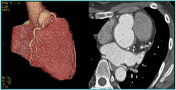
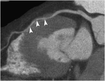
Usually, the left main coronary artery originates from the left posterior aortic sinus of Valsalva, passes to the left of and posterior to the pulmonary trunk, and usually bifurcates into the left anterior descending and the circumflex coronary arteries. The left anterior descending coronary artery runs in the anterior interventricular groove and the circumflex coronary artery courses in the left atrioventricular groove. The right coronary artery usually originates from the right anterior sinus of Valsalva, runs along the right atrioventricular groove, and usually gives rise to the posterior descending coronary artery near its termination. A “circle and half-loop” model has been introduced to illustrate the anatomical relationships among these coronary arteries. The circle consists of the right coronary artery and the circumflex coronary artery, whereas the half-loop is formed by the left anterior descending coronary artery and posterior descending coronary artery.
II. Clinical Importance of Coronary Arterial Anomalies
Overall, congenital coronary arterial anomalies are rare, occurring in 0.3–1% of healthy individuals.Reference Angelini, Velasco and Flamm1 Congenital coronary arterial anomalies are commonly sporadic, and may be either isolated or associated with other congenital cardiac abnormalities including bicuspid aortic valve.
The third most frequent cardiovascular cause of sudden death on the athletic field is the congenital anomaly of anomalous aortic origin of the coronary artery from the wrong aortic sinus of Valsalva; the anomalous aortic origin of the left main coronary artery origin from the right aortic sinus of Valsalva is the most lethal variant. Congenital coronary arterial anomalies are often incidentally found in asymptomatic individuals but life-threatening presentations occur in approximately 20% of cases; patients can present with myocardial infarction, arrhythmia, or even sudden cardiac death. Up to one-third of these cardiac-related deaths occur in young athletes and members of the military.
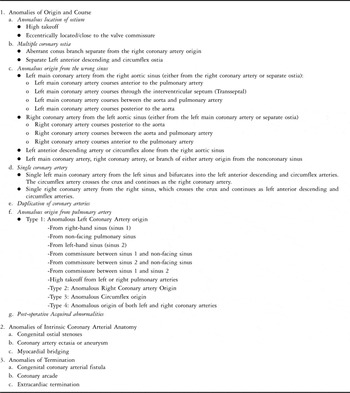
III. Classifications (Table 1)
We used a modified version of the classification system developed by Greenberg et alReference Greenberg, Fish and Spindola-Franco2 (Table 1). Coronary arterial anomalies may also be classified as either haemodynamically significant or haemodynamically insignificant. Anomalies that are haemodynamically significant are characterised by abnormalities of myocardial perfusion, which lead to an increased risk of myocardial ischaemia or sudden death.Reference Reagan, Boxt and Katz3 Haemodynamically significant congenital coronary arterial anomalies can include:
Congenital coronary arterial anomalies.

• anomalous origin of either the left or the right coronary artery from the pulmonary artery,
• anomalous aortic origin of the coronary artery from the wrong aortic sinus of Valsalva,
• myocardial bridging, and
• congenital coronary arterial fistula.
IV. Individual Coronary Arterial Anomalies (Summarised in Table 1)
1. Anomalies of Origin and Course
a. Anomalous location of the coronary ostium
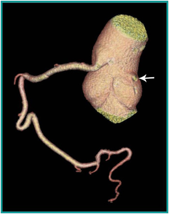
The coronary ostium in most individuals is usually located centrally in the sinus of Valsalva. In some patients, the ostium may be eccentrically located, and in others a coronary artery may arise close to the valve commissure. One or both ostia may have a high take-off and arise from the tubular aorta above the sinotubular junction (Fig 1). Vlodaver et alReference Vlodaver, Neufeld and Edwards4 reported that both coronary ostia were situated above the sinotubular junction in 6% of randomly selected hearts of adults. This anomaly is usually a benign finding. However, high take-off of a coronary ostium becomes significant and must be recognised in the following situations:
High “take-off” of the right coronary artery.

• during aortic cannulation for cardiopulmonary bypass,
• during placement of a catheter for the administration of cardioplegia during cardiopulmonary bypass
• if an aortotomy is necessary, because, if not recognised, the coronary artery could be transacted,
• during coronary arteriography because it may cause difficulty in cannulating the vessel(s).
b. Multiple coronary ostia
An aberrant coronary artery to the conus arising with a separate aortic origin distinct from the right coronary artery is particularly at risk for injury from ventriculotomy or other manoeuvres performed during cardiac surgery.Reference Van Geuns and Cademartiri5 Separate ostia of the left main and circumflex arteries may occur in a small percentage (0.41%) of individuals with otherwise normal anatomy.Reference Danias, Stuber, McConnell and Manning6 Although multiple coronary ostia represent a technical difficulty during coronary angiography, they may also allow alternate collateral sources of myocardial flow of blood in patients with proximal coronary arterial disease.Reference Greenberg, Fish and Spindola-Franco7
c. Anomalous aortic origin of the coronary artery from the wrong aortic sinus of Valsalva
Both coronary arteries may arise from the same aortic sinus with either a single ostium or two separate ostia. This finding is generally not significant if the anomalous coronary artery courses posterior to the aorta or anterior to the pulmonary artery, but it becomes important if it courses between the two great vessels, as this may lead to myocardial ischaemia and sudden death.
Four recognised patterns of anomalous aortic origin of the coronary artery from the wrong aortic sinus of Valsalva are:
• the right coronary artery arising from the left coronary sinus,
• the left main coronary artery arising from the right coronary sinus,
• the circumflex coronary artery or left anterior descending coronary artery arising from the right coronary sinus, and
• the left main or right coronary arteries, or a branch of either artery, arising from the non-coronary sinus.
Four common courses are described that depend on the anatomical relationship of the anomalous coronary artery to the aorta and the pulmonary trunk:
• interarterial, that is between the aorta and the pulmonary artery,
• retroaortic,
• prepulmonic, and
• septal, that is subpulmonic.Reference Ropers, Gehling and Pohle8
The diagnosis of anomalous aortic origin of the coronary artery from the wrong aortic sinus of Valsalva requires a high index of suspicion, and is particularly important, because surgical correction is feasible. The possibility of a coronary arterial anomaly should always be considered in a young athlete with a history of pain in the chest or syncope, particularly if the episode(s) are triggered by exercise. Transthoracic or transoesophageal echocardiography and magnetic resonance imaging can be used for diagnosis, and diagnostic coronary arteriography can ultimately be performed. Patients usually do not have abnormalities on electrocardiograms performed either at rest or with exercise, because myocardial ischaemia is episodic, thereby limiting the value of random screening.
1) Interarterial course of an anomalous coronary artery:
When there are two separate ostia, the right or left main coronary arteries may arise from the wrong sinus of Valsalva and course between the great vessels. The ostium of the anomalous coronary artery is frequently small and slit-like, with a take-off at an acute angle.Reference Virmani, Chun and Goldstein9 A similar situation may be found if there is a single coronary artery arising from the right aortic sinus and the left main or anterior descending arteries runs between the aorta and pulmonary artery, or if a single coronary artery arises from the left aortic sinus and the right coronary artery courses between the great vessels.
2) Pathophysiology:
Several mechanisms have been proposed to explain the pathophysiology of acute myocardial ischaemia in patients with an anomalous origin of the coronary artery from the wrong aortic sinus of Valsalva:Reference Angelini10, Reference Davis, Cecchin, Jones and Portman13
• “Flap-like closure” of the slit-like opening of the coronary orifice
• Acute angle of take-off and kinking of the coronary artery as it exits from the aorta
• Intramural segment of the proximal coronary artery
• Compression of the intramural segment by the aortic commissure
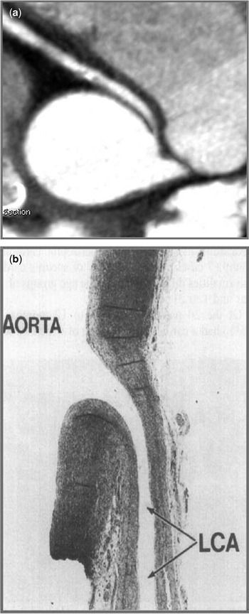
• Compression of the coronary artery as it courses between the aorta and the pulmonary artery, accentuated by exercise-related expansion of the pulmonary artery (Fig 2a)
Figure 2(a) Compression of the coronary artery as it courses between the aorta and the pulmonary artery, accentuated by exercise related expansion of the pulmonary artery. (b) Endothelial injury causes spasm of the coronary artery (LCA = left coronary artery).

• Spasm of the coronary artery as the result of endothelial injury (Fig 2b).

3) Clinical Presentation:
The true incidence of this anomaly is unknown, but estimates range from 0.1–0.3% of the general population.Reference Zeppilli, dello Russo and Santini11 Diagnosis is challenging because characteristic physical findings do not exist and physical examination is almost always normal. When symptoms are present, they most commonly include dyspnoea, pain in the chest, palpitations, dizziness, pre-syncope, or syncope during or just after exertion.Reference Liberthson, Dinsmore and Fallon12 The initial presentation may be a sudden death.Reference Davis, Cecchin, Jones and Portman13 Screening first-degree relatives with echocardiography has recently been suggested because of a potential genetic aetiology of this anomaly.Reference Brothers, Stephens and Gaynor14
4) Surgical Management:
a) Indications for surgery:
• signs or symptoms of myocardial ischaemia or ventricular arrhythmias
• asymptomatic patients with anomalous left main coronary artery from the wrong aortic sinus of Valsalva due to the high risk of sudden death
• management of asymptomatic patients with anomalous right coronary artery from the wrong aortic sinus of Valsalva is controversial. Documented ischaemia makes the decision to advise operation easier.
b) Surgical Techniques:
i. Unroofing of the Coronary Artery
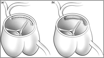
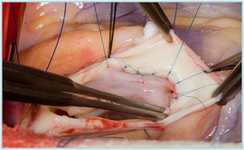
Unroofing of the coronary artery (Fig 3) is the procedure of choice for patients with anomalous aortic origin of the coronary artery from the wrong sinus of Valsalva with an interarterial and intramural course.Reference Frommelt, Frommelt, Tweddell and Jaquiss15 A transverse aortotomy is performed and the coronary arterial ostia are identified. The slit-like ostium is opened longitudinally starting at the anomalous coronary arterial ostium and continuing into the correct sinus. A segment of the common wall between the aorta and the coronary artery is excised, and the intimal surfaces are approximated with interrupted 8/0 sutures made of polypropylene (Fig 4). If the anomalous coronary arterial ostium is close to the commissure of the aortic valve, it may be necessary to take down the commissure, unroof the coronary artery, and then resuspend the commissure with a pledgeted suture.
(a and b). Unroofing is the procedure of choice for those with anomalous coronary artery with an interarterial and intramural course. To enlarge the often slit-like ostium, it is opened longitudinally starting at the anomalous coronary ostium and continuing into the correct sinus.

A segment of the common wall between the aorta and the coronary is excised, and the intimal surfaces are approximated with interrupted 8/0 polypropylene sutures.

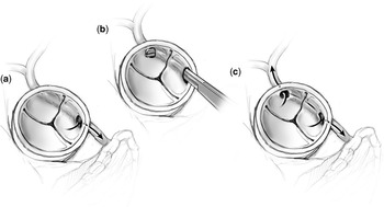
ii. Creation of a Neo-ostium (Fig 5)
For patients with an anomalous aortic origin of the coronary artery from the wrong sinus of Valsalva and an intramural course, the creation of a neo-ostium in the correct sinus of Valsalva is an alternative technique. A probe is passed through the intramural segment of the anomalous coronary artery into the correct aortic sinus of Valsalva. The coronary artery is opened at the location at which it exits the aorta and a neo-ostium is created. The intima is sewn to the aortic wall with interrupted sutures. This avoids take-down and re-suspension of the commissure of the aortic valve.
(a) Anomalous origin of the left main coronary artery from the right sinus. Creation of a neo-ostium is an alternative technique. (b) A probe is passed through the intramural segment of the anomalous artery into the correct sinus. The coronary artery is opened at the location where it exits the aorta and a neo-ostium (c) is created. This technique avoids the takedown and re-suspension of the commissure.

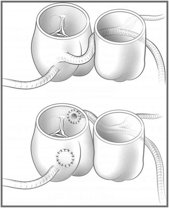
iii. Translocation and Re-implantation of the Coronary Artery (Fig 6)
When there are two separate coronary ostia with an interarterial but not intramural course, re-implantation of the coronary artery has been advocated. This procedure is similar to the arterial switch operation. The coronary artery is excised with a button of aortic tissue and usually re-implanted above the sinotubular junction in the correct sinus.
Coronary artery translocation with re-implantation. When there are two separate coronary ostia with an interarterial but not intramural course, coronary artery re-implantation has been advocated.

iv. Translocation of the Pulmonary Artery
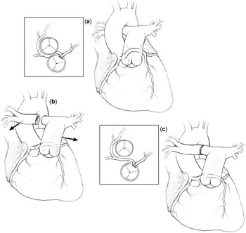
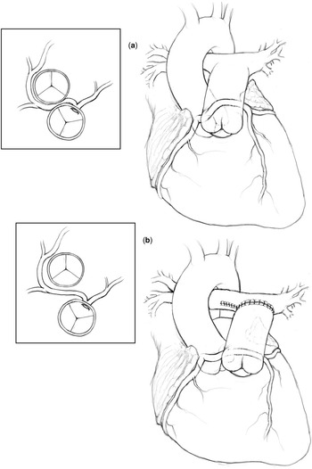
Compression of the anomalous coronary artery between the aorta and the pulmonary artery can be prevented by translocating the main pulmonary artery to the left pulmonary artery, or by translocating the right pulmonary artery and bifurcation anterior to the aorta (Fig 7), and thereby leaving the coronary circulation intact.Reference Rodefeld, Casey and Culbertson16 This technique may be used when the anomalous course is not intramural, but the ostia are close together.Reference Gulati, Reddy and Culbertson17
(a) Anomalous origin of left main coronary artery from the right aortic sinus. (b) Both branch pulmonary arteries are fully mobilised with the right pulmonary artery being transected and moved anterior to the aorta. (c) The right pulmonary artery is reattached. This moves the main pulmonary artery both anteriorly and leftward, relieving compression on the interarterial portion of the anomalous artery.

v. Translocation of the Pulmonary Artery with Patch Angioplasty of the Coronary Artery
Combining patch angioplasty of the coronary artery using pericardium with translocation of the main pulmonary artery to the left pulmonary artery (Fig 8) has also recently been proposed.Reference Alphonso, Anagnostopoulos and Nölke18
(a) Anomalous origin of left main coronary artery from the right aortic sinus. (b) The proximal pulmonary artery is translocated to the left pulmonary artery.

vi. Coronary Artery Bypass Grafting
Creating a bypass with a graft of a saphenous vein or internal mammary artery may be appropriate in an older adult, particularly if concomitant obstructive atherosclerotic disease of the coronary artery is present. However, concern about long-term patency of the graft in a young patient, because of competitive flow, makes bypass a less desirable option in children and young adults.
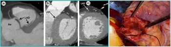
5) Transseptal Course of Anomalous Coronary Artery (Fig 9a–d):
Transseptal/subpulmonic left anterior descending artery: Computed tomography (a, b and c) and intraoperative pictures (d) showing the proximal portion of the left anterior descending artery completely surrounded by the myocardium of the interventricular septum. This anomalous pathway is mostly located within the upper, anterior interventricular septum.

In this anomaly, the proximal portion of the left main coronary artery or left anterior descending coronary artery is completely surrounded by the myocardium of the interventricular septum. This anomalous pathway is mostly located within the upper, anterior interventricular septum. Frequently, septal branches can be seen to arise from the anomalous vessel, a distinction that may help in differentiating an interarterial course from an intraseptal course on conventional angiography.
We have used coronary artery bypass grafting with the left internal mammary artery to treat this anomaly. In order to avoid competitive flow from left main coronary artery, we narrow and create a fixed stenosis of the left main until the internal mammary artery flow is greater than 15–18 millilitres per minute, in order to allow the internal mammary artery graft to mature.
6) Surgical results:
No data exist regarding the long-term rates of patency of the neo-ostia after the unroofing of an intramural coronary artery. Short- to mid-term results have overall been reassuring, with no reports of ostial stenosis by echocardiography. However, in a study by Romp et al, one patient developed severe aortic insufficiency requiring replacement of the aortic valve 44 months after the initial operation.Reference Romp, Herlong and Landolfo19
Davies et alReference Davies, Burkhart and Dearani20 reported the outcome in 36 patients with a median age of 47 years. Angina, shortness of breath, or syncope was present in 29 patients (81%), and 9 of 21 patients (43%) had an abnormal stress test. Coronary angiography or computed tomographic angiography demonstrated
• anomalous left main coronary artery arising from the right aortic sinus of Valsalva in 13 patients (36%),
• anomalous right coronary artery arising from the left aortic sinus of Valsalva in 21 patients (58%), and
• anomalous left anterior descending coronary artery arising from the right aortic sinus of Valsalva and travelling between the aorta and pulmonary artery in two patients (5%).
An intramural course was identified on pre-operative imaging in 34 patients (94%). Five patients (14%) had previous acute myocardial infarction related to the anomalous origin of the coronary artery. Operations included coronary artery bypass grafting in 14 patients and unroofing in 22 patients. There were no early deaths. The reported follow-up of these patients was 1.1 years mean, with a maximum follow-up of 14 years: chest pain recurred in one patient who had coronary artery bypass grafting while no recurrent symptoms were noted in the unroofing group.
Brothers et al evaluated children in the short term to mid-term after unroofing of the intramural segment of the coronary artery as treatment for anomalous aortic origin of the coronary artery. In this analysis by Brothers et al, subclinical evidence of post-operative myocardial ischaemia was found in half of the patients with anomalous right coronary artery from the wrong aortic sinus of Valsalva and in one of eight patients with anomalous left coronary artery from the wrong aortic sinus of Valsalva.Reference Brothers, McBride and Seliem21 This subclinical ischaemia was evident despite the documentation of patent coronary arterial neo-ostia by echocardiography and with the patients remaining asymptomatic during testing. Although re-implantation of the coronary artery has been used by some cardiologists, a study by Rinaldi et al reported that two patients required emergency bypass grafting.Reference Rinaldi, Carballido and Giles22 The long-term success of translocation of the pulmonary artery is not known.
d. Single Coronary Artery
A single coronary artery is a rare anomaly in which only one coronary artery arises from the aortic trunk via a single ostium. This single coronary artery supplies blood to the entire heart. It is rare and reported in approximately 0.024–0.066% of the population.Reference Angelini, Villason and Chan23 A single coronary artery can have many different patterns of distribution. It may follow the pattern of a normal artery, divide into two branches with distributions of the right and left coronary arteries, or have a distribution different from that of the normal coronary arterial tree. Single coronary artery is a very heterogeneous group of anomalies in which the single artery can arise from any of the three sinuses of Valsalva, and the pathways of the branches can vary greatly, taking retrocardiac, retroaortic, interarterial, intraseptal, and anterior courses.
e. Duplication of Coronary Artery
Duplication of the left anterior descending coronary artery is seen in 0.13–1% of the general population. This anomaly consists of
• a short left anterior descending coronary artery that courses and terminates in the anterior interventricular sulcus, and
• a long left anterior descending coronary artery that originates from either the left anterior descending coronary artery or the right coronary artery, enters the distal interventricular sulcus, and courses to the cardiac apex.
As the left anterior descending coronary artery is frequently bypassed surgically, it is important to recognise this anomaly before surgical revascularisation so that the surgical arteriotomy is correctly placed.Reference Sajja, Farooqi and Shaik24
f. Anomalous Origin of a Coronary Artery from the Pulmonary Artery (Adult Variant)
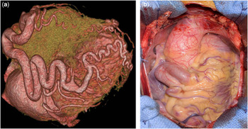
This is a rare congenital anomaly that is almost always fatal if not diagnosed and treated. The estimated incidence is 1 in 30,000 and 1 in 300,000. The most common variant is the anomalous origin of the left main coronary artery from the pulmonary artery. It is the most common cause of myocardial infarction in childhood. It is known also as the Bland–White–Garland syndrome, after Bland et al reported on both clinical and autopsy findings in an infant with this anomaly in 1933.Reference Bland, White and Garland25 Occasional reports document survival into adulthood of individuals with the left main coronary artery arising from the pulmonary artery.Reference Alexi-Meskishvili, Berger and Weng26 The majority of patients are subject to the risk of sudden death, chronic mitral regurgitation, and global ischaemic cardiomyopathy. A spectrum of coronary arterial collaterals exists, ranging from the “infantile type”, where there are no collaterals and no evidence of left-to-right flow, to the “adult type”, where there are well-established collaterals (Fig 10a and b) and generous left-to-right flow. Several surgical techniques have been employed including
(a) Adult variant of anomalous origin of left coronary artery from the pulmonary artery with coronary collateral circulation at its best. (b) Right coronary artery and branches are severely enlarged and redundant with a large loop of the artery overlying right atrium.

• aortocoronary bypass grafting using reversed saphenous vein or the left internal mammary artery,
• left subclavian artery to coronary artery transposition,
• intrapulmonary baffling, and
• direct aortic implantation.
Although no randomised trials comparing the respective options exist, successful surgical repair clearly depends upon the establishment of a two-coronary system perfused by the left heart. Direct coronary arterial translocation offers the advantage of avoiding late graft stenosis. If one is unable to mobilise a sufficient cuff of the pulmonary artery to reach the aorta, the options include
• a short interposition graft, possibly using polytetrafluoroethylene,
• construction of an intrapulmonary baffle, known as the Takeuchi procedure,Reference Takeuchi, Imamura and Katsumoto27 and
• bypass grafting.
Ultimately, surgical repair can restore or maintain left ventricular function, decrease ventricular dilation, relieve angina, protect the mitral apparatus, and prolong life.Reference Cochrane, Coleman and Davis28
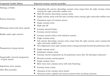
g. Coronary arterial anomalies seen in adults with congenital cardiac diseases (summarised in Table 2)
We advise pre-operative evaluation with a computed tomographic scan or magnetic resonance imaging of the chest for all patients with previously palliated or repaired conotruncal anomalies, conduits, and prior aortic root surgery in order to define the anatomy of the proximal coronary arteries and buttons, aortic root, and ascending aorta. It is important to remember the following coronary arterial variations when dealing with certain congenital cardiac defects, whether in children or adults.
Coronary arterial anomalies seen in adults with congenital cardiac diseases.

1) Conotruncal Anomalies:
a) Tetralogy of Fallot:
The incidence of a major coronary artery crossing the right ventricular outflow tract in the tetralogy of Fallot is between 5% and 12%. Pre-operative recognition of such anomalous coronary arteries is important when planning operations that involve the right ventricular outflow tract, including replacement of the pulmonary valve and replacement of conduits connecting the right ventricle to the pulmonary artery.
b) Common arterial trunk (Truncus Arteriosus):
In patients with a common arterial trunk, the abnormal origin and distribution of the coronary arteries are not uncommon and may have an unfavourable effect on surgical correction. Single origins of the left and the right coronary arteries have been reported, as has abnormally high ostial take-off above the sinuses of Valsalva, among others. Recognition and awareness of a “high take-off” of a coronary artery is particularly important when an aortotomy is planned for surgical procedures on the truncal valve.
c) Double outlet right ventricle:
In most patients with double outlet right ventricle, the coronary arterial orifices are rotated clockwise, as the observer looks from below. With a right-sided and anterior aorta, the coronary arterial pattern is similar to that seen in the complete transposition of the great arteries. The right coronary artery arises from the right posterior facing sinus and the left coronary artery arises from the left posterior facing sinus.
The reported variations include:
• single coronary artery,
• origin of the left anterior descending coronary artery from the right coronary artery, running anteriorly beneath the pulmonary valve across the right ventricular outflow tract.
d) Congenitally corrected transposition of great arteries:
In patients with congenitally corrected transposition of great arteries, the coronary arteries display the anatomy typical of the ventricle they supply, but in a reversed manner. In patients with a normal atrial arrangement, or atrial situs solitus, the right-sided coronary artery supplies the left ventricle and usually divides into an anterior descending coronary artery and a circumflex coronary artery, while the left-sided coronary artery encircles the orifice of the tricuspid valve orifice to supply the right ventricle and become the posterior descending artery. The right-sided coronary artery arises from the right posterior aortic sinus and passes directly in front of the ring of the pulmonary valve. The left-sided coronary artery arises from the left posterior aortic sinus. The anterior aortic sinus is the non-coronary sinus. The most common variation from this arrangement is for a single coronary artery to arise from the right-facing aortic sinus and divide into right and left main branches. A tendency towards early branching of the major coronary arteries exists in patients with congenitally corrected transposition of great arteries, with a short main stem that is often entrapped in right ventricular fat or muscle.
2) Post-operative Abnormalities:
The introduction of reparative operations involving manipulation, transfer, or reimplantation of the coronary arteries has defined a group of patients who are at increased risk of acute or chronic problems related to myocardial ischaemia. The patients at risk include those who have undergone the following previous operations:
• arterial switch operation,
• Ross operation,
• repair of the anomalous origin of the left main coronary artery from the pulmonary artery, and
• repair of the ascending aortic aneurysm.
a) Aneurysm of the coronary arterial button after the Ross procedure (Fig 11):
Coronary button aneurysm after Ross procedure.

A false aneurysm arising at the proximal suture line of a pulmonary autograft in the aortic position has been previously reported.Reference Metras, Kreitmann, Habib, Riberi, Yao and Ross29 In comparison to the aortic valve, the pulmonary valve lacks a discrete annulus. The pulmonary root is not fully supported in areas where it is in continuity with muscle. When translocated, this unsupported area becomes exposed to systemic arterial pressure, with the potential for dilatation. Therefore, we recommend one or more of the following preventative measures:
• The lower line of the suture of the autograft must be onto the annulus of the excised aortic valve to give it support and potentially prevent progressive dilatation in the future.
• Reinforce the proximal annular line of the suture with a fabric in patients who have completed their somatic growth.
• Avoid redundant right ventricular muscle when trimming the autograft. This muscle can potentially become a weak tissue at the proximal end of the autograft.Reference Shahid, Al-Halees and Khan30
• Consider the technique described by Ungerleider in which the pulmonary autograft is encased in a tube of dacron to prevent dilatation.Reference Slater, Shen and Welke31
b) The arterial switch population:
In the long term, the success of the arterial switch operation depends on the continued patency and adequate functioning of the coronary arteries. Intimal thickening of the proximal coronary arteries is common. Obstructive coronary arterial lesions are not rare. Compression of the anteriorly positioned proximal coronary arteries by the anterior pulmonary root after the LeCompte manoeuvre can also occur. This compression can be treated by the placement of a short interposition graft, possibly with polytetrafluoroethylene, in the right pulmonary artery, which effectively lifts the pulmonary root off of the proximal aorta. Sequential coronary arterial evaluation using coronary arterial angiography or multi-slice computed tomography is essential.
2. Anomalies of Intrinsic Coronary Arterial Anatomy
a. Myocardial Bridging
The phenomenon of myocardial bridging was described by Reyman in 1737.Reference Chen and Lin32 Myocardial bridging is caused by a band of myocardial muscle overlying a segment of a coronary artery. The involved coronary artery is called a tunnelled artery. It is most commonly localised in the middle segment of the left anterior descending coronary artery.Reference Tio, Van Gelder, Boonstra and Crijns33 The incidence of this anomaly is higher in women than in men. Myocardial bridging is seen in about 3–5% of hearts with hypertrophic cardiomyopathy. Some discrepancy exists between the prevalence of myocardial bridging at angiography, 0.5–2.5%, and at pathological analysis, 15–85%.Reference Amoroso, Battolla and Gemignani34 Four possible reasons for the discrepancy are as follows:
• Careful reading of coronary angiograms: Irvin stressed the necessity of looking for myocardial bridging after observing an increase of frequency from 1.7–9.7% at the second review of previously examined angiograms.Reference Irvin35
• The pressure generated by the myocardial bridge necessary to cause systolic compression during injection of the dye into the coronary arteries may need to be greater than under normal physiological conditions.Reference Channer, Bukis, Hartnell and Rees36
• Angiography detects only the deep myocardial bridges, which represent 25% of cases, and which are significantly larger and more likely to interfere with the coronary arterial flow of blood.Reference Ferreira, Trotter, Konig, Decourt, Fox and Olsen37
• Myocardial bridging often occurs without overt symptoms, so that patients are rarely referred for coronary angiography.Reference Mohlenkamp, Hort, Ge and Erbel38
In some cases, however, myocardial bridging is responsible for angina pectoris, myocardial infarction, life-threatening arrhythmias, and even deathReference Shahid, Al-Halees and Khan30. According to Fereira et alReference Irvin35, two different types of muscular bridges can be identified:
• The superficial type of myocardial bridge crosses the coronary artery transversely towards the apex of the heart, at an acute angle or perpendicular.
• The deep type of myocardial bridge crosses the left anterior descending coronary artery and surrounds it with a muscular bundle that arises from the right ventricular apical trabeculae and crosses the coronary artery transversely, obliquely, or helically, before terminating in the interventricular septum. Only deep muscular bridges can twist the coronary artery and compromise its diastolic flow.
Systolic compression of the right coronary artery by myocardial bridges is not usually seen due to the higher systolic pressure in the right coronary artery in comparison to the right ventricle. However, in patients with pulmonary arterial hypertension, the phenomena of myocardial bridges involving the right coronary artery can be observed during angiography.Reference Gurewitch, Gotsman and Rozenman39
The standard of reference for diagnosing myocardial bridges is coronary arterial angiography, at which a typical “milking” effect and a “step down–step up” phenomenon induced by systolic compression of the tunnelled segment of coronary artery may be seen.Reference Chen and Lin32 In contrast, multidetector-row computed tomography clearly shows the intramyocardial location of the involved coronary arterial segment (Fig 12).Reference Slater, Shen and Welke31 When a long segment of the vessel demonstrates systolic compression to less than 25% of its diastolic diameter, ischaemia may be revealed by
Myocardial Bridging demonstrated clearly on multidetector-row computed tomography.

• exercise,
• thallium 201 myocardial perfusion scanning, or
• exercise echocardiography.Reference Noble, Bourassa, Petitclerc and Dyrda40
As most coronary arterial flow of blood takes place during diastole, it is not clear how systolic compression alone results in myocardial ischaemia. In some symptomatic patients, coronary arterial compression may extend into early diastole, and excessive myocardial demand for oxygen may be present as the result of associated left ventricular hypertrophy.Reference Tio, Van Gelder, Boonstra and Crijns33 Although muscular bridges are a congenital anomaly, the symptoms of ischaemia may not develop until middle age.
1) Indications for intervention:
• Symptoms that are refractory to medical treatment,
• Inducible ischaemia that has been demonstrated unequivocally.
2) Options for treatment:
• Coronary arterial stenting, which is rarely done to treat myocardial bridging
• Coronary artery bypass grafting, particularly if concomitant obstructive atherosclerotic disease of the coronary artery is present
• Unroofing of the bridged coronary arterial segment, taking care to stay on the left ventricular side, in order to avoid right ventricular fenestration, has resulted in relief of symptoms and the normalisation of myocardial perfusion.Reference Grondin, Bourassa and Noble41
We prefer to adopt the surgical treatment because the placement of stent(s) in the coronary artery in patients with myocardial bridging has been complicated by intimal proliferation. Formation of thrombus, restenosis, and compression of the stent are also matters of concern. However, with the questionable maturation of coronary artery bypass grafts in this situation due to the non-fixed obstructive mechanism of myocardial bridges and its dynamic nature, we prefer unroofing as the surgical procedure of choice.
3) Surgical results:
Xiao-hong et alReference Xiao-hong, Shui-yun and Jian-ping42 described the results in 35 patients who underwent surgery for myocardial bridges. In their study, the angiographic prevalence of myocardial bridging was 1.3%. Eleven patients had only isolated myocardial bridges and their coronary angiograms revealed myocardial bridging in the middle segment of left anterior descending coronary artery causing systolic compression of at least 75%. The mean age of the patients was 48.4 years. Surgical myotomy was performed in three patients and coronary artery bypass grafting in eight patients. The acute clinical rate of success was 100% with respect to the absence of myocardial infarction, death, or other major complications in the hospital. All of the patients were followed clinically. The median follow-up was 35.3 months. During follow-up, none of the patients sustained a myocardial infarction or other major adverse cardiac events including death and revascularisation of the involved vessel.
3. Anomalies of Termination
a. Congenital Coronary Arterial Fistula
The first report of a coronary arterial fistula by Krause appeared in 1865, while the first successful surgical correction was in 1947 by Biorck and Crafoord.Reference Fernandes, Kadivar and Hallman43 Congenital coronary arterial fistulas are uncommon abnormal vascular communications between the coronary arteries and other cardiovascular structures. A limited number of cases are described in the literature. It is reported that 0.1–0.2% of all patients who undergo selective coronary arterial angiography are diagnosed with a coronary arterial fistula.Reference Said, El Gamal, Werf and Van der Werf44 Abnormal communication can occur between a coronary artery and a cardiac chamber, coronary sinus, superior caval vein, or a pulmonary vein close to the heart. Coronary arterial fistulas can be either isolated or associated with other congenital cardiac diseases. The sites of communication in decreasing order of frequency include:
• right ventricle (39%),
• right atrium (33%),
• pulmonary artery (20%), and
• left ventricle (2%).
Coronary arterial fistulas most commonly involve the right coronary artery (60%), but can involve both coronary arteries (5%).Reference Neufield, Lester and Adams45 They can be congenital or acquired, and can arise from the side of the main coronary vessel or its termination. As discussed elsewhere in this Supplement, coronary arterial fistulas are an important component of
• pulmonary atresia with intact ventricular septum, and
• the variant of hypoplastic left heart syndrome with aortic atresia and mitral stenosis.
Acquired coronary arterial fistula can occur as a result of
• inflammation,
• atherosclerosis,
• trauma, and
• collagen vascular disease.Reference Abe, Kamata, Nakanishi, Morishita and Komatsu46
The pathophysiology of the coronary arterial fistulae is based on the flow of blood through the fistula into a low-pressure right cardiac chamber, which causes myocardial ischaemia
• due to a steal of the flow of coronary arterial blood, and
• by imposing a volume load on the left ventricle due to a left-to-right shunt.
In the presence of a high flow of blood in the coronary artery, an increased risk of premature atherosclerosis exists due to the shear-induced intimal damage. In addition, aneurysmal dilation of the coronary artery or the fistula can occur due to high flow. The majority of cases are asymptomatic or have small shunts that required no intervention. However, in the presence of a large left-to-right shunt, the symptoms may include dyspnoea, fatigue, or rarely congestive heart failure. Congestive heart failure is more common in infants and patients older than 40 years of age due to large left ventricular volume overload. Myocardial ischaemia due to a steal of the flow of coronary arterial blood is an uncommon symptom. In 5–10% of cases, there is associated with bacterial endocarditis.
Diagnosis can be achieved by echocardiography with colour Doppler. However, coronary arterial angiography, with the assessment of a left-to-right shunt by oximetric data, is helpful. Accurate measurement of the shunt, however, can be difficult in the setting of coronary fistulae.
1) Indications for intervention:
• Presence of symptoms
• Evidence of right ventricular enlargement from an important left-to-right shunt.
2) Options for treatment:
In general, implantation of a coil to occlude the fistula is performed as during a cardiovascular catheterisation; surgery is reserved for situations when closure with a coil cannot be successfully or safely performed. Pre-operative angiographic delineation of the entry and exit points of the fistula, as well as the relationship of the fistula with the distal coronary artery, is of utmost importance. Surgical options will depend on the following factors:
• anatomical location of the fistula: lateral versus terminal fistula,
• presence of coronary arterial ectasia or aneurysm,
• status of the coronary artery distal to the fistula.
Cardiopulmonary bypass is typically used in the following circumstances:
• The coronary arterial fistula opens into the posterior aspect of the heart, for example, into the inflow of the right ventricle or into the coronary sinus.
• Need to open the fistula or the coronary artery.
• Use of an intracardiac approach.
Surgical options include:
a) Ligation or division
b) Transarterial (transcoronary) closure with aneurysmorrhaphy
c) Transcardiac chamber closure
d) Tangential arterioplasty
e) Coronary artery bypass graft.
a) Ligation or Division (Fig 13)
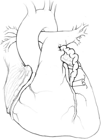
Epicardial suture ligation of the fistula.

This technique is best suited for a terminal fistula, when a large coronary artery communicates with a cardiac chamber or the pulmonary artery. The site of communication must be adequately exposed. Then, if temporary closure of the fistula with the disappearance of the underlying thrill does not show any electrocardiographic changes, then the fistula is permanently ligated or divided. This technique can be performed without cardiopulmonary bypass.
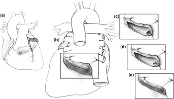
b) Transarterial (Transcoronary) closure with aneurysmorrhaphy (Fig 14)
(a) Diagram of a fistula between the circumflex artery and the right ventricle. (b) Approach to the fistula through the circumflex artery aneurysm. (c–e) Transarterial closure of the fistulous connection.

If a large aneurysm is present, it is possible to perform an aneurysmorrhaphy simultaneously with the closure of the fistula. Before establishing a cardiopulmonary bypass, the site of origin of the fistula should be confirmed by digital pressure. After the bypass is begun, care must be taken to avoid excessive runoff through the fistula. A short period of aortic cross clamping is required, during which a longitudinal incision is made in the aneurysm of the fistula. The fistula is oversewn from within the aneurysm, which is then tailored during closure of the aneurysm.
c) Transcardiac chamber closure (Intracardiac repair)
An intracardiac approach is indicated if
• an important area of the myocardium is supplied by the involved coronary artery distal to the origin of the fistula,
• external identification of the fistula is difficult, or
• multiple fistulae exist.
Delivery of cardioplegia helps in the identification of the orifice of the fistula as well as the security of the repair.
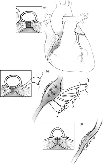
d) Tangential arterioplasty (Fig 15)
(a–c) Tangential arterioplasty as described by Cooley and Ellis in 1962.

This technique was first described by Cooley and Ellis in 1962 for lateral fistulae. This can be performed without cardiopulmonary bypass through the use of multiple horizontal mattress sutures that are passed underneath coronary artery to close the fistula.
e) Coronary artery bypass
If the flow of an important distal coronary artery, such as the posterior descending coronary artery, is compromised by the closure of the fistula, coronary artery bypass can be used in association with closure of the fistula.
3) Post-operative management:
It is important that the post-operative management of a patient with a coronary arterial fistula includes a period of anti-coagulation with warfarin for 3–6 months because of the low flow of blood in the dilated segment of the native coronary artery.
4) Surgical results:
Shuiyun et alReference Shuiyun, Qingyu and Shengshou47 described the outcome in 52 patients ranging in age from 9 months to 58 years. Of them, 16 patients had associated cardiac lesions. The site of origin of the fistula was the right coronary artery in 37 patients (71.2%), and the left coronary artery in 15 patients (28.8%). The sites of drainage of the fistula were
• the right ventricle in 22 patients (42.3%),
• right atrium in 16 patients (30.8%),
• left ventricle in six patients (11.5%),
• left atrium in three patients (5.8%), and
• pulmonary artery in five patients (9.6%).
Cardiopulmonary bypass was used in all patients with no mortality. An arteriotomy was performed on the anomalous coronary artery and the proximal opening of a fistula was closed within the vessel in 10 patients. Closure of the distal opening of a fistula draining into a cardiac chamber or pulmonary artery was performed in 26 patients. In 16 patients, both the proximal and distal openings were closed. Two and three distal openings of a fistula were found in six and three patients, respectively. No residual shunt was found before the patients were discharged from the hospital. Forty patients were followed for a mean period of 3.14 years. No clinical symptoms were found in those patients during follow-up.
b. Coronary Arcade
Coronary arcade is a rare communication between the right and the left coronary arteries, in the absence of coronary artery stenosis, which is large enough to be identified angiographically.Reference Kruskal and Hartnell48 Although the heart in the adult normally contains multiple small interconnecting vessels between the two coronary arteries, they are not usually seen at angiography. However, when these direct anastomoses are large enough to be identified angiographically, they can be differentiated from collateral vessels. The communication in coronary arcade is a prominent straight connection between the two unobstructed major arteries, often at or near the level of the crux. In contrast, collateral vessels are tortuous vascular connections between the patent vessel and the obstructed vessel.Reference Voci, Patel, Trivedi, Patel, Burris and Ruby49
c. Extracardiac Termination
Connections may exist between the coronary arteries and extracardiac vessels such as:
• bronchial arteries,
• internal mammary arteries,
• pericardial arteries,
• anterior mediastinal arteries,
• superior and inferior phrenic arteries,
• intercostal arteries, and
• the oesophageal branches of the aorta.Reference Greenberg, Fish and Spindola-Franco2
Moberg demonstrated connections between the bronchial arteries and the coronary arteries in all patients, regardless of age, and independent of the presence of atherosclerosis.Reference Moberg50 These pathways become functionally significant only when a pressure gradient exists between the two arterial systems. This condition is generally associated with atherosclerotic disease of the coronary arteries, which causes flow of blood from the bronchial artery to the coronary arteries.