A2-kilogram child had a pacemaker implanted by a sub-xyphoideal approach with the generator located under the rectus sheath. Days later, the battery eroded the abdominal wall and the peritoneum. The whole system was removed and a new one implanted inside the pericardium on an emergent basis.
Case report
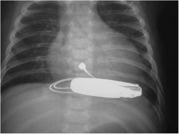
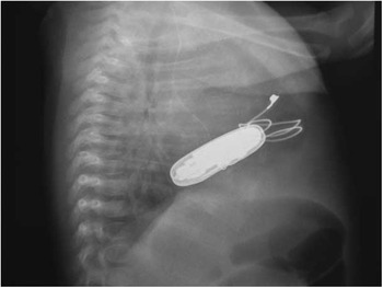
A lupic mother delivered a 2-kilogram female child with complete heart block. The low rate – 70 beats/minute on isoproterenol – gave way to congestive heart failure. Prompt surgery was scheduled and an epicardial monopolar pacemaker was implanted by a subxyphoid approach. The generator was located in an abdominal pouch developed between the rectus muscle and its posterior fascia. After 10 days, a dehiscence in the abdominal wall produced the extrusion of the generator, as well as the bowel. Emergent surgery was carried out, removing the whole system – epicardial lead plus battery, reducing the eventration and closing the abdominal wall. Thereafter, a new system was implanted with the generator placed between the heart and the diaphragm in an intrapericardial manner (Figs 1 and 2). After 1 year, the child is in good condition, with no signs of caval compression or abdominal dehiscence. This simple, bail-out procedure sorted out a dramatic problem in a 2-kilogram newborn.
X-ray, front view. Generator size slightly smaller than cardiac silhouette.

X-ray, lateral view. Bulky battery between sternum and posterior mediastinum.