Eosinophilic myocarditis is a rare, potentially lethal cardiac disease with a diversity of possible causes. Eosinophilic myocarditis was first reported 1942 as a result of sulphonamide treatment.
In general, if adequate treatment is initiated soon, the prognosis is good. However, eosinophilic myocarditis is often discovered post-mortem. Studies reported eosinophilic myocarditis in up to 0.5% of unselected autopsy series.Reference Ginsberg and Parrillo 1 According to the literature, autoimmune diseases, parasitic infections, hypersensitivity to drugs and substances, as well as vaccinations are found to be the main reasons causing eosinophilic myocarditis.Reference Ginsberg and Parrillo 1 – Reference Enko, Tada and Ohgo 5 Eosinophilic myocarditis can present clinically as an acute or chronic condition, as in the Churg–Strauss syndrome, hypereosinophilic syndrome, or eosinophilic leukaemia.Reference Spry, Take and Tai 6 The spectrum of clinical presentation in patients with eosinophilic myocarditis shows a vast variety including acute pericarditis, acute coronary syndrome, dilated cardiomyopathy, cardiogenic shock, acute heart failure, and sudden cardiac death.Reference Bilinska, Bilinska and Grzybowski 3 , Reference Galiuto, Enriquez-Sarano and Reeder 7 – Reference Sohn, Park and Chung 9 Diagnosis is exclusively proven by endomyocardial biopsy with histological detection of a typical inflammatory cell infiltration in the biopsy specimen. BrocklingtonReference Brocklington and Olsen 4 suggested three stages of eosinophilic myocarditis: acute necrotising phase, thrombotic phase, and endomyocardial fibrosis phase. The characteristic histopathology of eosinophilic myocarditis is a mixed cellular infiltrate, containing a variable amount of eosinophilic cells within the myocardium. Infiltrates vary as being either perivascular or interstitial. Apparently, there is no relationship between peripheral eosinophilic count, the extent of eosinophilic infiltrate, and clinical symptoms. In general, prednisolone therapy is used to suppress the inflammatory reaction and inotropic support; in addition, anti-failure treatment is performed including afterload reduction with angiotensin-converting enzyme inhibitors. Inconstantly, a therapy with warfarin to prevent thromboembolic events is recommended.
Case presentation
A week before admission to our hospital, the 15-year-old adolescent boy presented to outpatient clinics of various hospitals for sore throat, which was treated with ibuprofen symptomatically. He suffered from persistent chest pain, shortness of breath, and malaise 3 days before the acute presentation. On the night before admittance to our clinic, a syncope with loss of consciousness occurred.
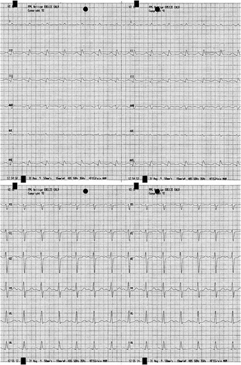
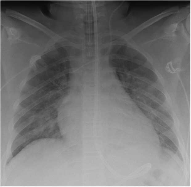
Initial blood examination revealed elevated cardiac enzyme troponin I (1.96 micrograms per litre). The electrocardiogram (see Fig 1) showed noticeable constant sinus tachycardia and unspecific repolarisation changes. The chest X-ray revealed signs of pulmonary venous congestion, as well as cardiomegaly (see Fig 2).
Electrocardiogram on admission shows constant sinus tachycardia (heart rate 150 per minute) and unspecific repolarisation changes.

Chest X-ray on admission shows a large cardiac silhouette and noticeable bilateral pulmonary oedema.

Echocardiography showed a reduced myocardial function with diffuse hypokinesia of the left ventricle and an ejection fraction of 44% (shortening fraction of 20%), as well as minimal pericardial effusion.
Owing to clinical deterioration with pulmonary oedema, the patient was admitted to our intensive care unit, intubated, ventilated, and started on catecholaminic support with dobutamine (8 micrograms per kilogram per minute) and adequate diuretic therapy.
The medical history of the patient showed a pertussis infection at the age of 1 year and a current allergy against polls. In his spare time, he played tennis, although in the week previous to admission he felt too tired to engage in any physical activity. He even felt the need to sleep at noon. Except for an uncle who suffered from pulmonary sarcoidosis, there was no family history of any chronic diseases. All vaccinations were up to date and the patient had not travelled abroad in the previous year. The other medical history was unremarkable.
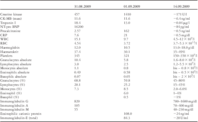
Blood examination (see Table 1) initially revealed a normal eosinophil count. There was elevated C-reactive protein (7.6 milligrams per decilitre) and highly elevated cardiac enzymes Troponin I. N-terminal pro-B-type natriuretic peptide was initially elevated at 16,200 picograms per millilitre and showed a rise to a maximum of 30,300 picograms per millilitre during follow-up.
Laboratory findings on admission (31.08.2009), 2 days after admission (01.09.2009), and 2 weeks after admission.

CK-MB = creatine kinase-MB; CRP = C-reactive protein; NT-pro BNP = N-terminal pro-B-type natriuretic peptide; RBC = red blood cells; WBC = white blood cells
Within the following days, C-reactive protein showed a marked rise to a maximum of 21 milligrams per decilitre and the eosinophil count raised to slightly elevated values (580 cubic millimetres). Eosinophilic cationic protein 108 nanograms per millilitre and Immunoglobulin E 84.3 units per millilitre were elevated during the clinical course (see Table 1).
Serology testing revealed negative results for cytomegalovirus, coxsackievirus, influenza A/B, H1N1, parainfluenza 1–3, parvovirus B19, adenovirus, picornavirus, chlamydia pneumoniae, legionella pneumophilia, toxoplasmosis, amobiasis, toxocara canis, pneumocystis jirovecii, and mycoplasma pneumoniae. Cultures of the blood or sputum and stool probes revealed no pathogenic germs or parasites.
The patient deteriorated clinically, as shown by echocardiographic findings (ejection fraction 37%, fractional shortening 16%), and the cardiac support with catecholamines was increased by epinephrine 0.1 micrograms per kilogram per minute and milrinone 0.25 micrograms per kilogram per minute. Empiric antibiotic therapy was started with cephalosporines and aminoglycosides. To confirm the presumed diagnosis of myocarditis, we performed a cardiac catheter examination with endomyocardial biopsies. Isochronical therapy with immunoglobulins was started.
The left ventricle showed severe systolic and diastolic impairment, the left ventricular end-diastolic pressure was 22 millimetres of mercury, and the Cardiac Index was 3.97 litres per minute per square metre despite the support with epinephrine 0.1 microgram per kilogram per minute, dobutamine 8 micrograms per kilogram per minute, and milrinone 0.25 microgram per kilogram per minute.
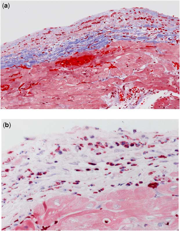
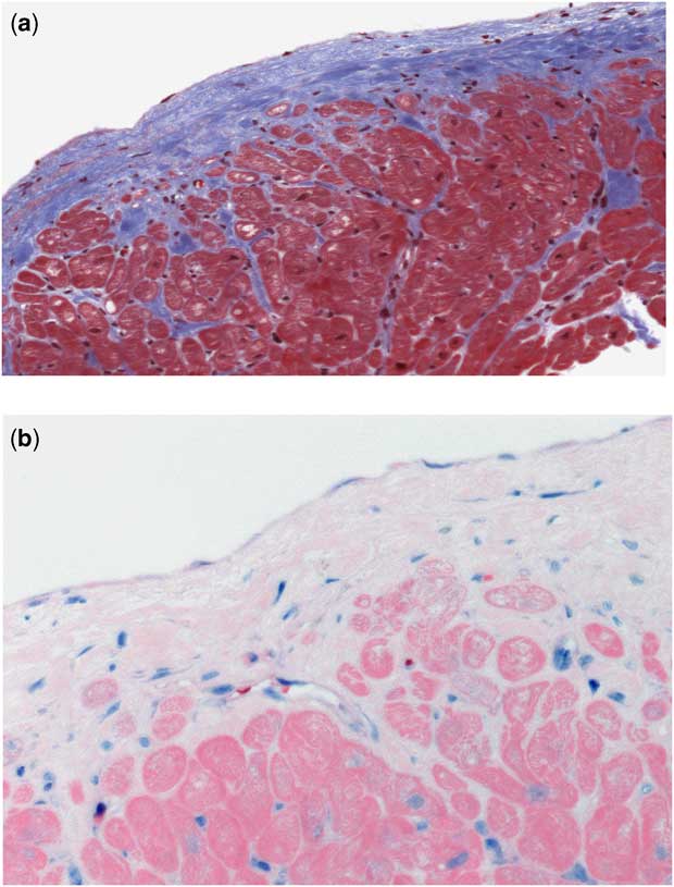
The histology of the endomyocardial biopsies showed an infiltration of the endomyocardium with eosinophil leucocytes (see Fig 3), thereby confirming the diagnosis of eosinophilic myocarditis. We started a therapy with prednisolone 5 milligrams per kilogram per day for 3 days and further reduced the dose to 1 milligram per kilogram per day.
( a and b ): The right-ventricular endomyocardial biopsy specimen showing a marked eosinophilic interstitial infiltration associated with interstitial oedema. (Upper image Masson trichrom staining, and lower image Giemsa staining) Special thanks to Professor Kandolf, Tübingen/Germany.

After initiation of the immunosuppressive therapy, there was rapid clinical improvement in the patient's condition. The patient was extubated within 2 days and catecholamine therapy was weaned during the following week. In parallel, the electrocardiogram showed normalisation of the repolarisation changes, and echocardiographic impairment to normal findings, as did troponin I and C-reactive protein within 1 week.
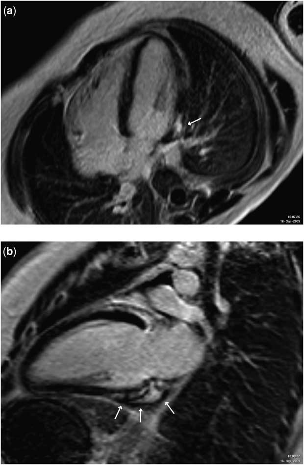
Magnetic resonance imaging examination (see Fig 4) performed 2 weeks after admission showed persistent myocardial oedema and late enhancement. Eosinophilic cationic protein was also markedly elevated.
( a and b ): Late enhancement in the basal inferolateral segment (indicated by white arrows) 3 weeks after the onset of disease and 2 weeks of corticotherapy.

Therefore, we decided to continue immunosuppressive therapy with prednisolone at a dose of 1 milligram per kilogram per day. To prevent thromboembolism, the patient was warfarinised. The patient was discharged home 3 weeks after the initial presentation. Low-dose anti-inflammatory therapy was continued at a dose of 0.5 milligram per kilogram per day prednisolone until cardiac catheter was performed 5 months after the initial presentation. Histology of the endomyocardial biopsy revealed normalisation of the myocarditis (see Fig 5).
( a and b ): Five months after prednisolone therapy. Control biopsy shows a marked decline of interstitial oedema and inflammatory cell infiltration. (Upper image Masson trichrome staining, and lower image Giemsa staining) Special thanks to Professor Kandolf, Tübingen/Germany.

The haemodynamics at the time of the second catheter investigation showed markedly improved results with slightly elevated left ventricular end-diastolic pressure only (13 millimetres of mercury). Echocardiography showed normalised systolic and diastolic function. Thereafter, prednisolone therapy was weaned and stopped 6 months after the onset of disease. Follow-up echocardiographic examination remained unremarkable and so was the clinical course of the patient.
Discussion
This is a unique case of eosinophilic myocarditis occurring in childhood, which presented with severe cardiac failure and showed several diagnostic pitfalls. In general, eosinophilic myocarditis is rarely recognised clinically, as there is no correlation between the severity of the disease and peripheral eosinophilic blood count. Similarly, in our case the diagnosis was made by endomyocardial biopsy only. Owing to the focal distribution of the inflammatory process, the diagnosis of eosinophilic myocarditis can be missed in endomyocardial biopsy.Reference Bilinska, Bilinska and Grzybowski 3 In those cases of negative testing in endomyocardial biopsy for eosinophilic myocarditis, however, with clinically suspected eosinophilic myocarditis, repeat endomyocardial biopsy has to be considered.
The accurate incidence of eosinophilic myocarditis is unknown. Case series of unselected autopsy reports showed an incidence between 0.04% and 0.5%. In explanted hearts of patients undergoing heart transplantation, the incidence (7.2–7.4%) of eosinophilic myocarditis is much higher. Our institution is a large tertiary referral centre with about 6000 outpatients, 550 heart catheter examinations, and about 500 cardiac surgeries in patients with congenital heart defects per year including a paediatric cardiac transplant and assist programme. As per institutional standard, every patient with cardiomyopathy or clinically suspected myocarditis will receive endomyocardial biopsy. This is the first case of eosinophilic myocarditis diagnosed in our institution for more than 10 years.
Clinically, hypersensitivity myocarditis is indistinguishable from myocarditis resulting from other causes. Differentiation, however, is crucial to initiate the appropriate treatment with corticosteroids, which are contraindicated in many forms of viral myocarditis.Reference Mason, O'Connell and Herskowitz 10
In our case, many clinical features were similar to that of common viral myocarditis, for example cardiac failure, elevated cardiac enzymes, and increased systemic inflammatory markers. Markers such as hypereosinophilia, cationic protein, or Immunoglobulin E level elevations, which could lead to the diagnosis of an eosinophilic myocarditis, were lacking at the onset of disease in our patient.
Patients with parasitic infections or hypereosinophilic syndromes typically have markedly elevated peripheral eosinophilia. Patients with eosinophilic myocarditis resulting from a hypersensitivity mechanism may present with normal or mildly elevated peripheral eosinophil counts. In all, 50% of the patients have no peripheral eosinophilia at the onset of disease. In those cases in whom hypereosinophilia is found, the levels are often only slightly elevated. This phenomenon is explained to be caused by the migration of peripheral blood eosinophils into the tissue, while bone marrow cannot respond immediately with increased production. In our reported case, eosinophil count was 490 cubic millimetres at admission and increased only one time slightly at day 3 after admission to 580 cubic millimetres. This fact again may demonstrate the importance of frequent and repeated white blood count examinations in patient with myocarditis and initially absent hypereosinophilia.
Unfortunately, in our patient there was no identifiable cause for eosinophilic myocarditis. Despite the variety of causes described in the literature, the causative agent for eosinophilic myocarditis often remains cryptic. Avoiding the administration of possibly triggering agents is as important as an immediate beginning with immunosuppressive therapy; the outcome seems to correlate with the initiation time of therapy.
Many case reports were published since the first reports of the positive effect of immunosuppressive therapy on the course of eosinophilic myocarditis with positive outcomes. In general, the discussion of immunosuppressive therapy for myocarditis has been controversial. The Myocarditis Treatment Trial demonstrated no improvement in the cardiac function or 5-year survival rates with steroid use, although patients with eosinophilic myocarditis were not included in the study.Reference Mason, O'Connell and Herskowitz 10
The pathological basis of myocardial damage in myocarditis is postulated to be due to toxic eosinophil granula release, autoimmune and/or type III hypersensitivity immune complex-mediated reaction. Corticosteroid therapy aims to stop toxic granula release and the subsequent immunological destruction of the myocardium.
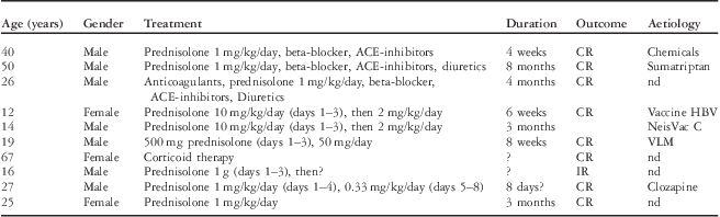
Despite many reports showing the benefit and dramatical improvements after initiation of corticotherapy, a general recommendation for dosage and duration of treatment in eosinophilic myocarditis remains unclear. The dosages reported vary from initial doses of 1 milligram per kilogram per day up to 10 milligrams per kilogram per day of prednisolone. The treatment duration ranged from 6 weeks to 8 months (see Table 2). Initially, we decided to treat our patient with 5 milligrams per kilogram per day prednisolone for 3 days and reduced the dosage to 1 milligram per kilogram per day. Therapy control in our patient was mainly managed by echocardiography and magnetic resonance imaging – 2 weeks after initiation of corticotherapy – and eosinophilic cationic protein. Owing to the fact that the high levels of eosinophilic cationic protein and the magnetic resonance imaging showed persistent myocardial inflammation activity (see Fig 4), we continued corticotherapy at the same dosage for 1 month. Reduction to 0.5 milligram per kilogram per day was necessary because of the severe side effects of steroid therapy, such as hypertension and weight gain. We performed a control endomyocardial biopsy 5 months after the onset of disease because of the initial impairment. On the basis of the potential risk of thromboembolism due to potential endocardial damage, an adjuvant anticoagulation therapy with warfarin is recommended. With the therapeutic regime outlined above, our patient recovered completely without thromboembolic complications.
Immunosuppressive therapy with prednisolone.2,5,7–9,11,12

ACE = angiotensin-converting-enzyme inhibitor; CR = complete recovery; HBV = hepatitis B virus; IR = incomplete recovery; nd = not determined; VLM = visceral larva migrans
Conclusion
We present a rare case of acute eosinophilic myocarditis in an adolescent with cardiac failure, diagnosed by endomyocardial biopsy alone. No identifiable cause was determined. The patient recovered completely and without thromboembolic complications with the use of an initial high-dose prednisolone therapy followed by a maintenance therapy with prednisolone for 5 months. In addition, anticoagulation with warfarin was performed. General recommendations for the duration and dosage of corticotherapy and adjuvant anticoagulant therapy are lacking. On the basis of the literature and our experience, the benefit of early endomyocardial biopsy and corticotherapy in eosinophilic myocarditis seems indisputable.