Introduction
The ocular complications associated with diabetes are the leading cause of new cases of blindness in adults between the ages of 20 and 74 years in the United States (Centers for Disease Control and Prevention, 2011). The characteristic diabetic pathologies of the retina, namely macular edema and neovascularization, are typically found after protracted periods, whereas electrophysiological defects of the retina associated with diabetes have been identified soon following the onset of hyperglycemia (Shirao & Kawasaki, Reference Shirao and Kawasaki1998). Thus, it has been suggested that defects in the electroretinogram (ERG) may be useful in diagnosing and predicting progression of diabetic retinopathy (Bresnick et al., Reference Bresnick, Korth, Groo and Palta1984).
Across 50 years of research in patient populations and animal models of diabetes, studies define dysfunction in the inner retina by delayed timing and amplitude of oscillatory potentials (OPs) and reductions in b-wave amplitude (Yonemura, Reference Yonemura1978; Levin et al., Reference Levin, Kwaan, Dobbie, Fetkenhour, Traisman and Kramer1982; Bresnick et al., Reference Bresnick, Korth, Groo and Palta1984; Bresnick & Palta, Reference Bresnick and Palta1987; Coupland, Reference Coupland1987; Lovasik & Spafford, Reference Lovasik and Spafford1988; Juen & Kieselbach, Reference Juen and Kieselbach1990; Hardy et al., Reference Hardy, Fisher, Heath, Foster and Scarpello1995; Shirao & Kawasaki, Reference Shirao and Kawasaki1998). Altered function of the outer retina has also been identified by changes in activation kinetics of the a-wave (Phipps et al., Reference Phipps, Yee, Fletcher and Vingrys2006). Similarly, retinal pigment epithelium (RPE) dysfunction has been observed through reduced c-wave amplitudes (Pautler & Ennis, Reference Pautler and Ennis1980; MacGregor & Matschinsky, Reference MacGregor and Matschinsky1986; Holopigian et al., Reference Holopigian, Seiple, Lorenzo and Carr1992; Phipps et al., Reference Phipps, Fletcher and Vingrys2004; Phipps & Feener, Reference Phipps and Feener2008; Wong et al., Reference Wong, Bui and Vingrys2011). These findings indicate that nearly every cell type in the outer retina is affected by diabetes (reviewed in Shirao & Kawasaki, Reference Shirao and Kawasaki1998; Tzekov & Arden, Reference Tzekov and Arden1999). Notably, the electrophysiological defects appear early and progress throughout the duration of the disease, classifying them as one of the first characteristics of diabetes identifiable by an objective in vivo measure.
Aldose reductase (AR) is the primary enzyme in the polyol metabolism pathway whereby glucose is converted to sorbitol (Hers, Reference Hers1962). Under normal blood glucose concentrations, AR has low substrate affinity for glucose compared to hexokinase. However, under hyperglycemic conditions, AR activity is elevated (Barski et al., Reference Barski, Tipparaju and Bhatnagar2008). As a byproduct of this reaction, nicotinamide adenine dinucleotide phosphate (NADPH) is converted to NADP+, reducing the availability of NADPH. Both animal studies and clinical trials have been conducted to determine the efficacy of AR inhibitors in reducing the occurrence of diabetes-related visual defects. Findings have, however, been mixed. A number of studies demonstrated positive outcomes in ERG testing as a result of pharmacological inhibition of AR (MacGregor & Matschinsky, Reference MacGregor and Matschinsky1985; Funada et al., Reference Funada, Okamoto, Fujinaga and Yamana1987), while others report no improvement in diabetes-induced alterations in retinal electrophysiology after treatment (Biersdorf et al., Reference Biersdorf, Malone, Pavan and Lowitt1988; Matsui et al., Reference Matsui, Nakamura, Ishikawa, Matsuura and Kobayashi1994; Hotta, Reference Hotta1995; Hotta et al., Reference Hotta, Koh, Sakakibara, Nakamura, Hamada, Hara, Takeuchi, Inukai, Kasama, Fukasawa and Kakuta1995a,Reference Hotta, Koh, Sakakibara, Nakamura, Hamada, Naruse, Sasaki, Mizuno, Matsubara, Kakuta, Fukasawa and Sakamotob, Reference Hotta, Koh, Sakakibara, Nakamura, Hara, Hamada, Fukasawa, Kakuta and Sakamoto1997; Ashizawa et al., Reference Ashizawa, Yoshida, Sugiyama, Akaike, Ohbayashi, Aotsuka, Abe, Fukushima and Matsuura1997).
In this study, we addressed both the impact of diabetes on the ERG and the role of AR in the pathogenesis of diabetes-induced defects in the ERG. In contrast to previous studies, which attempted to target AR by pharmacological inhibition, we directly assessed the contribution of AR by genetic inactivation of the enzyme. Streptozotocin (STZ) was utilized to induce type 1 diabetes in wildtype (WT) and AR −/−(AR KO) C57BL/6J mice. Diabetic and nondiabetic control mice were maintained for 6 months, and ERG analyses were subsequently performed to document the defects induced by diabetes and to determine if the absence of AR prevents the ERG reductions observed in diabetic animals.
Materials and methods
Animals
Male C57BL/6J WT and AR −/− mice (Ho et al., Reference Ho, Chung, Law, Ko, Tam, Brooks, Knepper and Chung2000) were randomly assigned to diabetic or nondiabetic groups, with half of each group designated for ERG analysis. Diabetes was induced by five sequential daily intraperitoneal injections of a freshly prepared solution of STZ in 0.1 m citrate buffer (pH 4.5) at 60 mg/kg body weight. Insulin was given as needed to prevent ketosis without preventing hyperglycemia and glucosuria (0–0.2 units of neutral protamine Hagedorn (NPH) insulin, subcutaneously, 0–3 times per week). Between 10 and 12 animals were assigned to each group. Survival rates varied between 83.3 and 100% (WT nondiabetic: 10/10; WT diabetic: 10/12; AR −/− nondiabetic: 10/11; AR −/−diabetic: 10/12) up to 6 months. Glycohemoglobin was measured by Bio-Rad Total Glycated Hemoglobin Assay (Bio-Rad Laboratories, Hercules, CA) every 2–3 months and just before animals were sacrificed. Food consumption and body weight were measured weekly. Treatment of animals was in compliance with the ARVO Resolution on Treatment of Animals in Research, as well as institutional guidelines. Animals were maintained for 6 months in order to investigate the long-term effects on electrophysiological changes associated with diabetes.
ERG testing
After overnight dark adaptation, mice were anesthetized (ketamine, 80 mg/kg; xylazine, 16 mg/kg), the cornea was anesthetized (1% proparacaine HCl), and the pupils were dilated (1% tropicamide, 2.5% phenylephrine HCl, 1% cyclopentolate). Mice were placed on a temperature-regulated heating pad throughout each recording session. The protocols used to record ERG components generated by the outer neural retina or the RPE have been previously described (Samuels et al., Reference Samuels, Sturgill, Grossman, Rayborn, Hollyfield and Peachey2010). In brief, responses of the outer retina were recorded with a stainless steel electrode referenced to a needle electrode placed in the cheek in response to strobe flash stimuli (ranging from −3.6 to 2.1 log cd s/m2) presented in the dark. Photopic stimuli (−0.8 to 1.9 log cd s/m2) were superimposed on a steady rod-desensitizing adapting field (20 cd/m2) after 7 min of light adaptation. Responses were differentially amplified (0.3–1500 Hz), averaged, and stored using a UTAS E-3000 signal averaging system (LKC Technologies, Gaithersburg, MD). The amplitude of the a-wave was measured 8 ms after flash onset from the prestimulus baseline. The amplitude of the b-wave was measured from the a-wave trough to the peak of the b-wave or, if no a-wave was present, from the prestimulus baseline. OPs were filtered from the averaged responses at 4.3 cds/m2, and peak amplitudes and latencies were measured as previously described (Hancock & Kraft, Reference Hancock and Kraft2004).
Components of the dc-ERG, generated by the RPE, were recorded with an Ag/AgCl electrode bridged to the corneal surface with Hanks’ balanced salt solution in response to a single light stimulus (2.4 log cd/m2) presented for 7 min. Responses were differentially amplified at dc-100 Hz; gain = 1000× (DP-301; Warner Instruments, Hamden, CT), digitized at 20 Hz, and stored using LabScribe Data Recording Software (iWorx, Dover, NH). Approximately 10 min of recording included a 30-s baseline, the four light-evoked waveform components, and 2 min of poststimulus recording time (see Fig. 2A). The amplitude of the c-wave was measured from the prestimulus baseline to the peak of the c-wave. The amplitude of the fast oscillation (FO) was measured from the c-wave peak to the trough of the FO. The amplitude of the light peak (LP) was measured from the FO trough to the asymptotic value. The amplitude of the off response was measured from the LP asymptote to the peak of the off response.
Results
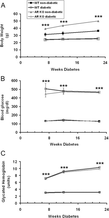
All nondiabetic mice had blood glucose levels below 150 mg/dl (Fig. 1B). STZ-induced diabetes affected WT and AR −/− mice similarly, both in blood glucose and glycated hemoglobin levels. Diabetic mice were hyperglycemic for the duration of the 6-month experiments and displayed significantly higher blood glucose and glycated hemoglobin levels at all time points (Fig. 1B and 1C). Both genotype and diabetes status had a significant effect on body weight. We found that nondiabetic AR −/− mice were significantly larger than WT mice throughout the duration of the experiment (Fig. 1A), and both groups of diabetic mice weighed significantly less than their nondiabetic counterparts (Fig. 1A). However, neither group lost weight during the course of the experiment.
Loss of AR does not significantly alter glycemia. Body weight (A), blood glucose (B), and glycated A1c hemoglobin (C) were measured in each group of mice at 7, 14, and 22 weeks following treatment (nondiabetic = citrate buffer; diabetic = STZ). For each group, n ≥ 4. Statistical analysis was performed with repeated measures ANOVA and post hoc Bonferroni test. ***P ≤ 0.0001.

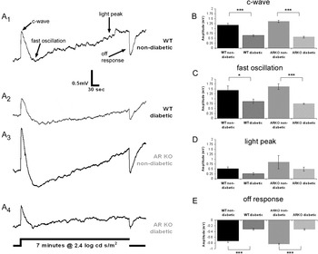
After 22 weeks of diabetes, ERG studies were conducted. In WT nondiabetic mice, the 7-min light stimulus evoked all four major components of the dc-ERG (Fig. 2A1), each of which was reduced in amplitude in WT diabetic mice (Fig. 2A2). AR −/− nondiabetic mice (Fig. 2A3) generated consistently larger dc-ERGs as compared to WT nondiabetic mice. Similar to WT diabetic mice, AR −/− diabetic mice also displayed reductions in each dc-ERG waveform (Fig. 2A4). Although AR −/− nondiabetic responses were larger, we did not find a significant difference in amplitude for any component when comparing WT and AR −/− nondiabetics. Importantly, there were no significant differences between WT diabetics and AR −/− diabetics. The amplitude of the c-wave was significantly decreased in WT diabetic as compared to WT nondiabetic mice (P < 0.001) and in AR −/− diabetic as compared to AR −/− nondiabetic mice (Fig. 2B; P < 0.001). We similarly found significant amplitude reductions in the FO and off response from WT diabetic compared to the WT nondiabetic mice (Fig. 2C and 2E). These amplitude reductions were also seen in AR −/− diabetic as compared to AR −/− nondiabetic animals. Our recordings revealed a general trend for reductions in the LP amplitude of the diabetic groups as compared to the nondiabetics; however, these reductions were not statistically significant (Fig. 2D).
Loss of AR does not inhibit diabetes-induced reductions in components of the dc-ERG. (A) Representative dc-ERG tracings for each group recorded in response to a 7-min light stimulus. (B–E) Amplitude of the major components of the dc-ERG. For each group, n ≥ 4. Statistical analysis was performed with one-way ANOVA and post hoc Tukey’s test. *P ≤ 0.01, ***P ≤ 0.0001.

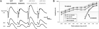
Strobe flash ERGs were recorded to determine how the neural retina is affected by loss of AR and to ensure that reductions in the dc-ERG waveforms are not due to reduced photoreceptor activity (Samuels et al., Reference Samuels, Sturgill, Grossman, Rayborn, Hollyfield and Peachey2010). As seen in Fig. 3A, characteristic strobe flash ERGs were generated by mice of all genotypes and diabetic states. The amplitude of the a-wave was not significantly affected by either diabetic status or genotype (Fig. 3B). In comparison, b-waves of both WT and AR −/− diabetic mice were smaller in amplitude as compared to their nondiabetic controls (Fig. 3B). Nondiabetic AR −/− mice generated slightly greater ERG responses as compared to WT nondiabetics; however, responses from AR −/− diabetic mice were reduced to a similar extent as WT diabetics. Interestingly, we did not observe a significant change in b-wave latency [data not shown, P = 0.13 by one-way analysis of variance (ANOVA)]. These data are consistent with previous findings demonstrating reductions in b-wave but not a-wave parameters (Phipps et al., Reference Phipps, Yee, Fletcher and Vingrys2006).
Loss of AR does not alter the differences in strobe flash ERG responses between normal and diabetic mice. (A) Averaged strobe flash ERG responses recorded at increasing light intensities for each group. (B) Luminance response function of a- and b-wave amplitude. For each group, n ≥ 4. Statistical analysis was performed using a one-way ANOVA followed by post hoc Tukey’s test. *P ≤ 0.01, **P ≤ 0.001.

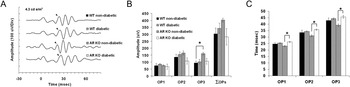
We digitally filtered OPs from the strobe flash ERGs and assessed peak amplitude and latency for each OP wavelet at 4.3 cd s/m2 (Fig. 4A). Reductions in OP amplitude and prolonged latencies are the most common ERG anomaly found as a result of diabetes (Yonemura et al., Reference Yonemura, Aoki and Tsuzuki1962; Hancock & Kraft, Reference Hancock and Kraft2004; Phipps et al., Reference Phipps, Fletcher and Vingrys2004) and have been proposed to be predictive of progression to retinopathy in humans (Bresnick et al., 1984). However, most animal studies have focused on rat models of diabetes, and only a few reports have found any significant differences in these parameters in diabetic mice (Barile et al., Reference Barile, Rachydaki, Tari, Lee, Donmoyer, Ma, Rong, Buciarelli, Wendt, Horig, Hudson, Qu, Weinberg, Yan and Schmidt2005; Zheng et al., Reference Zheng, Du, Miller, Gubitosi-Klug, Kern, Ball and Berkowitz2007). In our analysis, we did not observe significant differences in either peak latency or amplitude between WT diabetic or AR −/− diabetic mice and their corresponding control. The only significant difference identified between any group was found in the AR −/− nondiabetic as compared to its diabetic counterpart. Specifically, the AR −/− wavelets peaked faster (Fig. 4C) and were of greater amplitude (OP2) than the AR −/− diabetic (Fig. 4B). This finding matches the larger amplitudes found in both the dc-ERG components and the b-wave amplitude. These findings indicate that in this model of diabetes, the OP generators retain normal function at this time point.
Diabetes does not significantly affect OP wavelets of WT or AR KO mice at 22 weeks. (A) Representative OP wavelets in response to a 4.3 cd s/m2 strobe flash. The peak of the first wavelet is denoted by a closed arrow. (B) Peak amplitude of each wavelet and the summed amplitudes (ΣOP). (C) Latency of each wavelet. For each group, n ≥ 4. *P ≤ 0.05.

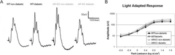
We also recorded the cone ERG. Despite a slightly greater amplitude observed in the AR −/− nondiabetic group, cone-dominant light-adapted ERG responses were not significantly different between groups (Fig. 5), indicating that diabetes did not affect cone function at this time point. Cone responses have been shown to exhibit reductions in hyperglycemic zebrafish (Alvarez et al., Reference Alvarez, Chen, Reynolds, Waghorne, O’Connor and Kennedy2010); however, no single animal model of type 1 diabetes recapitulates all aspects of the disorder identified in patients, and we do not observe significant defects in either rod or cone function within this model of type 1 diabetes.
Loss of AR does not alter differences in light-adapted ERG responses between normal and diabetic mice. (A) Representative ERG tracings recorded in response to a 1.9 log cds/m2 strobe flash superimposed on a steady rod-desensitizing (20 cd/m2) adaptation field. (B) Intensity response function for each group (for each group, n ≥ 4).

Discussion
This study presents two novel findings. It is the first comprehensive investigation of in vivo RPE electrophysiology in an animal model of diabetes. Our work demonstrates that after 6 months of diabetes, mice exhibit profound defects in outer retinal function manifested by reductions in most components of the dc-ERG. We also confirm reports of disrupted inner retina processing evidenced by reductions in b-wave amplitude. In addition, our findings indicate that AR does not play a key role in the mechanisms underlying these diabetes-induced retinal electrophysiology abnormalities. Diabetic mice lacking AR had decreases in bipolar cell and RPE function comparable to those in WT diabetic mice. In fact, control AR −/− mice generally displayed larger amplitude ERG waveforms than WT mice, yet ERG waveform amplitudes in diabetic AR −/−mice were reduced to similar levels as WT diabetics. As no change in a-wave amplitude or latency was found between any groups, this data set suggests that defects observed in the dc-ERG components of diabetic mice (RPE function) are not secondary to photoreceptor dysfunction.
Reductions in c-wave amplitude have been observed in diabetic rats as early as 2 weeks following the onset of diabetes (Pautler & Ennis, Reference Pautler and Ennis1980; MacGregor & Matschinsky, Reference MacGregor and Matschinsky1985). The later dc-ERG components had not previously been measured in diabetic animals. Importantly, the amplitudes of the FO, dark peak voltage and LP voltage of the electrooculogram (EOG), the clinical correlate of the dc-ERG, are all known to be reduced in diabetic patients as compared to controls (Schneck et al., Reference Schneck, Shupenko and Adams2008), and these parameters are affected independent of retinopathy status. Schneck et al. (Reference Schneck, Fortune and Adams2000, Reference Schneck, Shupenko and Adams2008) also reported that glucose severely impacts the FO component of the EOG in both diabetic and normoglycemic individuals. The EOG demonstrates a change in potential across the eye in response to light, and much like the dc-ERG, it measures the functional status of the RPE-photoreceptor complex in humans (Arden & Constable, Reference Arden and Constable2006).
A preponderance of data exists to support the idea that a structural insult to the RPE as a result of hyperglycemia may underlie alterations observed in the ERG of diabetic mice (reviewed in Simo & Hernandez, Reference Simo and Hernandez2009; Xu & Le, Reference Xu and Le2011). The RPE is a polarized monolayer that acts to separate the fenestrated choriocapillaris from the neural retina. It maintains this barrier function by the formation of tight junctions and adherent junctions, both of which can be altered in diabetes (Vinores et al., Reference Vinores, Derevjanik, Ozaki, Okamoto and Campochiaro1999; Wang et al., Reference Wang, Xu, Elliott, Zhu and Le2010; Xu & Le, Reference Xu and Le2011; Xu et al., Reference Xu, Song, Fu, Zhu and Le2011). Studies demonstrating the overexpression of tight junction proteins, increased transepithelial resistance (TER), and decreased dextran diffusion across the RPE of animal models of diabetes suggest that the RPE may employ a buffering mechanism to combat diabetic hyperglycemia (Villarroel et al., Reference Villarroel, Garcia-Ramirez, Corraliza, Hernandez and Simo2009a,Reference Villarroel, Garcia-Ramirez, Corraliza, Hernandez and Simob). Thus, stress to the RPE and its structural integrity in response to diabetes might underlie our observed reductions in the dc-ERG. Experiments using in vitro cell culture systems reveal that high glucose, chronic oxidative stress, and cytokine treatment each lead to decreases in the TER and reductions in tight junction proteins (Xu et al., Reference Xu, Song, Fu, Zhu and Le2011).
While establishment of a membrane potential and maintenance of the outer blood–retinal barrier is a key component of RPE function, another critical role of the RPE is to buffer the ion composition of the subretinal space, a requirement for proper photoreceptor function (Steinberg, Reference Steinberg1985; Strauss, Reference Strauss2005). Each dc-ERG component is generated by the movement of ions across the polarized epithelium, as a secondary response to photoreceptor activity (Wu et al., Reference Wu, Peachey and Marmorstein2004b). The c-wave is generated by hyperpolarization of the apical membrane (Steinberg et al., Reference Steinberg, Schmidt and Brown1970; Schmidt & Steinberg, Reference Schmidt and Steinberg1971), which occurs in response to the decrease in subretinal [K+] resulting from photoreceptor activation by light (Steinberg, Reference Steinberg1985; Wu et al., Reference Wu, Marmorstein, Kofuji and Peachey2004a,Reference Wu, Peachey and Marmorsteinb) and a negative polarity slow PIII [resulting from Kir4.1 activity in Muller glia (Steinberg & Miller, Reference Steinberg and Miller1973; Witkovsky et al., Reference Witkovsky, Dudek and Ripps1975; Oakley & Green, Reference Oakley and Green1976; Kofuji et al., Reference Kofuji, Ceelen, Zahs, Surbeck, Lester and Newman2000)]. The FO is generated by a restoration in subretinal [K+] and the delayed hyperpolarization of the basal RPE membrane mediated by a [Cl−] conductance (Linsenmeier & Steinberg, Reference Linsenmeier and Steinberg1982; Griff & Steinberg, Reference Griff and Steinberg1984). The LP reflects the depolarization of the RPE basal membrane by a [Cl−]-based conductance (Linsenmeier & Steinberg, Reference Linsenmeier and Steinberg1982; Gallemore & Steinberg, Reference Gallemore and Steinberg1989, Reference Gallemore and Steinberg1993; Fujii et al., Reference Fujii, Gallemore, Hughes and Steinberg1992). The off response is generated at light offset by an unknown mechanism, which, in the mouse, changes polarity in a luminance-dependent fashion (Wu et al., Reference Wu, Peachey and Marmorstein2004b; Samuels et al., Reference Samuels, Sturgill, Grossman, Rayborn, Hollyfield and Peachey2010). These findings imply that alterations in expression, localization, and/or activity of a variety of ion channels can underlie the diabetes-induced RPE dysfunction observed here. While specific ion conductances definitively altered by diabetes and underlying the reductions in the dc-ERG have not yet been elucidated, as noted above, Schneck et al. (Reference Schneck, Fortune and Adams2000) have demonstrated that increased blood glucose levels strongly affected FO amplitude of patient EOGs, which is generated by photoreceptor-driven changes in RPE cell [Cl−] concentration.
Structural and/or functional damage to the RPE likely factors into our findings of reductions in dc-ERG components. It is likely that other metabolic abnormalities may also contribute. Alterations in timing and amplitude of the b-wave and OPs associated with diabetes have been shown to be reversible by pharmacological treatments including docosahexaenoic acid (DHA)/lutein (Arnal et al., Reference Arnal, Miranda, Johnsen-Soriano, Alvarez-Nolting, Diaz-Llopis, Araiz, Cervera, Bosch-Morell and Romero2009; Yee et al., Reference Yee, Weymouth, Fletcher and Vingrys2010) and acetyl-l-carnitine (Lowitt et al., Reference Lowitt, Malone, Salem, Kozak and Orfalian1993). Other treatments such as lipoic acid (Johnsen-Soriano et al., Reference Johnsen-Soriano, Garcia-Pous, Arnal, Sancho-Tello, Garcia-Delpech, Miranda, Bosch-Morell, Diaz-Llopis, Navea and Romero2008), topical ciliary neurotrophic factor (CNTF) (Aizu et al., Reference Aizu, Katayama, Takahama, Hu, Nakagawa and Oyanagi2003), propionyl-l-carnitine (Hotta et al., Reference Hotta, Koh, Sakakibara, Nakamura, Hamada, Hara, Fukasawa, Kakuta and Sakamoto1996), angiotensin-converting-enzyme (ACE) inhibition (Bui et al., Reference Bui, Armitage, Tolcos, Cooper and Vingrys2003), protein kinase C (PKC) inhibitors (Nakamura et al., Reference Nakamura, Kato, Hamada, Nakayama, Chaya, Nakashima, Naruse, Kasuya, Mizubayashi, Miwa, Yasuda, Kamiya, Ienaga, Sakakibara, Koh and Hotta1999), and induction of heat shock proteins (Biro et al., Reference Biro, Palhalmi, Toth, Kukorelli and Juhasz1998; Nanasi & Jednakovits, Reference Nanasi and Jednakovits2001) have each also been shown to prevent delays and reductions in the OPs but not the b-wave. Comparison of results obtained using pharmacological therapies to those obtained using genetic deletion of the same enzyme likely will prove informative to identify the mechanisms by which therapies do and do not act.
With regard to AR, it was previously reported that administration of the AR inhibitor, sorbinil, or dietary supplementation with myo-inositol arrested the progressive deterioration of the c-wave in diabetes; however, these treatments did not prevent the initial c-wave defect (Pautler & Ennis, Reference Pautler and Ennis1980; MacGregor & Matschinsky, Reference MacGregor and Matschinsky1985; MacGregor et al., Reference MacGregor, Rosecan, Laties and Matschinsky1986). These results suggest that two distinct insults may occur; an early reduction in c-wave amplitude, followed by a further decline if intervention is not taken. The reports evaluated the ERG at early time points following the onset of diabetes (2–19 weeks) in rat and rabbit models of diabetes. In comparison, our study assessed the mouse ERG at a single time point, 6 months following the onset of diabetes. Notably, the c-wave reduction noted here is comparable to the initial defect observed in the previous reports (Pautler & Ennis, Reference Pautler and Ennis1980; MacGregor & Matschinsky, Reference MacGregor and Matschinsky1985; MacGregor et al., Reference MacGregor, Rosecan, Laties and Matschinsky1986) and the later decline is absent. This disparity may reflect differences between experimental protocols (species, method of diabetes induction). An alternate possibility is that the secondary reduction in c-wave amplitude in diabetic rats is due in part to formation of anterior cataracts (Pautler & Ennis, Reference Pautler and Ennis1980), which is greatly lessened in diabetic mice.
A major clinical interest in ERG stems from the possibility that changes in retinal function might predict susceptibility to diabetic retinopathy. That remains an exciting area of clinical and translational research. We have shown here that dc-ERG defects are easily identified in diabetic mice, and current studies will define the time course over which these defects developed. We have also shown that AR does not appear to contribute to the ERG changes associated with diabetes. Our results, coupled with previous studies that demonstrated positive effects of AR inhibitors on retinal function, raise a possibility that these benefits might have been mediated by off-target effects of the drugs.
Acknowledgments
We would like to thank Gwen M. Sturgill-Short for technical assistance and reading of the manuscript. This study was supported by the Department of Veterans Affairs CY10-035 (I.S.S.), Foundation Fighting Blindness, Research to Prevent Blindness, National Institute of Heath grants R01 EY005856 (J.M.P.), and R01 EY00300 (T.S.K.).