INTRODUCTION
Study cases of abnormalities in elasmobranchs are mostly focused on partial or complete albinism and leucism (e.g. Joseph, Reference Joseph1961; Nakaya, Reference Nakaya1973; Teixeira & Araújo, Reference Teixeira and Araújo2002; Ben Souissi et al., Reference Ben Souissi, Golani, Merji, Ben Salem and Capapé2007; Ball et al., Reference Ball, Jones, Lynghammar, Noble and Griffiths2013) and on morphological body deformities related, for instance, to incomplete fin-head fusion in batoids (skates and rays) and skeletal abnormalities of the vertebral column, recorded since 1642 (Driggers et al., Reference Driggers, Hannan, Hoffmayer and Jensen2012). Most published information on prenatal abnormalities of elasmobranchs were produced during the last 30 years and, there are a few studies if compared to land vertebrates, probably due to the relative unavailability of specimens of sharks and rays until recent times, the nature of these abnormalities, resulting in prenatal death, and, in a general view, the remarkable increase of interest to investigate other biological and anatomical parameters.
Several elasmobranch taxa were reported exhibiting some skeletal anomaly during embryonic development (Ford, Reference Ford1930; Gopalan, Reference Gopalan1971; Goto et al., Reference Goto, Taniuchi, Kuga and Iwata1981; Heupel et al., Reference Heupel, Simpfendorfer and Bennett1999; El Kamel et al., Reference El Kamel, Mnasri, Boumaïza and Capapé2009; Delpiani et al., Reference Delpiani, Antoni, Barbini and Figueroa2011; Galván-Magaña et al., Reference Galván-Magaña, Escobar-Sánchez and Carrera-Fernández2011; Hevia-Hormanzábal et al., Reference Hevia-Hormazábal, Pastén-Marambio and Alonso2011; Mejia-Falla et al., Reference Mejía-Falla, Navia and Muñoz2011; Driggers et al., Reference Driggers, Hannan, Hoffmayer and Jensen2012; Guida et al., Reference Guida, Walker and Reina2013; Muñoz-Osorio et al., Reference Muñoz-Osorio, Mejía-Falla and Navia2013; Wagner et al., Reference Wagner, Rice and Pease2013). Brazilian studies on elasmobranch prenatal body abnormalities are restricted to some case reports based on few specimens (Gomes et al., Reference Gomes, Santos and Medina1991; Rosa et al., Reference Rosa, Gomes and Gadig1996; Teixeira & Araújo, 2002; Mancini et al., Reference Mancini, Casas and Amorim2006; Bornatowski & Abilhoa, Reference Bornatowski and Abilhoa2008). The most comprehensive Brazilian data available were provided by Ribeiro-Prado et al. (Reference Ribeiro-Prado, Oddone, Gonzalez, Amorim and Capapé2008) describing different anomalies in six batoid species from southern Brazil.
The sharpnose shark genus Rhizoprionodon Whitley, Reference Whitley1929, is one of the most abundant small coastal predatory sharks in the tropical to temperate areas around the world, having at least seven known species (Ebert et al., Reference Ebert, Fowler and Compagno2013). On the Brazilian coast, the two recorded species, Rhizoprionodon porosus (Poey, Reference Poey1861) and Rhizoprionodon lalandii (Müller & Henle, Reference Müller and Henle1839) are an important elasmobranch resource caught by the small to medium scale fisheries along the coastline (Motta et al., Reference Motta, Gadig, Braga and Namora2005). Despite several studies concerning the biology and population parameters of these species in Brazil (e.g. Ferreira, Reference Ferreira1988; Lessa, Reference Lessa1988; Motta et al., Reference Motta, Gadig, Braga and Namora2005, Reference Motta, Namora, Gadig and Braga2007; Andrade et al., Reference Andrade, Silva-Júnior and Vianna2008), no previous published data reports any chromatic or morphological abnormality and, even in global scale, there is only one published paper describing anomaly in a sharpnose shark Rhizoprionodon acutus (Rüppell, Reference Rüppell1837) from India (Gopalan, Reference Gopalan1971). Thus, the present paper reports the second worldwide record of abnormalities in this genus, based on two abnormal conjoined embryos of Caribbean sharpnose shark, R. porosus and vertebral deformities in three embryos of the Brazilian sharpnose shark, R. lalandii, both commercially and ecologically important small shark species of the Brazilian coast, western South Atlantic, with a discussion on the possible relationship of the major human threats to these anomalies.
MATERIALS AND METHODS
The two conjoined embryos of Rhizoprionodon porosus were removed from a pregnant female caught by artisanal fisheries using monofilament gillnets at Ilhéus, south Bahia State, north-east Brazil (~14°45′S–38°59′W) in May 2000. The three R. lalandii were from two pregnant females, caught by the small coastal gillnets in Praia Grande, São Paulo State, south-east Brazil (~24°01′S–46°26′W), one from a female in 2007 and two from another in 2009. For both species, only the abnormal embryos were donated by the fishermen to researchers. After the species identification, according to Figueiredo (Reference Figueiredo1977), the specimens' total length (TL in mm) was measured, preservation was in 10% formalin and 70% alcohol, they were submitted to digital photographs, examined under light stereomicroscope (200 ×), radiographed in ventral and lateral views, eviscerated to gross visualization of the pericardial and abdominal organs (R. porosus), and deposited at the UNESP Elasmobranch Collection (R. porosus—UNESP/CLP 0025; R. lalandii—UNESP/CLP 0026.1, 0026.2 and 0026.3).
RESULTS
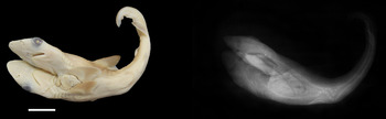
The conjoined Rhizoprionodon porosus were mid-term female embryos (Figure 1A), measuring 182 mm TL, and presented general aspect agreeing to an embryo of this species at this developmental stage, with dermal denticles completely formed along the entire body surface. External morphology showed the bifurcation occurring at the pectoral fin origins, resulting in two complete heads (both had well developed eyes, a nasal aperture pair, similar distributional patterns of the ampulary system pores and five pairs of gill slits), two pectoral fin pairs, two first and second dorsal fins, all structures similarly developed in both embryos, except the upper caudal fin lobe, which the left one was less developed than the right. Ventrally, the duplication reached the cloacal region, with one genital aperture, one female pelvic fin pair, one anal fin (distinguished from the second dorsal by its proportional dimensions and presence of the Rhizoprionodon diagnostic pre-anal dermal ridges) and one lower caudal lobe.
External morphology and radiography of the Rhizoprionodon porosus conjoined twin embryos from Ilhéus, Bahia, north-east Brazil; white scale bar = 2 cm.

The skeletal structures associated with cephalic region, pectoral and dorsal fins duplications (chondrocranium, gill arches, pectoral girdles, first and second dorsal fins skeleton) were formed and also duplicated. The vertebral column was entirely duplicated (Figure 1), reaching the tip of the caudal fin. The number of precaudal vertebrae was the same for both axes (=69). At the pelvic girdle and cloacal area, both presented scoliosis (Figure 1). In the right axis the curvature was located slightly anterior to those in the left axis, it was ‘U’ shaped and involved at least eight vertebrae. In the left axis the scoliosis was ‘V’ shaped, with up to six vertebrae (respectively below and above in the Figure 1). The vertebral centra, except at the curvature zone, presented normal aspect, with concave ends and separated by the symmetrical intervertebral discs.
A visceral gross analysis showed duplication in all main organs. Each pericardial cavity had one heart, more developed in the left embryo. Abdominal cavity with bilobed livers and complete digestive tracts, but, unlike the pericardial cavity, it was more developed in the right embryo. Concerning the reproductive apparatus, both were females. The right embryo had the left oviduct less developed and the left embryo had the right oviduct smaller, resulting in a pattern where the two smaller oviducts were positioned along the join line between the two abdominal cavities.
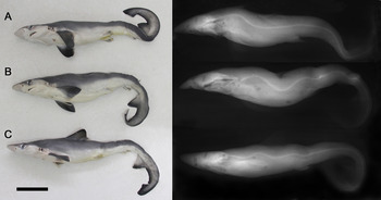
The three Rhizoprionodon lalandii were near-term male embryos, between 273 and 276 mm TL (Figure 2), all presenting kyphosis and lordosis. Despite the column deformities, which affected the general body aspect, no additional external abnormality was detected in the head and fins morphology. One specimen (Figure 2A) developed a kyphosis at the pectoral fin level, and a less accentuated lordosis above the vertebral abdominal area below to the first dorsal fin. Vertebral axis at the caudal peduncle suffered one-turn column twist, rolling down to the right side, with helicoidal aspect. The total precaudal vertebrae number was 83. The specimen B in the same figure had a small lordosis, followed by a kyphosis at the level of the pectoral fins which continued to form a deep lordosis below the first dorsal fin, resulting in an external deep concavity on the shark back. Posterior to that region there was a sequence of slight kyphosis and lordosis and a half-twist at pre-caudal point. In the specimen C, from the same pregnant female of B, the general pattern was similar to its relative, but had less angular curvatures at the axis, and the post-pelvic area exhibited a long curvature initiated at the second dorsal fin level, forcing down its caudal fin. For all R. lalandii specimens, the external dorsal surface reflected those deformities by humpbacks (kyphosis) and depressions (lordosis). The number of precaudal vertebrae was 86 in both B and C specimens.
External morphology and radiography of the three abnormal embryos of Rhizoprionodon lalandii from São Paulo coast, south-east Brazil; black scale bar = 5 cm.

DISCUSSION
Most data on conjoined elasmobranch embryos describe dicephalic twins, and not all descriptive studies focused on the exact degree of axial bifurcation (Ford, Reference Ford1930; Goto et al., Reference Goto, Taniuchi, Kuga and Iwata1981; Bejarano-Álvarez et al., Reference Bejarano-Álvarez, Galván-Maganã and Ocho-Baez2011; Delpiani et al., Reference Delpiani, Antoni, Barbini and Figueroa2011; Wagner et al., Reference Wagner, Rice and Pease2013). The general aspect of the abnormal Rhizoprionodon porosus embryos here studied resembles the dicephalic Galeorhinus galeus (Linnaeus, Reference Linnaeus1758) analysed by Delpiani et al. (Reference Delpiani, Antoni, Barbini and Figueroa2011), having two completely separated heads, duplication of the vertebral column and both dorsal and caudal fins, a single pelvic fin pair, anal fin, and ventral caudal lobe, but differs mainly by the two pairs of pectoral fins and a more posterior fusion of the trunk of R. porosus. Unfortunately these authors did not provide a pericardial and abdominal description for comparative purposes.
Despite the abnormalities, the embryos of two Brazilian Rhizoprionodon species show general external morphology accordingly with the embryonic developmental stages found for those species for the same geographical areas where the pregnant females were captured. Size at birth of R. porosus from north-east Brazil was estimated about 330–370 mm TL (Mattos et al., Reference Mattos, Broadhurst, Hazin and Jonnes2001), corroborating the mid-term developmental stage for the specimen here studied (182 mm TL). Likewise, the specimens of R. lalandii here studied are regarded as near term, since, the newborns from southern Brazil have about 300–320 mm TL (Motta et al., Reference Motta, Namora, Gadig and Braga2007).
The vertebral counts also fit with previous observations for both species (Springer, Reference Springer1964). In several cases, the vertebral number verified in abnormal individuals is according to that found in healthy ones of the same species (Heupel et al., Reference Heupel, Simpfendorfer and Bennett1999; Delpiani et al., Reference Delpiani, Antoni, Barbini and Figueroa2011), as herein noted for Rhizoprionodon. However, some alterations of the external morphology of embryos recorded in some species reflect differences in the skeleton, as observed by Wagner et al. (Reference Wagner, Rice and Pease2013) in a dicephalic Carcharhinus leucas (Müller & Henle, Reference Müller and Henle1839) having two very enlarged heads and a less developed posterior trunk. These authors found vertebral counts of 88–89 versus the reported range of 208–227 in normal specimens (Garrick, Reference Garrick1982). Delpiani et al. (Reference Delpiani, Antoni, Barbini and Figueroa2011) also reported smaller vertebral counts in anomalous Galeorhinus galeus.
The conjoined twins of Rhizoprionodon porosus are parapagus dicephalus, a type of anomaly reported for several vertebrate taxa, from fish to mammals (Kamiya et al., Reference Kamiya, Miyazaki and Shiraga1981; Spencer, Reference Spencer2000; Kaufman, Reference Kaufman2004; Kompaje, Reference Kompanje2005; Galván-Magaña et al., Reference Galván-Magaña, Escobar-Sánchez and Carrera-Fernández2011; Petrescu-Mag et al., Reference Petrescu-Mag, Păsărin, Hoha, Hărşan and And Odagiu2011). In such deformity, the embryos have two heads with separate braincases, they are attached laterally, with the posterior region fused in different degrees, and present the same gender (Spencer, Reference Spencer2000), as observed in the embryos of this study, both females. This malformation occurs in early embryonic development, but there is much controversy whether it is the result of fusion of two embryonic discs (Spencer, Reference Spencer2000) or fission of a single disc (Kaufman, Reference Kaufman2004). Although the genetic and chemical bases of this alteration are not yet known, hypotheses about exposure to substances and genetic factors have been raised (Kaufman, Reference Kaufman2004; Petrescu-Mag, Reference Petrescu-Mag, Păsărin, Hoha, Hărşan and And Odagiu2011).
The post-natal survival rate in anomalous embryos is expected to be very low for deformities involving the axis bifurcation, and the only reported exception, to date, refers to one free swimming dicephalic cownose ray, Rhinoptera steindachneri Evermann & Jenkins, Reference Evermann and Jenkins1891, caught alive in the Gulf of Mexico (Castro-Aguirre & Torres-Villegas, Reference Castro-Aguirre and Torres-Villegas1979). In reference to anomalies on the vertebral column of non-bifurcated embryos, as herein studied for Rhizoprionodon lalandii, survival of newborns has been recorded for some species in the wild (Ford, Reference Ford1930; Hoenig & Walsh, Reference Hoenig and Walsh1983; Heupel et al., Reference Heupel, Simpfendorfer and Bennett1999) and for an impressive number (~35%) of captive grey nurse shark, Carcharias taurus (Rafinesque, Reference Rafinesque1810) (Anderson et al., Reference Anderson, Huber and Berzins2012; Tate et al., Reference Tate, Anderson, Huber and Berzins2013). In these cases, the pre-pelvic curvatures of the axis (kyphosis/lordosis/scoliosis), although severe, seem to be less harmful for swimming purposes than helicoidal twists of the axis at post-pelvic area in the caudal peduncle. The caudal twist found in R. lalandii, a small coastal species, would prevent the animal from surviving long after birth, due to its compromised swimming capabilities for gill ventilation and movements for hunting, avoiding predators, searching for sexual partners, as the same verified for embryos of other sharks (Pastore & Prato, Reference Pastore and Prato1989; Mancini et al., Reference Mancini, Casas and Amorim2006; Muñoz-Osorio et al., Reference Muñoz-Osorio, Mejía-Falla and Navia2013). Thus, it is expected that the typology of the abnormality, associated with the species habitat, should be regarded as determinants for the post-natal survival period in abnormal individuals.
There are several causes referred as natural and anthropogenic agents implicated in the elasmobranch abnormalities, like parasite infection, mechanical injuries, physiological diseases, deficient nutrition, congenital abnormality and environmental conditions, but these assumptions may not always be conclusive in all cases (Rosa et al., Reference Rosa, Gomes and Gadig1996; Heupel et al., Reference Heupel, Simpfendorfer and Bennett1999; Mancini et al., Reference Mancini, Casas and Amorim2006; Ribeiro-Prado et al., Reference Ribeiro-Prado, Oddone, Gonzalez, Amorim and Capapé2008; Escobar-Sánchez et al., Reference Escobar-Sánchez, Galván-Magaña, Downton-Hoffman, Carrera-Fernández and Alatorre-Ramírez2009; Delpiani, et al., Reference Delpiani, Antoni, Barbini and Figueroa2011; Wagner et al., Reference Wagner, Rice and Pease2013). Parasitic infestation seems to be excluded as a direct agent in prenatal elasmobranch anatomical anomalies.
Two major worldwide human threats imposed on elasmobranchs are the depletion of large portions of individuals from the wild population by the fisheries and the physical/chemical degradation of marine habitats (Dulvy et al., Reference Dulvy, Fowler, Musick, Cavanagh, Kyne, Harrison, Carlson, Davidson, Fordham, Francis, Pollock, Simpfendorfer, Burgess, Carpenter, Compagno, Ebert, Gibson, Heupel, Livingstone, Sanciangco, Stevens, Valenti and White2014).
The fishing pressures can reduce the genetic (nucleotides and haplotypes) diversity in an overexploited elasmobranch population due to loss of the reproductive stock, which implicates in bottleneck, expansion or selection process (Pereyra et al., Reference Pereyra, Garcia, Miller, Oviedo and Domingo2010). Although not conclusively, the lower genetic variability for Rhizoprionodon lalandii along its geographical distribution (western Atlantic, from Caribbean to South Brazil (Figueiredo, Reference Figueiredo1977)) was detected at the São Paulo coast (Mendonça et al., Reference Mendonça, Oliveira, Gadig and Foresti2013) a region under remarkable fishing pressure (Mendonça & Miranda, Reference Mendonça and Miranda2008).
Concerning the environmental degradation, the elasmobranch high trophic level makes them exposed to accumulation of several contaminants (Gelsleichter & Walker, Reference Gelsleichter, Walker, Carrier, Musick and Heithaus2010). The coast of São Paulo is under strong anthropogenic impact regarding the discharge of a large number of pollutants (Bicego et al., Reference Bícego, Taniguchi, Yogui, Montone, Silva, Lourenço, Martins, Sasaki, Pellizari and Weber2006; Araújo et al., Reference Araújo, Moreira, Morais, Davansso, Garcia, Cruz and Abessa2013). Lacerda et al. (Reference Lacerda, Paraquetti, Marins, Rezende, Zalmon, Gomes and Farias2000) showed that R. lalandii presented higher tissue concentration of mercury among the small coastal shark species, suggesting biomagnification according to the body growth. Anomalous embryos of R. lalandii here studied are from the same area where these genetic and ecotoxicological studies were performed.
Additionally, Motta et al. (Reference Motta, Namora, Gadig and Braga2007) examined 137 embryos from 47 females between 1996 and 2002 from the same location where these anomalous embryos were recently sampled, and no abnormal individual was verified. Thus, the increase of recorded anatomical anomalies on elasmobranchs, seems to be not only a reflection of the academic interest in such research or the employment of advanced methodological tools to access embryological–genetic issues, but also because of the increase of litters having malformed embryos produced by genetic or physiologically affected parents. The direct influence of the high fishing mortality experienced by the elasmobranchs in recent times, and marine pollutants in its embryonic malformation was not conclusively presented to date, but can be assessed in the near future, through more detailed studies on abnormal embryos linked to the embryological, genetic and ecotoxicological approaches, aiming to establish public policies for the management of the several worldwide elasmobranch populations.
ACKNOWLEDGEMENTS
The authors thank Caroline C.T. Augusto, Catia Banjai and Priscila M. Dolphine for assistance with the radiographic procedures; Aline F. Pasquino and Mariana R.O.G. Gonçalves, for the general comments on the text; Ricardo O. Vasques, Renata Guedes, Silmara Loiola and the fishermen from Ilhéus (Bahia) and Praia Grande (São Paulo) for donating the embryos; and São Paulo's Research Support Foundation (FAPESP, #2010/18448-0) and the Brazilian Council for Scientific Research (CNPq, # 04600/2012) for their grants to C.M.H.S. and O.B.F.G., respectively.