Introduction
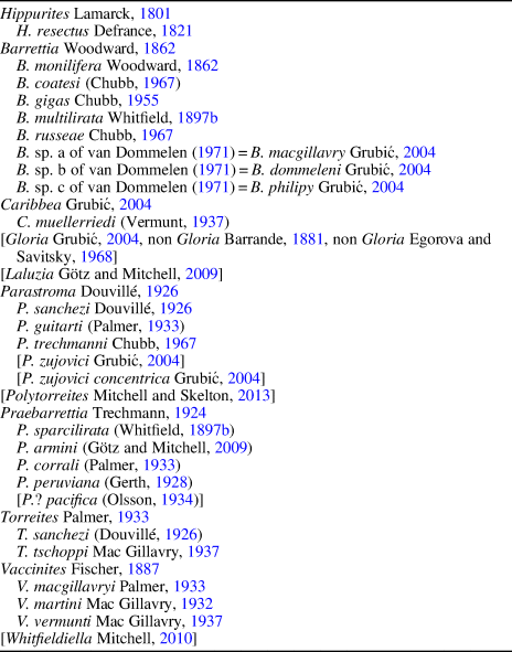
The rudist family Hippuritidae is a major and characteristic component in most Upper Cretaceous rudist-bearing outcrops of the Caribbean Province. Seven genera with 22 species are recognized (Table 1). Genera Barrettia Woodward, Reference Woodward1862, Parastroma Douvillé, Reference Douvillé1926, and Praebarrettia Trechmann, Reference Trechmann1924, with multiple-folds, and Caribbea Grubić, Reference Grubić2004 are exclusive to the American Tethys, whereas Hippurites Lamarck, Reference Lamarck1801, Torreites Palmer, Reference Palmer1933, and Vaccinites Fischer, Reference Fischer1887 occur on both sides of the Atlantic, with different species in Torreites and Vaccinites and with one cosmopolitan species in Hippurites.
American hippuritid rudist taxa. Taxa not widely recognized, controversial, or under discussion, are indicated within brackets.

The Upper Cretaceous rudist fauna of Chiapas is known mainly after Alencáster (Reference Alencáster1971) and older papers by Müllerried (Reference Müllerried1931a, Reference Müllerried1931b, Reference Müllerried1933, Reference Müllerried1934, Reference Müllerried1936). More recent papers (Alencáster and Michaud, Reference Alencáster and Michaud1991; Alencáster and Pons, Reference Alencáster and Pons1992; García-Barrera et al., Reference García-Barrera, Avendaño Gil, Omaña, Alencáster, Masse and Skelton1998; Mitchell, Reference Mitchell2013; Pons et al., Reference Pons, Vicens and García-Barrera2017) added some complementary taxonomic data to the knowledge of this fauna. Pons et al. (Reference Pons, Vicens, Martínez, García-Barrera, Nieto, Oviedo and Avendaño-Gil2016a) provided an inventory of rudist occurrences in the Upper Cretaceous of the Chiapas Central Depression, related their location to a lithostratigraphic framework and paleogeographic reconstruction, identified the successive rudist assemblages, and discussed their age based on the co-occurrence of other fossils (planktic and benthic foraminifers, ammonites, and inoceramid bivalves). Nevertheless, with the exception of the Plagioptychidae (Pons et al., Reference Pons, Vicens and García-Barrera2017), current and detailed systematic descriptions of these rudist species remains undone.
The main aim of this paper is the identification and description of the hippuritid rudist species occurring in the Campanian and Maastrichtian of this area, based mainly on the specimens collected by the authors, but also on a revision of previous material maintained in museum collections, and taking into account the recent taxonomic and biostratigraphic advances on the knowledge of the Upper Cretaceous Caribbean rudist faunas. The stratigraphic conclusions on the Upper Cretaceous of the Chiapas Central Depression by Pons et al. (Reference Pons, Vicens, Martínez, García-Barrera, Nieto, Oviedo and Avendaño-Gil2016a) are reprised herein. Several American multiple-fold hippuritid taxa have been revised to help fit Chiapas’ specimen determinations within a correct taxonomic framework.
Materials and methods
Localities and stratigraphy
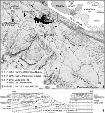
Specimens in this study were collected at five localities in the Chiapas Central Depression (Fig. 1) corresponding to two different lithostratigraphic units and to three of the four rudist assemblages distinguished by Pons et al. (Reference Pons, Vicens, Martínez, García-Barrera, Nieto, Oviedo and Avendaño-Gil2016a). Coincidence with the localities of Müllerried is also indicated when appropriate; although never published by Müllerried, the numbers and names of his fossil localities were registered in the collection and were subsequently used and indicated on a map by Alencáster (Reference Alencáster1971). These localities are:
Studied fossil localities: (1) on a map of the Chiapas Central Depression (shaded topographic relief map from Google Maps); (2) on a depositional model (modified from Pons et al., Reference Pons, Vicens, Martínez, García-Barrera, Nieto, Oviedo and Avendaño-Gil2016a, fig. 2).

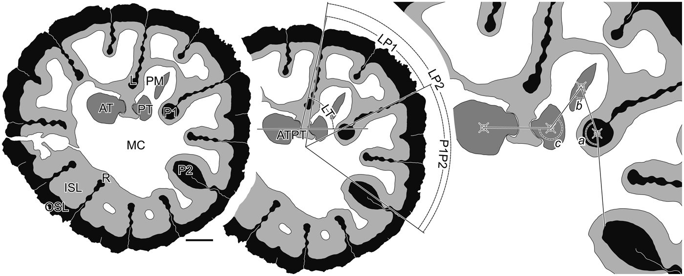
Praebarrettia sparcilirata (Whitfield, Reference Whitfield1897b) from the upper part of Ocozocoautla Formation, drawings of transverse section of RV, close to the commissure, indicating the main internal characters and angular measures, PUAB 81791. a = angle defined by the tip of P2 and the centers of P1 and PM; AT = anterior tooth; ATPT = axis defined by the centers of AT and PT; b = angle defined by the centers of P1, PM, and PT; c = angle defined by the centers of PM, PT, and AT; ISL = inner shell layer; L = ligament ridge; LP1 = portion of shell circumference between L and P1; LP2 = portion of shell circumference between L and P2; LT = angle defined by L and the ATPT; MC = main body cavity; OSL = outer shell layer; P1 = first pillar (formerly S or Sp); P1P2 = portion of shell circumference between P1 and P2; P2 = second pillar (formerly E or Ep); PM = posterior myophore; PT = posterior tooth; R = in-fold of the OSL (ray). Scale bar = 10 mm.

(1) P-0754, Rancho el Comiteco Quarry (16°46'38.31”N, 93°08'34”W), and (2) P-1316, on the road to Paraíso del Grijalva (16°15'15.94”N, 92°38'15.5”W) in the uppermost part of the Suchiapa Formation; the latter corresponds to Müllerried's locality 30, at Vega del Paso. Both correspond to the lower rudist assemblage, although their age is early and middle Campanian, respectively. Co-occurring rudists are: Antillocaprinidae indet., Radiolites acutocostata (Adkins, Reference Adkins1930), Chiapasella sp., and Potosites tristantorresi Alencáster and Pons in Pons et al., Reference Pons, Vicens, Pichardo, Aguilar, Oviedo, Alencáster and García-Barrera2010 in the former; and Alencasteria macrotubularis Mitchell, Reference Mitchell2013, Stellacaprina? macgillavryi (Alencáster, Reference Alencáster1971), Plagioptychus antillarum (Douvillé, Reference Douvillé1927), Mitrocaprina sp., Alencasterites mooretownensis (Trechmann, Reference Trechmann1924), Bournonia bailei Chubb, Reference Chubb1967, and Thyrastylon? nicholasi (Whitfield, Reference Whitfield1897a) in the latter.
(3) P-0743, Campo de Tiro (16°46'55.41”N, 93°22'52.49”W) and (4) P-1358, Los Chamaquitos (16°46'35.58”N, 93°22'44.9”W), both along the Cuesta NW of Ocozocoautla corresponding to Mullerried locality 6, Ocozocoautla; and (5) P-0753, km 178.2, on the road (MEX145) from Ocozocoautla to Las Choapas (16°53'22.27”N, 93°25'35.72”W). All three, in the upper part of the Ocozocoautla Formation, correspond to the middle assemblage, early Maastrichtian. Co-occurring rudists are: Plagioptychus muellerriedi Alencáster, Reference Alencáster1971, Biradiolites cardenasensis Böse, Reference Böse1906, Chiapasella radiolitiformis (Trechmann, Reference Trechmann1924), Huasteca ojanchalensis (Myers, Reference Myers1968), Thyrastylon? nicholasi, and Trechmannites rudissimus (Trechmann, Reference Trechmann1924), in the first two. Only B. cardenasensis occurs in the last.
No hippuritid rudists were found in the upper assemblage, late Maastrichtian.
Methods.—Specimens collected and used for this study were sectioned and polished, and acetate peels were prepared, at the laboratories of the Universitat Autònoma de Barcelona and the Universidad Nacional Autónoma de México; thin sections were produced at the former. Larger specimens were cut at the laboratory of the Instituto Mexicano del Petróleo.
Anatomical conventions
Abbreviations used in text, figures, and tables are: a = angle defined by the tip of P2 and the centers of P1 and PM; ADC = anterodorsal cavity; AT = anterior tooth; at = socket of the AT; ATPT = axis defined by the centers of AT and PT; b = angle defined by the centers of P1, PM, and PT; c = canal at the ISL; c = angle defined by the centers of PM, PT, and AT; ISL = inner shell layer; L = ligament ridge; LP1 = portion of shell circumference between L and P1; LP2 = portion of shell circumference between L and P2; LT = angle defined by L and the ATPT; LV = left valve; MC = main body cavity; OSL = outer shell layer; p = pores; P1 = first pillar (formerly S or Sp); P1P2 = portion of shell circumference between P1 and P2; P2 = second pillar (formerly E or Ep); PM = posterior myophore; pm = cavity of the PM; PT = posterior tooth; pt = socket of the PT; R = in-fold of the OSL (ray); RC = radial canal; RV = right valve; T = inter-ray tabula; VC = vertical canal. All transverse cross sections are figured in standard orientation, i.e., in adapical view and with the dorsal side up, unless indicated. In cross-sectional drawings, the OSL is represented in black, RVs ISL in pale gray, and LVs ISL in dark gray. Synonymy lists are not exhaustive; only those references relevant to the discussion presented herein are included; the reader should consult Steuber (Reference Steuber2002) for more complete lists to that date.
Repositories and institutional abbreviations
FCMP = Museo de Paleontología, Facultad de Ciencias, Universidad Nacional Autónoma de México, Mexico City, Mexico; IGM = Colección Nacional de Paleontología, Instituto de Geología, Universidad Nacional Autónoma de México, Mexico City, Mexico; NHMUK = The Natural History Museum (formerly British Museum [Natural History]), London, UK; NMNH = National Museum of Natural History, Smithsonian Institution, Washington, DC, USA; PUAB = Paleontological Collections of the Universitat Autònoma de Barcelona, Bellaterra, Spain; UWI = University of West Indies, Geology Museum, Mona, Kingston, Jamaica.
Systematic paleontology
Family Hippuritidae Gray, Reference Gray1848
Genus Barrettia Woodward, Reference Woodward1862
Type species
Barrettia monilifera Woodward, Reference Woodward1862 from the lower limestone of the Back Rio Grande Formation, Back Rio Grande, Blue Mountain Inlier, Jamaica.
Diagnosis
Large multiple-fold hippuritid rudist; ISL of the LV canaliculated; OSL of the LV with specialized pore-canal system, horizontal radial canals near the apical central part, originally opening to the commissural margin, are substituted outward by aligned vertical canals opening above rays, pores polygonal; rays of the RV nonmoniliform, submoniliform, or moniliform; ISL of the RV between rays, vesicular; OSL of RV compact, thin; myocardinal elements, as seen in transverse cross section of RV close to commissure, disposed with PM ventral to axis ATPT and close to knob of P1.
Remarks
Dechaseaux and Coogan (Reference Dechaseaux, Coogan and Moore1969) provided a rather general diagnosis, referring to the form and size of the RV, number of rays, form of the pillars, portion of the valve between L and pillars, cellular and tabular internal layer, and form and oscula of the LV. Van Dommelen (Reference van Dommelen1971) introduced, as additional diagnostic characters, the presence of canals in the ISL of the LV and considered the development degree of constrictions in the rays of the RV (category 2 to 4) and the relative position of the myocardinal elements with the ligament ridge (type A or C). Mitchell (Reference Mitchell2010) considered, besides the canals in the ISL of the LV, the myocardinal pillar arrangement (range values of angles a, b, and c), the structure of the ISL of the RV, and the multiple reticulate pores of the LV. The pattern of the pore-canal system of the LV is introduced as a diagnostic character herein; also, the ‘multiple reticulate pores’ of Mitchell (Reference Mitchell2010) are reinterpreted within this pattern.
Barrettia monilifera Woodward, Reference Woodward1862
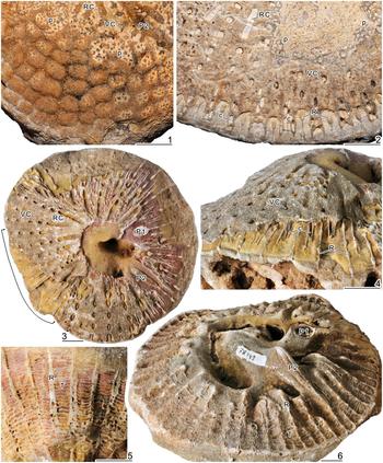
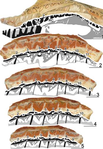
Figures 3.1–3.3, 4, 5
Reference Woodward1862 Barrettia monilifera Woodward, p. 372–378, pl. 20, figs. 1–4, pl. 21, fig. 5 (all figures are drawings, figs. 2, 5 = type specimen).
Reference Douvillé1894 Barrettia monilifera; Douvillé, p. 110, pl. 17, fig. 6 (photograph of another slice of the type specimen).
Reference Alencáster1971 Barrettia monilifera; Alencáster, p. 57, pl. 11, figs. 2–5, pl. 12, figs. 1–6.
Reference Alencáster1971 Barrettia multilirata; Alencáster, p. 61, pl. 15, fig. 1.
Reference Chubb1971 Barrettia monilifera; Chubb, p. 208, text-fig. 7, pl. 51, figs. 1–3, pl. 52, figs. 2, 3 (text-fig. 7 and pl. 52, fig. 2 = drawing and photograph, respectively, of Woodward, pl. 20, fig. 2 specimen counterpart).
Reference Mitchell2010Barrettia monilifera; Mitchell, p. 10, text-figs. 3D, 4, 6A–D, 8A, 9 (4 = photograph of Woodward specimen, pl. 20, fig. 2, pl. 21, fig. 5; with complete synonymy list).
Reference Munujos, Pons and Vicens2016 Barrettia sp.; Munujos et al., fig. 9D–F.
Reference Pons, Vicens, Martínez, García-Barrera, Nieto, Oviedo and Avendaño-Gil2016aBarrettia monilifera; Pons et al., table 2.
Reference Pons, Vicens and Schmidt-Effing2016b Barrettia monilifera; Pons et al., p. 216, fig. 3.1–3.6.
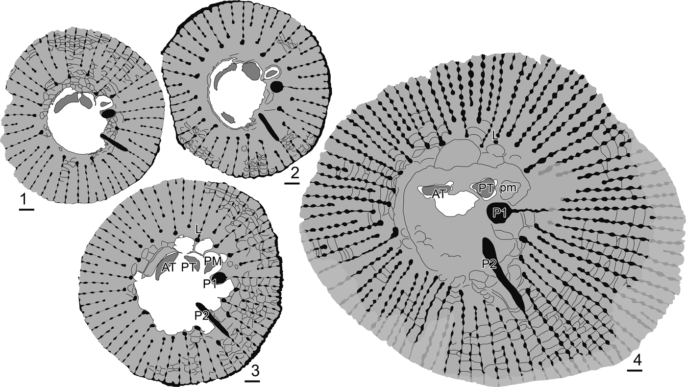
Barrettia monilifera Woodward, Reference Woodward1862 (1–3) and B. gigas Chubb, Reference Chubb1955 (4) from the upper part of Suchiapa Formation, middle Campanian, road to Paraíso del Grijalva (P-1316), drawings of transverse sections of RVs: (1) PUAB 78333; (2) PUAB 78332; (3) PUAB 78316; (4) FCMP 14/100. AT = anterior tooth; L = ligament ridge; P1 = first pillar (formerly S or Sp); P2 = second pillar (formerly E or Ep); PM = posterior myophore; pm = cavity of the posterior myophore; PT = posterior tooth. Scale bars = 10 mm.

Barrettia monilifera Woodward, Reference Woodward1862 from the upper part of Suchiapa Formation, middle Campanian, Road to Paraíso del Grijalva (P-1316): (1) IGM 2156, detail of LV outer surface, slightly weathered at right and upper parts; (2) IGM 2155, detail of weathered LV; (3–5) PUAB 78330, upper view of LV, oblique view of both valves in the weathered area indicated, and outer surface detail of the weathered RV, respectively; (6) PUAB 78448, oblique commissural view of RV, upper surface of pillars and rays outward inclined. c = canal at the ISL; p = pores; P1 = first pillar (formerly S or Sp); P2 = second pillar (formerly E or Ep); R = in-fold of the OSL (ray); RC = radial canal; T = inter-ray tabula; VC = vertical canal. Scale bars = 10 mm.

Barrettia monilifera Woodward, Reference Woodward1862 from the upper part of Suchiapa Formation, middle Campanian, road to Paraíso del Grijalva (P-1316): (1) PUAB 78449, radial cross section and drawing at the commissure between both valves; (2–5) PUAB 78331, seriated outward tangential cross sections and drawings at the commissure between valves. c = canal at the ISL; p = pores; RC = radial canal; VC = vertical canal; numbers indicate the position of rays. Scale bars = 10 mm.

Holotype
Specimen NHMUK PI OR 42861, cut into several parts, from the lower limestone member of the Back Rio Grande Limestones, Blue Mountain Inlier, Jamaica (Woodward, Reference Woodward1862, pl. 20, fig. 2, longitudinal section, and pl. 21, fig. 5, transverse section). Douvillé (Reference Douvillé1894) plate 17, figure 6 is a photograph of a different transverse section of Woodward's type specimen, sent by the author to the École National Supériéure des Mines de Paris, France. Mitchell (Reference Mitchell2010) provided a full account of the complex history of the several fragments of the type specimen and their figures by various authors.
Diagnosis
“A Barrettia with an advanced myocardinal-pillar arrangement, a short ligamental ray, a moderate ray density, moderately large round beads, and thin, irregular inter-ray partitions; pores of left valve on raised domes over each bead” (Mitchell, Reference Mitchell2010, p. 10).
Occurrence
Road to Paraíso del Grijalva (P-1316), uppermost part of Suchiapa Formation, middle Campanian. Several localities of El Viejo Formation, in Costa Rica (Pons et al., Reference Pons, Vicens and Schmidt-Effing2016b). The species is abundant in Jamaica. Chubb (Reference Chubb1971) considered that the material identified as B. monilifera from Cuba, Puerto Rico, St. Croix, and elsewhere from Jamaica could belong to several other species of Barrettia. Van Dommelen (Reference van Dommelen1971) proposed informal names for three of them, which were formally named by Grubić (Reference Grubić2004). Mitchell (Reference Mitchell2010) repeated Chubb's (Reference Chubb1971) consideration.
Description
Right valves are conical-cylindrical. Those of adult specimens are predominantly cylindrical, reaching 600 mm in height with diameters between 110 and 140 mm. Those of young specimens are conical, to 150 mm in height with diameters between 55 and 110 mm. Most specimens grew singly although a few bouquets with two to four specimens have also been collected. The external surface is abraded in nearly all specimens; regular rounded costae (~4 per 10 mm), and wider grooves have only been observed in a few young conical shells. Transverse cross sections close to the commissure show 60–70 in-folds of the outer shell layer into the inner shell layer; the outer part of the outer shell layer is only rarely and partially preserved. The in-folds are moniliform. Their beads are slightly radially elongated and the constrictions between beads are slightly longer than the beads themselves. Up to eight beads are counted in longer in-folds. Length of the ligament ridge (the in-fold with a circular knob pointing against the posterior tooth) is similar to those of longer secondary pillars. The first pillar has an elliptical knob (~7 per 10 mm in diameter) and a 35 mm long moniliform peduncle. The second pillar has a long parallel-sided knob (to 30 mm long) and a shorter moniliform peduncle. The secondary pillars each have a knob larger than the beads. As clearly seen in disarticulated valves, all in-folds are raised and outwardly inclined on the commissural surface. The inner shell layer between contiguous in-folds is vesicular; a double row of vesicles appears, more or less clearly between the contiguous rays, in transverse cross sections. Left valves are very low cones. Well-preserved left valves, with polygonal pores on radially-arranged raised domes, are rare; those, more common, with various degrees of abrasion provide as much information about structure as could be obtained through numerous differently-oriented sections. The outer shell layer develops normal hippuritid radial canals in the apical central part of the valve; evidently, canals opened to the commissural margin at early growth stages but, with growth, they became open to the distal knob of longer rays in the RV. Outward, radial canals are substituted by radially-aligned, very short vertical canals opened to each raised bead of the rays. The appearance of new (shorter) radial canals and their substitution by radially-aligned vertical canals also occurs in coincidence with the appearance of each new ray at the RV. Vertical (in fact, outwardly inclined) canals are circular in deep transverse cross section but wider than long near the outer surface, or even appear double below the raised domes with pores. The inner shell layer is well developed at the inter-rays and crossed by very fine, radially-oriented pallial canals. The myocardinal structure, in transverse cross section, fits with van Dommelen's (Reference van Dommelen1971) type C, and the angles a, b, and c are within the range indicated by Mitchell (Reference Mitchell2010). PM is elliptical and radially elongated, and PT is slightly smaller than AT.
Materials
Forty-five specimens, most with conjoined valves, left ones more or less abraded, from P-1316 road to Paraíso del Grijalva, uppermost part of Suchiapa Formation: PUAB 78305, 78307, 78328–78331; FCMP 13/01, 13/03–13/17, 13/19–13/21, 13/72–13/86, 13/90–13/95, 13/108, 14/08, 14/16, 14/47, 14/65, 14/76, 14/80, 14/99, and 15/53. The upper slices of some FCMP specimens are maintained and registered as PUAB 78303, 78304, 78306, 78311–78317, 78332, 78333, and 78448–78450.
Remarks
Van Dommelen (Reference van Dommelen1971) considered all specimens with moniliform rays (Moniliform Branch) as Barrettia monilifera sensu lato, recognizing as B. monilifera, B. multilirata Whitfield, Reference Whitfield1897b, and B. gigas Chubb, Reference Chubb1955 only those specimens more or less identical to the corresponding holotype. Mitchell (Reference Mitchell2010) revised the diagnostic characters of B. monilifera, B. multilirata, and B. gigas.
Barrettia monilifera at its type locality—the lower limestone member of the Back Rio Grande Formation at the Blue Mountain Inlier, Jamaica—is considered middle Campanian (Mitchell, Reference Mitchell2010). In Chiapas, as in several Jamaican localities, it occurs together with B. gigas and Parastroma trechmanni Chubb, Reference Chubb1967.
Barrettia gigas Chubb, Reference Chubb1955
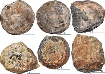
Figures 3.4, 6
Reference Whitfield1897b Barrettia monilifera; Whitfield, p. 233, pls. 27–32.
Reference Trechmann1922 Barrettia cf. monilifera; Trechmann, p. 510, pl. 20, figs. 1, 2 (non pl. 19, fig. 2a, b = Parastroma trechmanni).
Reference Chubb1955 Barrettia gigas; Chubb, p. 9.
Reference Chubb1956 Barrettia gigas; Chubb, p. 7.
Reference Alencáster1971 Barrettia gigas; Alencáster, p. 63, pl. 13, figs. 1, 2, 4 (non fig. 3 = Parastroma trechmanni).
Reference Alencáster1971 Barrettia multilirata; Alencáster, p. 61, pl. 15, fig. 1.
Reference Chubb1971 Barrettia gigas; Chubb, p. 210, pl. 52, fig. 1, pl. 53, figs. 1–3.
2010 Whitfieldiella gigas; Mitchell, p. 13, figs. 3E, F, 8C, 9, 12 (with complete synonymy list).
Reference Pons, Vicens, Martínez, García-Barrera, Nieto, Oviedo and Avendaño-Gil2016a Whitfieldiella gigas; Pons et al., table 2.
Reference Pons, Vicens and Schmidt-Effing2016b cf. Whitfieldiella gigas; Pons et al., p. 218, fig. 4.
Barrettia gigas Chubb, Reference Chubb1955 from the upper part of Suchiapa Formation, middle Campanian, road to Paraíso del Grijalva (P-1316): (1–3) FCMP 13/40, upper view of LV, apical view of RV, and side view of both valves, respectively; (4) FCMP 14/100, commissural view of RV, LV weathered, transverse cross section of the RV in Fig. 3.4; (5, 6) FCMP 13/41, RV, apical and side views, respectively. AT = anterior tooth; P1 = first pillar (formerly S or Sp); P2 = second pillar (formerly E or Ep); PM = posterior myophore; PT = posterior tooth; arrows indicate the apex. Scale bars = 100 mm.

Holotype
Designated by Chubb (Reference Chubb1955), specimen NMNH PAL9665/1, from the middle Campanian, Haughton Hall Formation, Green Island Inlier, Hannover, Jamaica (Whitfield, Reference Whitfield1897b, pl. 27, fig. 1, pl. 28, fig. 1).
Diagnosis
“A very large species with a short ligamental ray, full moniliform rays, well-developed inter-ray partitions and relatively large and widely spaced beads” (Mitchell, Reference Mitchell2010, p. 13, as a member of Whitfieldiella Mitchell, Reference Mitchell2010).
Occurrence
Road to Paraíso del Grijalva (P-1316), uppermost part of Suchiapa Formation, middle Campanian. The species is found in Jamaica, in the Haughton Hall and Stapleton Limestones of the Green Island and Sunderland Inliers, respectively; these limestones were placed in the ‘upper Middle Campanian’ by Mitchell (Reference Mitchell2010). The species was also reported from Costa Rica, in Cuajiniquil, Santa Rosa National Park (Pons et al., Reference Pons, Vicens and Schmidt-Effing2016b).
Description
All specimens grew as isolated individuals. RVs are conical-cylindrical with a flat or slightly convex, eccentric base; diameters at the commissure range 180–300 mm, with a diameter at the base of 140 mm; height range is 160–300 mm; diameter of the body cavity is ~90–100 mm; the outer shell layer, rarely preserved because of weathering, is 1 mm thick; moniliform in-folds number 88 in a specimen 250 mm in diameter; at the outer surface, the spacing of rays ranges 12–18 per 100 mm; length of the beads ranges 2–3 mm, their length slightly greater than their width, and bead centers are separated 4–6 mm; knobs of the rays are larger than the beads; up to 19 beads were counted in a 106 mm long ray; length of L is similar to that of longer rays; the knob of P1 is circular with a diameter to 14 mm; that of P2 is much elongated (56 per 8 mm) with nearly parallel sides. On the commissural surface, all rays are raised and outwardly inclined. The inner shell layer between contiguous rays is composed of regularly spaced (~3 mm) simple concave tabulae. LVs are very low cones. Their surfaces are strongly weathered. The pores are not preserved; only a few vertical canals of the outer shell layer, above the rays, and pallial canals of the inner shell layer, in between, were preserved in a specimen; also the knob of P1 of the RV and the myocardinal elements of the LV currently appear as voids because of dissolution. The angles of the myocardinal-pillar elements were measured in the transverse cross section of a RV: a = 165°; b = 73°; and c = 176°.
Materials
Six specimens, all RVs, from P-1316 road to Paraíso del Grijalva, uppermost part of Suchiapa Formation: FCMP 13/40, 13/41, 13/122, 13/123, 14/100, and 15/42. The last two are transversally cross-sectioned close to the commissure; although strongly weathered, the first one preserves part of the left valve.
Remarks
Barrettia gigas was proposed by Chubb (Reference Chubb1955) for specimens identified as B. monilifera by Whitfield (Reference Whitfield1897b) and as B. cf. monilifera by Trechmann (Reference Trechmann1922), and were subsequently figured by Chubb (Reference Chubb1971). Chubb's description of the species focused on the shell form, size, number and development of rays, relative positions of L, P1 ( = S), and P2 ( = E), and body cavity, and noted that only the cardinal apparatus of the left valve was known.
At first look, Barrettia gigas differs from B. monilifera, the type species, mainly in size and form of the RV: very large, with a nearly flat young part and relatively low (when compared with the large diameter), cylindrical adult shell, versus being smaller in diameter, with high conical young part and high cylindrical adult shell, respectively. The two species differ in the number of in-folds, clearly moniliform in both but with slightly different size and spacing of beads. Also, in the location of PM: although slightly ventral to the ATPT axis in both, B. gigas presents a larger angle a and smaller angle b than B. monilifera.
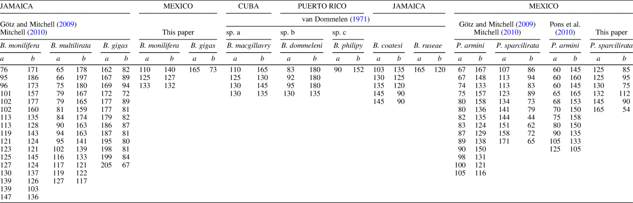
Mitchell (Reference Mitchell2010) proposed placing this species within the genus Whitfieldiella based on differences with other species of Barrettia mainly concerning the a and b angle ratio but also a purported particularly fine vesicular structure in the ISL of the RV and unique pore pattern of the LV. Angles a and b were measured on all figured specimens of all known species of Barrettia and Praebarrettia (Table 2). The scatter plot (Fig. 7) shows the ratio between a and b angles for all figured specimens of the species B. coatesi (Chubb, Reference Chubb1967), B. ruseae, B. monilifera, B. multilirata, van Dommelen's (Reference van Dommelen1971) sp. a, sp. b, and sp. c ( = Grubić’s, [2004] B. macgillavry, B. dommeleni, and B. philipy, respectively), and B. gigas, all of them sharing common characters, indicates that all values fit a characteristic regression line, differ from that of other groups of multiple-fold hippuritid species having other characters in common and, thus, corresponding to different genera (i.e., Praebarrettia or Pironaea Meneghini, Reference Meneghini1868). Although with some overlaps, this ratio differs for each species within each genus. The size of the vesicular structure in the ISL of the RV shows a relatively wide variation among different Barrettia spp., as does the number of rays and the number and development of constriction within each ray. The raised pustules with irregular pores separated by imperforate bands are a common character of Barrettia spp., although they are rarely well preserved.
Scatter plot of pillar-myocardinal arrangement, as indicated by values of angles a (P2-P1-PM) and b (P1-PM-PT), on specimens of American multiple-fold hippuritid species (see Table 2).

Measurements (in °) of angles a (P2-P1-PM) and b (P1-PM-PT) on specimens of American multiple-fold hippuritid species of the genera Barrettia and Praebarrettia. Geographic information and sources of data are indicated. Angles on figured specimens presented by van Dommelen (Reference van Dommelen1971) and Pons et al. (Reference Pons, Vicens, Pichardo, Aguilar, Oviedo, Alencáster and García-Barrera2010) were measured by the authors for this paper.

Whitfieldiella is herein considered a junior synonym of Barrettia. The ratio between angles a and b is only one of the many shell characters shared by all species of Barrettia and the variation among different species concerning this character is not more significant than that of other characters.
Barrettia gigas occurs together with B. monilifera and Parastroma trechmanni in the same horizon in Chiapas, as well as in Jamaica where these species are considered to be middle Campanian (Mitchell, Reference Mitchell2010).
Barrettia cf. B. ruseae Chubb, Reference Chubb1967
Figure 8
Reference Chubb1967 Barrettia ruseae Chubb, p. 30.
Reference Chubb1971 Barrettia ruseae; Chubb, p. 213, pl. 55, fig. 1.
Reference van Dommelen1971 Barrettia ruseae; van Dommelen, p. 84, pl. 5, figs. 1, 2, pl. 6, figs. 1–3.
Reference Grubić2004 Barrettia ruseae; Grubić, p. 159, pl. 6, fig. 1 (drawing of Chubb's [1971, pl. 55, fig. 1] specimen).
Reference Pons, Vicens, Pichardo, Aguilar, Oviedo, Alencáster and García-Barrera2010 Barrettia cf. ruseae; Pons et al., p. 981, text-fig. 10.1–10.4.
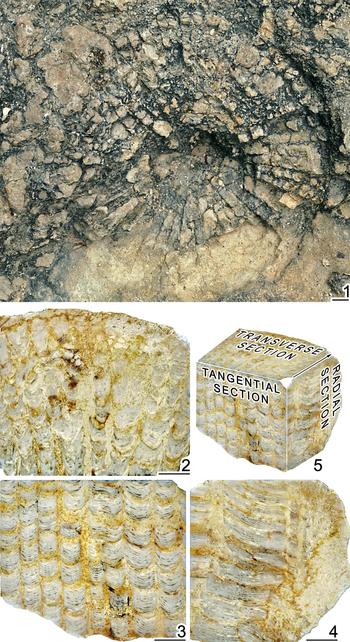
Barrettia cf. B. ruseae Chubb, Reference Chubb1967 from the upper part of Suchiapa Formation, lower Campanian, Rancho el Comiteco Quarry (P-745): (1) RV (not sampled), natural transverse cross section on outcrop; (2–5) PUAB 78296, transverse, tangential, and radial cross sections of RV, and their oriented reconstructions, respectively. Scale bars = 10 mm.

Holotype
The only specimen, UWI 1190, figured by Chubb (Reference Chubb2010, pl. 55, fig. 1), from the Clifton Limestone, SE of Lucea, Hannover, Jamaica.
Diagnosis
“Shell large, discoidal or saucer-shaped with a flat base, averaging 140 mm in diameter by 60 mm high, but can be higher; outer layer 1 mm thick; number of in-folds varies with diameter, their average distance apart, measured around the circumference, being about 5.5 mm; in-folds extremely attenuated, the beads seen in section being barely 0.5 mm wide and 2 mm long, each tapering into the thin tread-like line connecting them; longest in-folds penetrate 50 or 55 mm with 10 or 12 beads; distance between L and E pillars between 35 and 40 per cent of circumference; body cavity about one-fifth of diameter of shell; inner layer forms irregular vesicular tissue in body cavity and inter-radial areas. Left valve is unknown” (from original description; Chubb, Reference Chubb1967, p. 30).
Occurrence
Rancho el Comiteco Quarry (P-0754), uppermost part of Suchiapa Formation, early Campanian. In Mexico, the species has also been reported from the Temazcal limestones, San Luis Potosí (Pons et al., Reference Pons, Vicens, Pichardo, Aguilar, Oviedo, Alencáster and García-Barrera2010); in Jamaica, known only from the type locality.
Description
The natural (not sampled) cross sections on limestone surface indicate a Barrettia with an RV ~180 mm in diameter, 1 mm thick outer shell layer, with ~64 in-folds, 15 per 10 mm measured around the circumference. In-folds are long, L being the longest, submoniliform to moniliform with elongated beads. The body cavity is, consequently, quite small. Oriented sections on the few sampled shell fragments reveal the ISL between in-folds is formed by very small vesicles arranged in concave-upward microrhythm sets spaced 3.3–7.3 mm.
Materials
Natural cross sections on massive limestone surfaces were observed and photographed in the field. From the three shell fragments collected, one of them (PUAB 78296) was transversally, radially, and tangentially sectioned.
Remarks
Although the few characters observed in the field and the sampled shell fragments point to an attribution to Barrettia ruseae, this material has been left under open nomenclature. The structure of the ISL of the RV is figured herein for the first time.
Shell characters of Barrettia ruseae, as described by its author (Chubb, Reference Chubb1967), are considered more primitive than those of the other two Barrettia species reported from Chiapas, B. monilifera and B. gigas. At the type locality in Jamaica, and San Luis Potosí, Mexico, B. ruseae occurs, as in Chiapas, together with Vaccinites vermunti Mac Gillavry, Reference Mac Gillavry1937, being considered early Campanian everywhere.
Genus Parastroma Douvillé, Reference Douvillé1926
Type species
Parastroma sanchezi Douvillé, Reference Douvillé1926 from the beds with Barrettia in Sánchez Agramonte Domain, Arroyo Hondo, Camagüey Province, Cuba.
Diagnosis
Large to very large multiple fold hippuritid; OSL of the LV very thin, traversed by very short aligned vertical canals, each one communicating upward with a flat bubble traversed by grouped small polygonal pores; ISL of the LV finely canaliculated, developed between the canals; rays of the RV and L absent or reduced to the margin; only the knobs and the bases of P1 and P2 present, without the stems; vesicular ISL of the RV well developed, the folded tabulae forming concave vesicles limited by conical micro-folds; OSL of the RV compact and thin; myocardinal elements disposed with a large PM dorsal to the axis ATPT and close to P1, in transverse cross section of RV close to the commissure.
Remarks
Dechaseaux and Coogan (Reference Dechaseaux, Coogan and Moore1969) emphasized the vesicular wall, the effacement of rays in the AV, and the portion of shell circumference between P1 and P2. Van Dommelen (Reference van Dommelen1971) recognized the canals in the ISL of the LV, the submoniliform rays in young shells, and the Barrettia-like relative position of the myocardinal elements. The pattern of the pore-canal system of the LV and the ISL of the RV is introduced herein. Genus Parastroma appears to be closely related to the genus Barrettia, because the relative position of the myocardinal elements is identical, and the effacement of rays in the RV seems to be morphostructurally responsible of the differences in the ISL of the RV and the pattern of the pore-canal system of the LV.
Parastroma trechmanni Chubb, Reference Chubb1967
Figures 9–12
Reference Trechmann1922 Barrettia cf. monilifera; Trechmann, p. 510, pl. 19, figs. 2a, b (non pl. 20, figs. 1, 2 = B. gigas)
Reference Chubb1967 Parastroma trechmanni Chubb, p. 30.
Reference Alencáster1971 Barrettia gigas; Alencáster, p. 63, pl. 13, fig. 3.
Reference Alencáster1971 Barrettia multilirata; Alencáster, p. 61, pl. 13, fig. 5.
Reference Chubb1971 Parastroma trechmanni; Chubb, p. 214, pl. 56, figs. 1–3 (1 = same specimen by Trechmann, Reference Trechmann1922, pl. 19, figs. 2a, b).
Reference van Dommelen1971 Parastroma trechmanni; van Dommelen, p. 95, text-fig. 8C, pls. 16–21.
Reference Grubić2004 Parastroma trechmanni; Grubić, p. 168, pl. 10, figs. 1, 2 (new drawings of Trechmann's, 1922, pl. 19, figs. 2a, b specimen).
Reference Grubić2004 Parastroma zujovici Grubić, p. 166, pl. 8, fig. 2 (new drawing of van Dommelen's, 1971, pl. 17 specimen).
Reference Grubić2004 Parastroma zujovici concentrica Grubić, p. 167, pl. 9, fig. 1.
Reference Pons, Vicens, Martínez, García-Barrera, Nieto, Oviedo and Avendaño-Gil2016a Parastroma trechmanni; Pons et al., table 2.
Reference Pons, Vicens and Schmidt-Effing2016b Parastroma trechmanni; Pons et al., p. 216, fig. 3.7, 3.8.
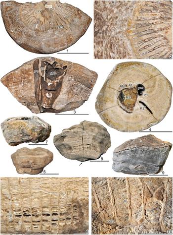
Parastroma trechmanni Chubb, Reference Chubb1967 from the upper part of Suchiapa Formation, middle Campanian, Road to Paraíso del Grijalva (P-1316): (1–3) IGM 2148, upper view of both valves showing commissural lip and weathered LV, detail as indicated, and natural radial section, respectively; (4–6) FCMP 13/34, transverse cross section of RV, lateral view, and detail of weathered outer surface as indicated, respectively; (7) FCMP 13/36, lateral view of RV; (8) FCMP 13/31, lateral view of RV; (9) FCMP 13/39, lateral view of RV; (10) PUAB 78491, detail of weathered outer surface of RV showing thin OSL and tangential section of rays and inter-ray vesicles. at = socket of the AT; c = canal at the ISL; LV = left valve; MC = main body cavity; OSL = outer shell layer; P1 = first pillar (formerly S or Sp); P2 = second pillar (formerly E or Ep); pm = cavity of the PM; pt = socket of the PT; R = in-fold of the OSL (ray); RV = right valve; T = inter-ray tabula; arrow indicates apex. Scale bars = 100 mm (1, 3–5, 7–9); 10 mm (2, 6, 10).

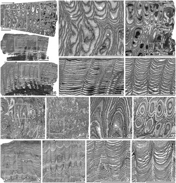
Parastroma trechmanni Chubb, Reference Chubb1967 from the upper part of Suchiapa Formation, middle Campanian, road to Paraíso del Grijalva (P-1316), thin sections: (1–7) PUAB 78494, transverse, tangential, and radial cross sections, respectively, and details as indicated; (8–15) PUAB 78495: (8–11) transverse cross sections, far and close to the outer margin, respectively, and details as indicated; (12–15) tangential and radial cross sections, respectively, and details as indicated. R = in-fold of the OSL (ray); arrows point outward. Scale bars = 10 mm.

Parastroma ?trechmanni Chubb, Reference Chubb1967 from the upper part of Suchiapa Formation, middle Campanian, road to Paraíso del Grijalva (P-1316), thin sections, PUAB 78493, transverse, tangential, and radial cross sections, respectively, and details as indicated. Arrows point outward. Scale bars = 10 mm.

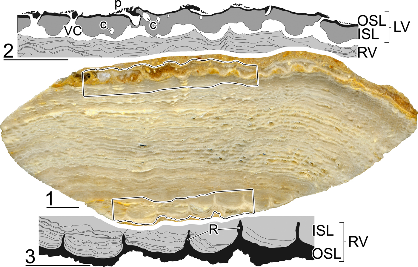
Parastroma trechmanni Chubb, Reference Chubb1967 from the upper part of Suchiapa Formation, middle Campanian, Road to Paraíso del Grijalva (P-1316), PUAB 78491, tangential cross section of both valves and drawings of details as indicated. c = canal at the ISL; ISL = inner shell layer; LV = left valve; OSL = outer shell layer; p = pores; R = in-fold of the OSL (ray); RV = right valve; VC = vertical canal. Scale bars = 10 mm.

Holotype
Right valve (NHMUK PI L 44200), from the middle Campanian Barrettia Limestone, Haughton Hall, Green Island Inlier, Hannover, Jamaica (Trechmann, Reference Trechmann1922, pl. 19, fig. 2a, b; Chubb, Reference Chubb1971, pl. 56, fig. 1).
Diagnosis
Shell large or gigantic; RV broadly conical, outer surface with numerous radial sulci; small MC; rays discernible near outer margin but lost inward between cavities formed by concentric wrinkling of ISL; hinge and myophores Barrettia-like (extracted from Chubb's, Reference Chubb1967, Reference Chubb1971 original description; LV was unknown). LV as stated in diagnosis of the genus.
Occurrence
Road to Paraíso del Grijalva (P-1316), uppermost part of Suchiapa Formation, middle Campanian. Barrettia beds of the Haughton Hall Limestone, Green Island Inlier (Chubb, Reference Chubb1971), and Glenbrook Limestone, Grange Inlier (van Dommelen, Reference van Dommelen1971), Jamaica.
Description
All specimens grew as isolated individuals. RVs are roughly cylindrical with a low conical (sometimes concave) base, with eccentric apex; diameters at the commissure range 200–320 mm, with a diameter at the base of ~170 mm; height to 300 mm; diameter of MC of ~70–120 mm; the OSL, rarely preserved because of weathering, is ~1 mm thick and ribbed; when not preserved, narrow radial furrows at the outer surface indicate the location of the base of the in-folds. The transverse section close to the commissure shows the knobs of P1 and P2 and the myocardinal elements; P1 and P2 are Barrettia-like, i.e., the knob of P1 is circular and that of P2 is much elongated with parallel sides; PM is much larger than the knob of P1, both appearing frequently as voids because of dissolution, because it sometimes also occurs to the other elements; only the base of the in-folds can be recognized near the outer margin; The ISL is formed by tabulae, very thin and close each other, which are folded and form concave vesicles limited by conical microfolds; vesicles are concentrically arranged, measuring to 15 mm tangentially and <5 mm radially; close to the outer shell margin, vesicles are radially arranged as if the in-folds of the OSL were present; the aligned conical microfolds limiting the vesicles measure ~1 mm in diameter.
One specimen (PUAB 78493) from the same locality and identical in external aspect to the other specimens described herein, revealed some differences in the structure of the RVs ISL (Fig. 11.1–11.6). The folded tabulae appear significantly smaller, <5 mm tangentially and 2 mm radially, and their pattern in transverse cross section is less regular, probably because micro-folding is more important. Beside the smaller size of the elements, no other differences were observed in radial and tangential sections.
PUAB 78491 preserves, covered by embedding fine sediment, a small part of the LV (Fig. 12.1–12.3): the OSL is very thin, its lower part is traversed by very short, aligned vertical canals, each one communicating upward with a flat bubble traversed by grouped small polygonal pores; the ISL develops below, between the canals, and is traversed by very thin radially oriented pallial canals; as observed in another specimen with a partially weathered left valve (Fig. 10.1, 10.2), the ISL forms continuous radial walls with fine, radially oriented pallial canals (inter-radial plates of van Dommelen, Reference van Dommelen1971) intercalated with the aligned vertical canals of the OSL.
Materials
Thirty-five more or less complete RVs, slightly or intensely weathered, a few of them showing remnants of the LV, from P-1316 road to Paraíso del Grijalva, uppermost part of Suchiapa Formation: FCMP 13/22, 13/23, 13/25–13/28, 13/31–13/39, 13/96, 13/97, 13/102–13/104, 13/124–13/126, 14/03, 14/08, 14/14 (→PUAB 78492), 14/17, 14/19, 14/22, 14/59, 14/89, 14/101, 14/129, 15/054, and 15/057 (→PUAB 78491). PUAB 78493–78495 are parts of FCMP 13/26, 14/89, and 13/102, respectively.
Remarks
The LV of Parastroma trechmanni was described by van Dommelen (Reference van Dommelen1971, p. 97), because OSL and ISL were indistinguishable in his specimens, as being formed by “vertical, radial, canaliferous plates, separated by radial spaces, marked by rows of pores excavated into the covering of the outer shell layer.” Van Dommelen (Reference van Dommelen1971, p. 99) assumed that P. trechmanni might be intermediate between P. sanchezi and P. guitarti (Palmer, Reference Palmer1933) because the structure of the ISL of the RV has numerous conical microfolds as in the former but, in contrast, these are arranged according to the concentric arrangement of the tabulae as in the latter. Grubić (Reference Grubić2004) misinterpreted as ‘capillary pseudocanals’ the dome-like sharp folds of the tabulae, mostly arranged one above another in the successive tabulae and appearing as small circles, arranged on irregular lines, in transverse cross section, as were already described in P. sanchezi by Thiadens (Reference Thiadens1936). Thus, he proposed P. zujovici and P. zujovici concentrica, based on specimens of P. trechmanni figured by van Dommelen (Reference van Dommelen1971) in which these ‘capillary pseudocanals’ become evident.
Parastroma trechmanni, in Chiapas as well as in Jamaica, occurs together with Barrettia monilifera and B. gigas, being considered middle Campanian.
Genus Praebarrettia Trechmann, Reference Trechmann1924
Type species
Barrettia sparcilirata Whitfield, Reference Whitfield1897b from Logie Green, Jamaica.
Diagnosis
Small to moderately large multiple-fold hippuritid; ISL of the LV not canaliculated; OSL of the LV only slightly specialized, with horizontal and vertical canals; rays of the RV nonmoniliform to submoniliform; ISL of the RV vesicular (simple); OSL of the RV noncompact, growth lamellae radially folded, with empty space between successive growth lamellae at down-folds; myocardinal elements disposed with PM dorsal to the axis ATPT, in transverse cross section of RV close to the commissure.
Remarks
Dechaseaux and Coogan (Reference Dechaseaux, Coogan and Moore1969) centered their diagnosis on the differences with Barrettia concerning number and development of rays, and easy identification of main folds, occupying >0.3 of shell circumference. Van Dommelen (Reference van Dommelen1971) remarked about the absence of canals in the ISL of the LV and the furrows at the outer surface of the LV, beside the development of the rays in the RV and the relative location of the myocardinal elements. Grubić (Reference Grubić2004) considered as relevant the presence of a particular capillary structure in the OSL of the RV; this character was described and morphostructurally reinterpreted by Pons et al. (Reference Pons, Vicens, Oviedo, Aguilar, García-Barrera and Alencáster2013) as a noncompact hippuritid shell structure and is herein considered a diagnostic character of the genus. Götz and Mitchell (Reference Götz and Mitchell2009) proposed the genus Laluzia Götz and Mitchell, Reference Götz and Mitchell2009 (type species L. armini Götz and Mitchell, Reference Götz and Mitchell2009) for specimens from the Cárdenas Formation, San Luis Potosí, Mexico. Laluzia is herein considered a junior synonym of Praebarrettia because both are identical in all diagnostic characters and the purported differences in the arrangement of myocardinal elements are due to different values of angles a and b. As evidenced in Figure 7, the ratio between angles a and b for L. armini and P. sparcilirata fit within the same regression line; thus, differences are specific, not generic, similarly because they occur among the diverse species of Barrettia, all fitting within another regression line.
Praebarrettia sparcilirata (Whitfield, Reference Whitfield1897b)
Figures 2, 13–15
Reference Whitfield1897b Barrettia sparcilirata Whitfield, p. 245, pls. 36, 37
Reference Trechmann1924 Praebarrettia sparcilirata; Trechmann, p. 395, pl. 23, figs. 3, 4.
Reference Müllerried1931b Pseudobarrettia chiapasensis Müllerried, p. 255, text-figs. 1.a, 1.b, 2.a, 2.b (nomen nudum).
Reference Alencáster1971 Praebarrettia sparcilirata; Alencáster, p. 70, pl. 14, figs. 1–6, pl. 15, figs. 2, 3.
Reference Chubb1971 Prebarrettia sparcilirata; Chubb, p. 215, pl. 57, figs. 1, 2 (new photo of Trechmann, Reference Trechmann1924, pl. 23, fig. 4), pl. 58, fig.1 (reproduction of Whitfield's, 1897b, pl. 37, specimen).
Reference van Dommelen1971 Prebarrettia sparcilirata s.l.; van Dommelen, p. 69, text-figs. 20, 21, pl. 3, fig. 3, pl. 4, figs. 1, 2 (with complete synonymy list).
Reference Grubić2004 Prebarrettia sparcilirata; Grubić, p. 153, pl. 3, fig. 1 (new drawing of Whitfield's, 1897b, pl. 37 specimen).
non Reference Philip and Jaillard2004 Prebarrettia sparcilirata; Philip and Jaillard, p. 45, pl. 3, figs. 1, 2 ( = Praebarrettia armini).
non Reference Pons, Vicens, Oviedo, Aguilar, García-Barrera and Alencáster2013 Praebarrettia sparcilirata s.l.; Pons et al., p. 742, text-figs. 19.1–19.10, 20.1–20.12, 21.1–21.6, 22.1–22.3, 23.1–23.8 ( = Praebarrettia armini).
Praebarrettia sparcilirata (Whitfield, Reference Whitfield1897b) from the upper part of Ocozocoautla Formation, lower Maastrichtian, drawings of RV transverse cross sections: (1–3, 8, 9) larger morphotype, Los Chamaquitos (P-1358), along the Cuesta NW of Ocozocoautla: (1) PUAB 81791; (2) PUAB 81793; (3) PUAB 81798; (8) PUAB 81792; (9) PUAB 81797; (4–7) larger morphotype, Campo de Tiro (P-0743) along the Cuesta NW of Ocozocoautla: (4) PUAB 78239; (5) IGM 2158; (6) IGM 2163, although not that of Alencáster (Reference Alencáster1971, pl. 14, fig. 5) with the same number; (7) PUAB 78229; (10–12) smaller morphotype, km 178.2, road (MEX145) from Ocozocoautla to Las Choapas (P-0753): (10) PUAB 753 370; (11) IGM 8182; (12) PUAB 753 377. AT = anterior tooth; L = ligament ridge; P1 = first pillar (formerly S or Sp); P2 = second pillar (formerly E or Ep); PM = posterior myophore; PT = posterior tooth; R = in-fold of the OSL (ray). Scale bars = 10 mm.

Praebarrettia sparcilirata (Whitfield, Reference Whitfield1897b) from the upper part of Ocozocoautla Formation, lower Maastrichtian, PUAB 81789, thin sections showing the noncompact structure of the OSL of the RV: (1, 2) transverse section, and detail as indicated; (3, 4) tangential section, and detail as indicated. ISL = inner shell layer; OSL = outer shell layer; R = in-fold of the OSL (ray). Scale bars = 10 mm.

Praebarrettia sparcilirata (Whitfield, Reference Whitfield1897b) from the upper part of Ocozocoautla Formation, lower Maastrichtian, stereo pairs: (1) IGM 2158, young specimen, upper view of LV; (2, 3) IGM 2160 and 2161, respectively, commissural view of disarticulated RV, broken myocardinal elements of the LV partly preserved inside the sockets of the RV. Scale bars = 10 mm.

Holotype
NMNH PAL9667, lowest beds of the Titanosarcolites Series, Logie Green, Jamaica.
Diagnosis
Praebarrettia with long submoniliform in-folds (rays), ending in an elliptical knob; L is the longest in-fold; P1 is nearly as long as L, has a large round knob at the end of a long stem; P2 has a long elliptical knob and a shorter stem; LP1 < P1P2 and LP2 ~1/3 of the shell circumference; in transverse cross section, AT is nearly twice as large as PT and PM is thin, radially long, embayed between L and P1, and aligned with an OSL in-fold in this position; angles: a > 105° and b < 115°.
Occurrence
Along the Cuesta NW of Ocozocoautla (P-0743 and P-1358) and km 178.2, road (MEX145) from Ocozocoautla to Las Choapas (P-0753); all in the upper part of the Ocozocoautla Formation, early Maastrichtian, Mexico. In Jamaica, in the lowest beds of the Titanosarcolites Series in the Logie Green area at the Central Inlier of Upper Clarendon, and in the Sunderland Inlier of St. James (Chubb, Reference Chubb1971). In Cuba, in Pinar del Rio, Santa Clara, and Ciego de Ávila provinces (Boissevain and Mac Gillavry, Reference Boissevain and Mac Gillavry1932; Mac Gillavry, Reference Mac Gillavry1937).
Description
RVs are conical-cylindrical. Adult specimens reach 150 mm in height with diameters between 60 and 107 mm. Young specimens are conical, to 30 mm in height and diameters between 50 and 60 mm. Most specimens grew singly although bouquets with two to four specimens also occur. The external surface bears regular round ribs, to 5 mm in width separated by narrower furrows. The thickness of the OSL can reach 5 mm in larger specimens. Many specimens appear covered by a red algae layer of variable thickness. Transverse cross sections close to the commissure show 10–20 in-folds of the outer shell layer into the inner shell layer. Longer in-folds are submoniliform, ending in an elliptical knob. L is the longest in-fold, reaching six constrictions in larger specimens. P1 nearly as long as L, with a large round knob at the end of a long stem. P2 has a long elliptical knob and a shorter stem. LP1 < P1P2 and LP2 is ~1/3 of the shell circumference. The OSL has a noncompact structure: growth lamellae are radially folded, up-folds are very narrow, whereas down-folds are wider; successive growth lamellae are in contact at up-folds, whereas an empty space is left between successive growth lamellae at down-folds. The ISL is mostly recrystallized; in very few specimens, it is possible to observe that, between contiguous in-folds, it is formed by simple concave tabulae. Specimens from P-0753 (km 178.2, road MEX145) are smaller, conical, reach a maximal diameter of ~60 mm; its OSL is much thinner (~2 mm), also the in-folds are much thinner and the stem of P2 longer. Although specimens with articulated valves occur, allowing the myocardinal elements to be observed in cross section, no well-preserved LV has been found. Instead, disarticulated RVs showing their commissural interiors are common, some with broken-off teeth inside their sockets. A specimen with a well-preserved LV is maintained in the IGM: the LV is low conical with a central apex; its outer surface presents radial furrows in correspondence with the in-folds of the RV, thus, in small number and deeper at the central part and in larger number and shallower outward; pores are irregular polygonal, ~1 mm in size. As observed in transverse cross section of RVs close to the commissure, AT is nearly twice as large as PT and PM is thin, radially long, embayed between L and P1, and aligned with an OSL in-fold in this position. Myocardinal elements in P-0753 specimens show identical characteristics, although are much weaker. The myocardinal structure corresponds with van Dommelen's (Reference van Dommelen1971) type C and Mitchell's (Reference Mitchell2010) angles: a > 105° and b < 115°.
Materials
Thirty specimens, mostly RVs, five with LV more or less preserved, nine are bouquets of two to four individuals: PUAB 78200, 78224, 78229, 78230, 78238, 78239 and FCMP 13/117, 13/118, 13/120, and 13/121, from P-0743, Campo de Tiro; PUAB 81789–81799, from P-1358, Los Chamaquitos; PUAB 370–377, from P-0753, km 178.2, road (MEX145) from Ocozocoautla to Las Choapas.
Remarks
Praebarrettia sparcilirata differs from P. armini in: (1) the development of the in-folds, submoniliform versus without constrictions; (2) the pattern of P1 and P2, very long submoniliform stem versus barely pedunculated P1 and very large elliptical knob with short stem versus barely pedunculated P2 (because of the OSL thinness these differences are less accentuated in the smaller morphotype); and (3) the PM location, embayed between L and P1 versus aligned with L and P1 tips, thus, angles a > 105° and b < 115° versus a < 105° and b > 115°. Both species comprise larger and smaller morphotypes, each differing, beside in size, in the thickness of the OSL and the robustness of the myocardinal elements. Philip and Jaillard (Reference Philip and Jaillard2004) described as P. sparcilirata a specimen from Paita (Peru) at the same locality and horizon from where Gerth (Reference Gerth1928) described Pironaea peruviana. Based on new observations of Philip and Jaillard's (Reference Philip and Jaillard2004) specimen, such as the pattern of the in-folds (without constrictions) and the location of the myocardinal elements (PM aligned with L and P1 tips, and angles a = 87°, b = 135°), beside the size, we propose ascribing it to Praebarrettia armini. Although the myocardinal elements of Gerth's (Reference Gerth1928) specimen of are not known, all other characters look very similar to those of Philip and Jaillard's (Reference Philip and Jaillard2004) specimen. If new material confirms that the two are conspecific, Praebarrettia armini would be a junior synonym of Pironaea peruviana. Gerth's (Reference Gerth1928) type specimen appears to be lost (van Dommelen, Reference van Dommelen1971, p. 49). The internal characters (mainly myocardinal elements) are insufficiently known for Praebarrettia corrali (Palmer, Reference Palmer1933) and Praebarrettia porosa Palmer, Reference Palmer1933; the former is characterized by the relative size and location of the three main in-folds (L, P1, P2), the latter probably corresponds to Praebarrettia sparcilirata. Praebarrettia torrei Lupu, Reference Lupu1975 and Praebarrettia sparcilirata albeari Lupu, Reference Lupu1975 were based on specimens from Pinar del Río, Cuba; as far as can be seen from Lupu's (Reference Lupu1975) schematic drawings, neither significantly differs from Praebarrettia sparcilirata. Praebarrettia thiadensi Grubić, Reference Grubić2004 was proposed for one of the specimens figured by Thiadens (Reference Thiadens1936, fig. 2) as Barrettia sparcilirata, because it has L and P1 very close to each other. Pseudobarrettia chiapasensis Müllerried, Reference Müllerried1931b was proposed “based on pathological anomaly in growth of AV pillar in individuals of a Barrettia-like hippuritid” (Dechaseaux and Coogan, Reference Dechaseaux, Coogan and Moore1969, p. N803), probably in a specimen of Praebarrettia sparcilirata from Ocozocoautla.
With the exception of Praebarrettia corrali, considered as “(?Santonian?) in Cuba and ?Puerto Rico” by van Dommelen (Reference van Dommelen1971, p. 68), all known Praebarrettia species are Maastrichtian.
Genus Vaccinites Fischer, Reference Fischer1887
Type species
Hippurites cornuvaccinum Bronn, Reference Bronn, Ersch and Gruber1831
Diagnosis
“Arête cardinale très développée, généralement longue, mince et lamelliforme. Piliers inégaux, le premier très variable, le deuxième toujours beaucoup plus allongé, le plus souvent pincé à la base ou même longuement et finement pédicule. Appareil cardinal, peu développé en profondeur par rapport à la longueur de la valve inférieure, disposé suivant une direction peu inclinée par rapport à l'axe de l'arête cardinale (inclinaison moyenne de 25° et rarement supérieure à 45°), ce qui a pour effet de rendre la cavité accessoire antérieure toujours très grande. Portion du pourtour occupée par les trois replis relativement faible, variant du 1/4 à 1/8 de la circonférence et pouvant même être réduite à 1/20. Pores réticulés, ou simplement denticulés, quelquefois subréticulés.” (Toucas, Reference Toucas1904, p. 65) [Ligament ridge much developed, generally long, thin, and lamelliform. Pillars unequal, the first very variable, the second always much more elongate, frequently pinched at the base or even long and finely pedunculated. Cardinal apparatus poorly developed in depth compared with the length of the lower valve, arranged following a moderately inclined direction with respect to the ligament ridge axis (mean inclination 25° and rarely >45°), consequently resulting in the anterior accessory cavity always becoming very large. Part of the circumference occupied by the three relatively small in-folds, ranging from 1/4 to 1/8 of the circumference or even reduced to 1/20. Pores reticulated, or merely denticulate, sometimes subreticulated.]
Remarks
Dechaseaux and Coogan (Reference Dechaseaux, Coogan and Moore1969) provided a reduced version of the description by Toucas (Reference Toucas1904), thus, with some characters missing (anterior accessory cavity and poor development in deep of cardinal apparatus) and with some inaccuracies (truncation of L and undulating inner margin of OSL). Bilotte (Reference Bilotte1981) maintained the opinion of Sénesse (Reference Sénesse1946) distinguishing the genera Vaccinites and Pseudovaccinites Sémesse. 1946 by having, respectively, polygonal and reticulate, and subreticulate or denticulate pores, a point of view not adopted by Dechaseaux and Coogan (Reference Dechaseaux, Coogan and Moore1969) but followed by van Dommelen (Reference van Dommelen1971). Because, in our opinion, the generic issue of historical Hippuritidae is not definitively solved, we use, as by Dechaseaux and Coogan (Reference Dechaseaux, Coogan and Moore1969), genus Vaccinites herein.
Vaccinites vermunti Mac Gillavry, Reference Mac Gillavry1937
Figure 16
Reference Vermunt1937 Vaccinites macgillavryi; Vermunt, p. 266, text-fig. 2 g, h.
Reference Mac Gillavry1937 Vaccinites inaequicostatus vermunti Mac Gillavry, p. 118, pl. 5, fig. 2.
Reference Chubb1967 Vaccinites eyrei Chubb, p. 29.
Reference Chubb1971 Vaccinites eyrei; Chubb, p. 206, pl. 50, figs. 1–3.
Reference van Dommelen1971 Pseudovaccinites inaequicostatus vermunti; van Dommelen, p. 25, text-fig. 4E, F.
Reference Alencáster1990 Vaccinites inaequicostatus vermunti; Alencáster, p. 64, pl. 2, fig. 6.
Reference Alencáster and Michaud1991 Vaccinites inaequicostatus vermunti; Alencáster and Michaud, p. 180, pl. 1, figs. 1–3, pl. 2, figs. 1, 2.
Reference Oviedo2006 Vaccinites vermunti; Oviedo, p. 77, text-fig. 58.1–3.
Reference Pons, Vicens, Pichardo, Aguilar, Oviedo, Alencáster and García-Barrera2010 Vaccinites vermunti; Pons et al., p. 983, text-figs. 12.1–12.8, 13.1–13.10.
Vaccinites vermunti Mac Gillavry, Reference Mac Gillavry1937 from the upper part of Suchiapa Formation, lower Campanian, Rancho el Comiteco Quarry (P-745), drawings of RV transverse cross sections: (1) IGM 4564; (2) IGM 5903; (3) IGM 8183; (4) IGM 8184; (5) IGM 8186; (6) IGM 8185; (7) IGM 8187. ADC = anterodorsal cavity; AT = anterior tooth; L = ligament ridge; P1 = first pillar (formerly S or Sp); P2 = second pillar (formerly E or Ep); PM = posterior myophore; PT = posterior tooth. Scale bars = 10 mm.

Holotype
Originally designated by Mac Gillavry (Reference Mac Gillavry1937) was specimen W 11 (Utrecht University Collection), figured by Vermunt (Reference Vermunt1937, text-fig. 2h), from locality H 787, 0.5 km S of San Diego de los Baños, Pinar del Río Province, Cuba.
Diagnosis
Vaccinites with relatively thick OSL of RV; commissural lip with grooves and tubercles, reflected also in transverse and tangential sections by the micro-folded growth lamellae; L very long, with rounded distal end, sometimes slightly inflated; P2 only slightly longer than P1, both with round to elliptical knob and thin stem; LP1 > P1P2; PM deeply embayed between L and P1; ISL of RV vesicular; LV with pustules and with small polygonal secondary pores grouped above larger primary pores (after Pons et al., Reference Pons, Vicens, Pichardo, Aguilar, Oviedo, Alencáster and García-Barrera2010).
Occurrence
Rancho el Comiteco Quarry (P-0754) and Río Suchiapa section, in the uppermost part of the Suchiapa Formation, early Campanian; also in Mexico, in the Temazcal limestones at San Luis Potosí State (Pons et al., Reference Pons, Vicens, Pichardo, Aguilar, Oviedo, Alencáster and García-Barrera2010); in San Diego de los Baños, Pinar del Río Province, Cuba (Vermunt, Reference Vermunt1937); in the Clifton Limestone, SE of Lucea, Hanover, Jamaica (van Dommelen, Reference van Dommelen1971).
Description
The material herein studied consists of incomplete RVs. Maximum diameter of larger specimens can reach 96 mm; the outer surface bears ribs roundly triangular in cross section, wider than limiting grooves. As observed in transverse cross sections, the thickness of the outer shell layer reaches 18 mm in larger specimens; the ISL is vesicular; L is long, lamellar, and develops a slightly inflated end in some specimens; P1 and P2 are pedunculated and have elliptical knobs; P2 is only slightly longer and more developed than P1; LP1 measures 55–65°, P1P2, 30–40°, and LP2 ~95°.
Materials
Fourteen specimens, all more or less incomplete RVs: PUAB 0754 384, 0754 390–0754 392; IGM 4564, 5903, 8183–8187; FCMP 12/308, from P-0754, Rancho el Comiteco Quarry; and IGM 4565, 4566, from Río Suchiapa section.
Remarks
Vaccinites vermunti was proposed by Mac Gillavry (Reference Mac Gillavry1937), as a subspecies of V. inaequicostatus Münster in Goldfuss, Reference Goldfuss1840 for specimens from southern Santa Clara, Cuba previously identified by Vermunt (Reference Vermunt1937) as V. macgillavryi Palmer, Reference Palmer1933. The subspecies status was maintained by van Dommelen (Reference van Dommelen1971), Alencáster (Reference Alencáster1990), and Alencáster and Michaud (Reference Alencáster and Michaud1991) and was changed by Oviedo (Reference Oviedo2006). Vaccinites eyrei Chubb, Reference Chubb1967, as pointed out by van Dommelen (Reference van Dommelen1971), shows no significant differences with V. vermunti and is considered a junior synonym. Details of the pore-canal system of the LV and the structure of ISL and OSL of the RV, as well as of the variability of L, P1, and P2, based on abundant and well-preserved specimens from the Temazcal limestones in San Luis Potosí, Mexico, were provided by Pons et al. (Reference Pons, Vicens, Pichardo, Aguilar, Oviedo, Alencáster and García-Barrera2010). Vaccinites macgillavryi and V. vermunti are similar in the pore pattern of the LV but differ in other characters: (1) size, smaller versus larger with vesicular RV's ISL, respectively; (2) distal end of L, often truncated versus round, sometimes slightly inflated; (3) pillars (P1, P2), lamellar, only slightly pinched at the base versus clearly differentiated into a round or elliptical knob and a stem; and (4) relative distances between main in-folds (L, P1, P2), LP1 = P1P2 versus LP1 > P1P2. Because characters in V. macgillavryi are more primitive than in V. vermunti, van Dommelen (Reference van Dommelen1971) considered that the latter can be considered derived from the former. This author also proposed another lineage, V. macgillavryi → V. martini Mac Gillavry, Reference Mac Gillavry1932, raising the question of the desirable, although not imperative, independent stratigraphic support for both lineages. The occurrence of V. macgillavryi in an older stratigraphic unit than V. vermunti was reported in Mexico by Oviedo et al. (Reference Oviedo, Pons, Pichardo-Barrón, Aguilar-Pérez, Alencáster, Díaz-Martínez and Rábano2007).
In Mexican and Jamaican localities, Vaccinites vermunti occurs together with Barrettia ruseae and is considered early Campanian. In the type locality of the species in Cuba, the age was indicated as “(?)Maastrichtian” by Vermunt (Reference Vermunt1937, p. 266); unfortunately, more recent data are not available.
Conclusions
With the present research, the knowledge of the hippuritid rudists from Chiapas, including their morphological characters (a), taxonomy (b), and stratigraphic distribution (c), is remarkably improved. Also, the taxonomic revision of other American multiple-fold hippuritid taxa led to some improvements (d), when incorporating the new observations on Chiapas’ specimens.
(a.1) Description and illustration, for the first time, of the pore-canal system of Barrettia monilifera, type species of the genus, through seriated tangential sections of the LV (images and corresponding interpretative drawings). The same is done also, for the first time, with other Barrettia species.
(a.2) Description and illustration, for the first time, of the shell structure of the RV of Barrettia ruseae through transverse, radial, and tangential sections.
(a.3) Description and illustration, for the first time, of the shell structure of the RV of Parastroma trechmanni, and its variability, through transverse, radial, and tangential sections of different specimens.
(a.4) Description and illustration, for the first time, of the pore-canal system of Parastroma trechmanni through seriated tangential sections of the LV (images and corresponding interpretative drawings). This is also the first time that this was done for other Parastroma species.
(a.5) Recognition and illustration of the noncompact structure of the OSL of the RV for the type species of the genus, Praebarrettia sparcilirata, as observed by Mac Gillavry (Reference Mac Gillavry1937) for P. corrali and described and interpreted by Pons et al. (Reference Pons, Vicens, Oviedo, Aguilar, García-Barrera and Alencáster2013) for P. armini. This character thus becomes a diagnostic character of the genus.
(b.1) First identification of the genus Parastroma in southern Mexico, with the species P. trechmanni. Some Chiapas’ specimens of P. trechmanni were previously misidentified as Barrettia gigas or B. multilirata.
(b.2) Rejection of previous identifications of Barrettia multilirata in Chiapas; the specimens previously misidentified as B. multilirata correspond, in fact, to B. gigas or to Parastroma trechmanni.
(c) The following species (identified in Chiapas) have stratigraphic significance: Barrettia ruseae and Vaccinites vermunti, lower Campanian; Barrettia monilifera, B. gigas, and Parastroma trechmanni, middle Campanian; and Praebarrettia sparcilirata, lower Maastrichtian.
(d.1) Amended diagnosis of the genus Barrettia, adding characters of the pore-canal system in the LV. Rejection of the genus Whitfieldiella, returning its type species B. gigas to its original genus Barrettia.
(d.2) Amended diagnosis of the genus Parastroma, adding characters of the shell structure of the RV and the pore-canal system of the LV.
(d.3) Amended diagnosis of the genus Praebarrettia, adding characters of the pore-canal system of the LV and the structure of the OSL of the RV. Rejection of the genus Laluzia, recognizing its type species L. armini as a Praebarrettia species. Discussion of the various species of genus Praebarrettia highlighted that, in several original descriptions, key characters fail to be unambiguously identified.
Acknowledgments
Field work in Chiapas was financed by the Spanish Ministerio de Economía y Competitividad, project CGL2011-25581. The Facultad de Ciencias of the UNAM, the Coordinación Técnica de Investigación of the SEMAHN, and the Museo de Paleontología ‘Eliseo Palacios Aguilera’ provided logistical support. M.J. Avendaño-Gil, I.E. Nieto, and A. Oviedo shared several field journeys with us. J. Alvarado, V. Romero, and A. Martínez allowed us to work on the Colección Nacional de Paleontología in their care and provided all needed help. I.E. Nieto proficiently assisted in preparation of specimens in the laboratory of the Museo de Paleontología of the Facultad de Ciencias of the UNAM. M. Aguilar provided needed facilities to cut the larger specimens at the Instituto Mexicano del Petróleo laboratories. J. Gallemí assisted with the English versions of the manuscript. Concerns and suggestions by S. Özer, two anonymous reviewers, M. Hautmann, and J. Jin helped to improve the manuscript significantly. All of these persons and institutions are gratefully acknowledged. The authors dedicate this paper to the memory of the eminent Mexican rudist researcher Gloria Alencáster, who recently passed away.