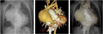
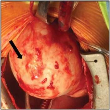
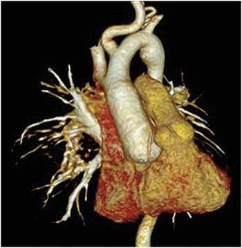
Significant widening of the superior mediastinum was found on chest radiography in a 10-month-old girl with a history of chronic cough. Physical findings included craniofacial (craniosynostosis, hypertelorism, bifid uvula) and skeletal (funnel chest, joint laxity, and club foot) manifestations consistent with type I Loeys–Dietz syndrome (LDS). Molecular genetic testing confirmed a TGFBR1 mutation. Chest radiography and contrast-enhanced CT demonstrated significant dilatation of the aortic root (Z-score+14) and ascending aorta along with a massive aortic aneurysm measuring 56×51 mm at its widest diameter (Fig 1). Elective repair of the aneurysm was determined on the basis of both ascending aortic dimensions exceeding the 99th percentile for age and gender and compression of the right main bronchus. She underwent uneventful aneurysm resection (Fig 2) and valve-sparing aortic root replacement. Follow-up CT-scan 6 months later demonstrated adequate graft reconstruction of the ascending aorta (Fig 3).
Imaging evaluation. Left panel: chest radiograph showing superior mediastinal widening. Middle panel: volume-rendered CT image showing a massive aortic root aneurysm and a dilated and elongated pulmonary artery. Right panel: CT image overlaid on the chest radiograph.

Intraoperative view from the median sternotomy showing the massive aortic root aneurysm (arrow).

Postoperative volume-rendered CT image shows the reconstructed aorta 6 months after surgical intervention.

Loeys–Dietz syndrome is a rare connective tissue disorder characterised by aggressive arterial disease with aneurysm formation at risk for rupture and/or dissection. Interestingly, dissection can occur at smaller aortic diameters than those observed in similar genetic conditions (i.e. the Marfan or the Ehlers–Danlos syndrome). Early surgical intervention with aneurysm resection and repair of the ascending aorta is generally advocated.Reference Williams, Loeys and Nwakanma 1 The risk for aortic dissection still exists, although surgical intervention provides an improved outcome with a lower risk for dissection. This in turn stresses the need for lifelong surveillance, relying on cross-sectional imaging to achieve this purpose.
Acknowledgements
The authors thank Dr Leonardo Flores and Dr Carmen Alaez of the National Institute of Genomic Medicine for their invaluable collaboration in establishing the specific genetic mutation and Dr Dinora Sandoval for precise CT reconstruction and CT/chest X-ray fusion.
Financial Support
This research received no specific grant from any funding agency, commercial or not-for-profit sectors.
Conflicts of Interest
None.
Ethical Standards
The authors assert that all procedures contributing to this work comply with the ethical standards of the relevant national guidelines on human experimentation and with the Helsinki Declaration of 1975, as revised in 2008, and has been approved by the institutional committee of the Ignacio Chavez National Institute of Cardiology.