Introduction
Cognitive abnormalities occur in bipolar disorder (BP) (Bora, Yucel, & Pantelis, Reference Bora, Yucel and Pantelis2010), more severe in attentional and memory domains and also present in euthymic patients.
White matter pathology has been reported by using magnetic resonance imaging (MRI) and diffusion tensor imaging (DTI) predominantly in fronto-temporal areas (Bruno, Cercignani, & Ron, Reference Bruno, Cercignani and Ron2008) and less frequently gray matter loss in the anterior cingulate and dorsolateral prefrontal cortex (Bruno, Barker, Cercignani, Symms, & Ron, Reference Bruno, Barker, Cercignani, Symms and Ron2004) areas where histopathological changes are known to occur (Beasley, Cotter, & Everall, Reference Beasley, Cotter and Everall2002). The association between cortical abnormalities and cognitive impairment in BP has received limited attention and the same applies the possibility that these associations may be different in patients with BPI who experience manic episodes and psychotic symptoms and those with BPII who do not.
We previously reported, using voxel-based morphometry (VBM) and magnetization transfer ratio (MTR) (Bruno, Papadopoulou, Cercignani, Cipolotti, & Ron, Reference Bruno, Papadopoulou, Cercignani, Cipolotti and Ron2006), correlations between IQ change from premorbid levels and abnormalities in the superior temporal, parahippocampal gyri, uncus, and the adjacent white matter particularly in BP II patients. In this exploratory study we investigate associations between cognition and cortical parameters using surface-based morphometry (SBM) (Fischl & Dale, Reference Fischl and Dale2000) that measures independently cortical area and thickness. These indices share a high heritability, but are determined by different genetic mechanisms (Panizzon et al., Reference Panizzon, Fennema-Notestine, Eyler, Jernigan, Prom-Wormley, Neale and Kremen2009) and may respond differently to disease-related factors. We predicted that cortical thinning would be present in fronto-temporal cortex (Rimol et al., Reference Rimol, Hartberg, Nesvag, Fennema-Notestine, Hagler, Pung and Agartz2010) and associated with cognitive impairment. We also explored possible differences in cortico-cognitive associations in BP subgroups.
Methods
Subjects
Thirty-six patients meeting Diagnostic And Statistical Manual-IV (DSM-IV) criteria for BP were included in the study. Twenty-five were recruited from inner-London psychiatric clinics and 11 from respondents to an advertisement in the Journal of the Manic-Depressive Fellowship. Exclusion criteria were previous or current Axis I comorbidity, history of neurological or systemic disease, head injury leading to unconsciousness, and previous history of substance abuse.
Imaging and neuropsychological findings in this cohort have previously been reported (Bruno et al., Reference Bruno, Barker, Cercignani, Symms and Ron2004, Reference Bruno, Papadopoulou, Cercignani, Cipolotti and Ron2006, Reference Bruno, Cercignani and Ron2008; Summers et al., Reference Summers, Papadopoulou, Bruno, Cipolotti and Ron2006). The study was approved by the Joint Research Ethics Committee of the National Hospital for Neurology and Neurosurgery and UCL Institute of Neurology. Written informed consent was obtained from all participants.
Clinical Assessment
Subjects were interviewed by a psychiatrist (S.B.) using the Structured Clinical Interview For DSM-IV (SCID) for DSM-IV Axis I Disorders. All met DSM-IV diagnostic criteria for BP and none had a manic/hypomanic episode at the time of the study. Depressive symptoms at the time of the study were assessed using the Beck's Depression Inventory (BDI).
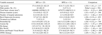
Information was collected about developmental milestones, education, employment, substance misuse, medical history, symptom onset, hospital admissions, exposure to medication and electroconvulsive therapy (ECT), and family history of psychiatric illness. Duration of illness was measured from symptom onset to the MRI date. Clinical details are given in Table 1.
Demographic and cognitive measures in BPI and BPII subgroups

Values are means (SD) [range] no. tested.
*The proportion of subjects in each BP group showing an IQ change greater than the mean IQ change for the sample as a whole was: BPI: 12 of 24 tested (50%) and BPII: 10 of 11 tested (90.9%); Fisher's exact = 0.03.
BP = bipolar disorder.
Neuropsychological Assessment
The battery used has previously been described (Bruno et al., Reference Bruno, Papadopoulou, Cercignani, Cipolotti and Ron2006; Summers et al., Reference Summers, Papadopoulou, Bruno, Cipolotti and Ron2006).
Premorbid IQ was estimated using the Revised National Adult Reading Test (NART).
Current IQ using the Wechsler Adult Intelligence Scale-Revised (WAIS-R). Measures of Full-Scale IQ were obtained pro-rating from four verbal (Vocabulary, Digit Span, Arithmetic, Similarities) and three nonverbal (Picture Completion, Picture Arrangement, Block Design) subtests.
IQ change was the difference between Premorbid and Current IQ. A difference ≥14 IQ points is considered to be outside the norm for the general population (Nelson & Willson, Reference Nelson and Willson1991).
Visual Memory was assessed using the Rey-Osterreich Complex Figure Test and the Doors and People Test: Shapes subtest (DPVR:Shapes) and Executive function using the Spatial Working Memory (SWM) test from the CANTAB battery and the SWM strategy measure (i.e., the number of errors) obtained (higher scores indicated worse performance).
MRI Data Acquisition
MRI was performed on a GE Signa 1.5 Tesla scanner using a standard quadrature head coil. T1-weighted volumetric images were obtained using an inversion recovery spoiled gradient-recalled (IR-SPGR) echo sequence with voxel size of 1.2 × 1.2 × 1.2 mm. A total of 124 axial contiguous slices were acquired. Other parameters were: echo time (TE) = 5.4 ms, repetition time (TR) = 15 ms, inversion time (TI) = 450 ms, field of view = 31 × 16 cm2, acquisition matrix 256 × 128, number of averages = 1, excitation flip angle = 15°, receive bandwidth = 15.63 kHz.
Image processing
Was performed by one of us (L.G.G.), blind to participant status, using Freesurfer 4.3.0 (http://surfer.nmr.mgh.harvard.edu/). This program generates maps of surface area and cortical thickness (Fischl & Dale, 2000). After skull stripping and white matter segmentation, the cortical surface of each hemisphere is inflated to an average spherical surface to locate the pial surface and the gray/white matter boundary. The distance between the two at each “vertex” (i.e., surface point) across the cortex is considered a measure of cortical thickness. Cortical maps are smoothed with 10-mm full-width half-maximum Gaussian kernel and aligned to a common surface template using a high-resolution surface-based averaging technique and 32 cortical parcellations are automatically generated. The only manual step is the correction of topological errors when the above steps had been completed. Total brain volume was also estimated using Freesurfer.
Measurement of cortical parameters
Cortical thickness, surface area, and volume of frontal and temporal regions were measured from the Desikan template, six frontal (superior, pars opercularis, caudal middle, rostral middle, caudal anterior cingulate, and rostral anterior cingulate) and six temporal (superior, middle and inferior, fusiform, temporal pole, and transverse) parcellations in each hemisphere were selected. Average thickness, surface area, and volume of the cortex for the regions covered by these parcellations were calculated for each hemisphere.
Statistical Analysis
Demographic, clinical, and cognitive variables
Age, gender, exposure to lithium, and total brain volume were compared between BPI and BPII subgroups using t tests or Fisher's exact tests. Linear regression models adjusted by age and gender, between subgroups, were used: (a) to compare the clinical variables (disease duration, number of hospital admissions, and BDI score) and cognitive scores; (b) to explore the associations between the clinical and cognitive variables.
Imaging variables
Stata 9.2 (Stata Corporation, College Station, TX) was used for the following statistical analyses: Linear mixed models, which allow repeated measurements within subjects, were used to explore: (a) associations between cortical parameters with gender, age, clinical variables, diagnosis, region, and side in all BP patients; (b) differences in cortical parameters between BP subgroups due to gender, age, and clinical variables with two-way interactions by region and side; (c) differences in cortical parameters between BP subgroups due to diagnosis, brain region and side, with two-way interactions (diagnosis by region, diagnosis by side, and region by side); (d) differences in cortical parameters in specific parcellations using the same model with two-way interaction (diagnosis by region), entering the six parcellations within that region (i.e., frontal or temporal). Age, gender, and total brain volume were used as covariates for these models.
To explore the associations between cognitive scores and cortical parameters the following linear mixed models were used: (a) two-way interactions by region and side to investigate associations between clinical variables and cognitive scores with cortical parameters in the whole group; (b) with two- and three-way interactions to investigate differences in the associations between cognitive scores and cortical parameters in BP. For these analyses, region (frontal and temporal) was entered as a within-subject effect and diagnosis, cognitive scores, gender, and age as between-subjects effects. Three separate models were created for average cortical thickness, surface area, and cortical volume. The same models were also used for side as a within-subject effect; (c) When there was a significant interaction between a cognitive variable and the frontal/temporal cortical parameters, the linear mixed model was repeated with the corresponding indicators and interaction terms for the six parcellations of that particular region (i.e., frontal or temporal).
Adjustment for multiple comparisons was performed using False Discovery Rate (FDR) correction across all tests of each linear mixed model. The level of significance was set at 0.05.
Results
Thirty-six BP patients (mean age, 39.1 years) comprising 25 BPI (mean age, 37.4 years; 10 males); and 11 BPII (mean age, 42.8 years; 3 males) were included. Two patients were unmedicated at the time of the study and information on medication was incomplete for four. The rest were prescribed mood stabilizers (10 lithium, 4 carbamazepine, and 1 lamotrigine) and/or antidepressants (11) and neuroleptics (5). Five patients (three BPI and two BPII) had ECT.
Sixteen patients scored in the normal BDI range (0–9), 11 in the mild/moderate range (10–30) and two in the severe range (above 30). BDI scores were not available in 7 patients.
There were no significant differences in age, gender, duration of illness, or BDI scores between the BPI and II patients, but BPII patients had fewer hospital admissions (p = .013). Demographic and cognitive data are given in Table 1.
The cognitive performance of this cohort has previously been reported (Summers et al., Reference Summers, Papadopoulou, Bruno, Cipolotti and Ron2006). There was no difference in premorbid IQ between BPI and BPII patients. IQ change was significantly greater in the BPII subgroup (p = .003), and there were more subjects in the BPII subgroup in whom IQ change was greater than the mean IQ change for the whole group (Fisher's exact = 0.03). There were no associations between clinical variables and cognitive scores.
There were no significant differences in total brain volume between BPI and BPII subgroups.
Group Differences in Cortical Parameters
There were no differences in frontal or temporal cortical thickness or area between BPI and II patients. There were no significant associations between cortical parameters and age, gender and clinical variables.
Associations Between Cortical Parameters and Cognition
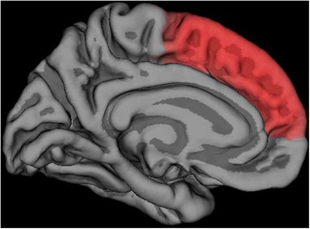
Premorbid IQ was significantly associated with frontal cortical volume in the whole group with an increase of 126.38 mm3 per IQ point (95% CI [32.66 to 220.09]; p = .008). The association with frontal cortical area (with an increase of 37.22 mm2 per IQ point (95% CI [1.80 to 72.63]; p = .039), rather than thickness (95% CI [−0.00562 to 0.00283]; p = .518) explained the link between premorbid IQ and cortical volume. Within the frontal cortex, the area (95% CI [6.04 to 30.92]; p = .048) and volume (95% CI [8.07 to 30.34]; p = .048) of the superior frontal parcellation were the main contributors to these associations (Figure 1).
Parcellation where cortical area and volume were associated with premorbid IQ in the whole BP group: superior frontal.

Current IQ, IQ change, memory and executive function scores, and clinical variables where not associated with frontal or temporal cortical parameters.
Associations of cortical parameters and cognition in BPI and BPII subgroups
There were no differences in the strength of associations between premorbid IQ, memory and executive functions, and frontal cortical parameters in BPI and BPII subgroups. The association between Current IQ and temporal cortical area was stronger in the BPII than in the BPI subgroup (mean difference of subgroup for area increase per IQ point = 56.86 mm2 per IQ point; 95% CI [5.60 to 108.12]; SD = 126.20 mm2 per IQ point; p = .030; effect size [ES] = 0.45) reaching statistical significance in BPII (with an increase of 58.46 mm2 per IQ point (95% CI [11.49 to 105.43]; p = .015), but not in BPI (95% CI [−19.11 to 22.31]; p = .879) patients. The area of the middle (95% CI [8.07 to 30.34]; p = .012) and inferior temporal (95% CI [17.76 to 40.02]; p = .005) parcellations accounted for the association.
The association between IQ change and temporal cortical area was stronger for the BPII subgroup, but did not reach statistical significance (mean difference of subgroup for area reduction per IQ point = 65.89 mm2 per IQ point; 95% CI [−138.39 to 6.61]; SD = 218.38 mm2 per IQ point; p = .075; ES = 0.30).
Discussion
The association between premorbid IQ and cortical area, and hence cortical volume, of the superior frontal cortex was the main finding of the study. No significant associations were present between cortical thickness and other cognitive scores as we had predicted. The association between current IQ and area of the middle and inferior temporal cortex was stronger for the BPII subgroup, but the association with IQ change failed to reach statistical significance. The main limitation of our study is the lack of a normal control group to compare the cognitive performance and cortical parameters of our patients. Our strict selection criteria may suggest that our results may not apply to patients with axis I co-morbidity and the high IQ of our patients may also suggest that our sample may not be fully representative.
Abnormal frontal gyrification related to IQ (McIntosh et al., Reference McIntosh, Moorhead, McKirdy, Hall, Sussmann, Stanfield and Lawrie2009), probably neurodevelopmental in origin (Fornito et al., Reference Fornito, Malhi, Lagopoulos, Ivanovski, Wood, Velakoulis and Yucel2007), has been regarded by some as a possible BP endophenotype (Penttila et al., Reference Penttila, Cachia, Martinot, Ringuenet, Wessa, Houenou and Paillere-Martinot2009) and the association of premorbid, rather than current IQ, and frontal cortical area in our patients is in keeping with these findings.
The lack of a control group prevented us from determining whether fronto-temporal cortical thinning reported by others (Lyoo et al., Reference Lyoo, Sung, Dager, Friedman, Lee, Kim and Renshaw2006; Rimol et al., Reference Rimol, Hartberg, Nesvag, Fennema-Notestine, Hagler, Pung and Agartz2010) was present in our patients, although the MTR reductions in superior and middle temporal gyrus and right anterior cingulate previously reported in this cohort (Bruno et al., Reference Bruno, Papadopoulou, Cercignani, Cipolotti and Ron2006) suggest that this may have been the case. Our results overlap with those we reported in patients with first episode schizophrenia (Gutierrez-Galve et al., Reference Gutierrez-Galve, Wheeler-Kingshott, Altmann, Price, Chu, Leeson and Ron2010) in whom the area of the temporal cortex was associated with estimates of premorbid and current IQ. The similar pattern and severity of cognitive impairment in schizophrenia and BP (Bora, Yucel, & Pantelis, Reference Bora, Yucel and Pantelis2009) and their overlapping genetic risk (Moskvina et al., Reference Moskvina, Craddock, Holmans, Nikolov, Pahwa, Green and O'Donovan2009) give some credence to the findings presented here.
We did not find any associations between cortical thickness and illness duration as reported by others (Lyoo et al., Reference Lyoo, Sung, Dager, Friedman, Lee, Kim and Renshaw2006), although these associations may have been detected in a larger sample.
In contrast with previous reports (Ha, Ha, Kim, & Choi, Reference Ha, Ha, Kim and Choi2009), we did not find cortical differences between BPI and BPII patients, but the associations between IQ and area of the temporal cortex were stronger in BPII patients. The significance of these findings is unclear, but group differences are unlikely to be due to residual depressive symptoms (similar in both groups) or to medication effects, as cognitive changes occur in unmedicated patients (Lopez-Jaramillo et al., Reference Lopez-Jaramillo, Lopera-Vasquez, Ospina-Duque, Garcia, Gallo, Cortez and Vieta2010) and current exposure to medication was similar in both subgroups. These associations may be driven by the lower IQ of the BPII patients and may not be specific to BP.
In summary, while premorbid IQ was associated with frontal cortical area and volume in the whole BP group, we failed to find associations between current cognitive performance and cortical thinning, as described in patients with Alzheimer's disease and other dementias (Du et al., Reference Du, Schuff, Kramer, Rosen, Gorno-Tempini, Rankin and Weiner2007). Our results raise the possibility that cortico-cognitive associations may be different in BPI and BPII patients and future studies in larger, more representative samples are needed to elucidate this further.
Acknowledgments
Dr. Gutiérrez-Galve was supported by grant from the Instituto de Salud Carlos III (FIS, CM07/00048). Dr. Stefania Bruno by a grant from the Brain Research Trust and Dr. Mary Summers by the Multiple Sclerosis Society. Our thanks are due to Professor Miller and other members of the NMR Research Unit of the UCL Institute of Neurology and to the MS Society for supporting the Unit. We are grateful to Ms. Kika Papadopoulou for the original collection of the neuropsychological data. We are grateful to all the subjects who took part in this study. The authors declare no conflict of interest.