INTRODUCTION
Toxoplasma gondii is an intracellular coccidian with a complex life cycle, belonging to the phylum Apicomplexa. Although T. gondii infects a wide range of intermediate hosts, such as mammal and bird species, as well as humans, the sexual cycle of this parasite occurs only in the feline intestinal epithelium, thereby yielding sporozoite-containing oocysts in the feces (Innes, Reference Innes1997; Montoya and Liesenfeld, Reference Montoya and Liesenfeld2004). In the body of the intermediate host, 2 asexual forms, namely acute lytic tachyzoites and latent bradyzoite tissue cysts, are found (Lyons et al. Reference Lyons, McLeod and Roberts2002). Infection of intermediate hosts with T. gondii occurs by ingestion of either tissue cysts (bradyzoites) or oocysts (sporozoites). After infection, bradyzoites and sporozoites rapidly differentiate into tachyzoites and disseminate throughout the body; this is referred to as an acute infection. Although the majority of tachyzoites are usually eliminated by the host's immune response, a small percentage of tachyzoites survive and differentiate into bradyzoites, primarily in the brain and muscle. The bradyzoites persist for the life of the host, and are essentially dormant and harmless (Carruthers and Suzuki, Reference Carruthers and Suzuki2007).
Stage conversion between tachyzoites and bradyzoites is a sporadic phenomenon, rather than occurring en masse (Ferreira da Silva Mda et al. Reference Ferreira da Silva Mda, Barbosa, Gross and Lüder2008). The co-existence of tachyzoites and bradyzoites in one vacuole was demonstrated in an in vitro experimental study (Bohne et al. Reference Bohne, Heesemann and Gross1993). Although such a vacuole, containing both tachyzoites and bradyzoites, has not yet been observed in animal tissue, latent cysts containing both SAG1-positive and -negative zoites have been observed in the mouse brain (Silva et al. Reference Silva, Gazzinelli, Silva, Ferro, Kasper and Mineo1998; Bohne et al. Reference Bohne, Hunter, White, Ferguson, Gross and Roos1998). The co-existence of latent cysts and tachyzoites in the brain has also been shown in animal and human studies (Ferguson et al. Reference Ferguson, Hutchison and Pettersen1989; Takashima et al. Reference Takashima, Suzuki, Xuan, Nishikawa, Unno and Kitoh2008). Thus, T. gondii stage conversion seems to be a gradual and progressive event.
Time-lapse analysis is a powerful tool for studying gradually progressing events. In the field of virology, for example, simultaneous tracking of capsid, tegument, and envelope proteins of herpes simplex virus-1 characterized how virus particles are produced, providing the first evidence that these three proteins are assembled in the trans-Golgi network of the host cell (Sugimoto et al. Reference Sugimoto, Uema, Sagara, Tanaka, Sata, Hashimoto and Kawaguchi2008). T. gondii stage conversion is also a gradually progressing event, like the assembly of a virus particle. However, to further understand this phenomenon, the behaviour of stage-converting zoites needs to be characterized over time. To date, T. gondii stage conversion and the detection of stage-specific antigens has been demonstrated utilizing immunohistochemistry (Bohne et al. Reference Bohne, Heesemann and Gross1993, Reference Bohne, Heesemann and Gross1994; Gross et al. Reference Gross, Bormuth, Gaissmaier, Dittrich, Krenn, Bohne and Ferguson1995; Lüder et al. Reference Lüder, Giraldo-Velásquez, Sendtner and Gross1999; Ferguson, Reference Ferguson2004). The necessity of a fixation procedure for immunohistochemistry makes it impossible to observe living parasites during stage conversion. To observe temporal changes in gene-expression patterns in stage-converting T. gondii zoites, Lyons et al. (Reference Lyons, Lyons, McLeod and Roberts2001) harvested mRNA from cell cultures infected with stage-converting zoites once every 24 h. They clearly described the stage conversion process in a mass of parasites in terms of the changes in stage-specific gene expression. However, even with this method, it is impossible to fully characterize the mechanism underlying the stage conversion of an individual parasite.
To identify the mechanism underlying the stage conversion of an individual parasite, it is necessary to ascertain the stage the observed parasite is in without killing it. That is, it is necessary to utilize live-cell imaging. Fluorescent proteins have proven to be powerful tools for live-cell imaging (Day and Schaufele, Reference Day and Schaufele2008). In the present study, a transgenic T. gondii line expressing stage-specific red and green fluorescent proteins (DsRed Express and GFP, respectively) was constructed. This transgenic parasite permits live imaging of the stage conversion in T. gondii.
MATERIALS AND METHODS
Mice
Female, 6 to 8-week-old, C57BL/6J and BALB/c mice were purchased from Oriental Yeast Co., Ltd (Tokyo, Japan). The C57BL/6J mice were infected with parasites and their organs excised after euthanasia by cervical dislocation. BALB/c mice were used for the production of polyclonal antibody. Experiments were performed in accordance with the Gifu University Animal Care and Use Committee guidelines.
Cells and parasites
The African green monkey-kidney cell line Vero was cultured in RPMI1640 medium supplemented with 7·5% FCS at 37°C in a 5% CO2 incubator. T. gondii tachyzoites of the following strains were propagated for routine use in Vero cells: T. gondii P(LK) HXGPRT-strain (Roos et al. Reference Roos, Donald, Morrissette and Moulton1994; Pfefferkorn and Borotz Reference Pfefferkorn and Borotz1994), GFP-expressing stable transgenic T. gondii (PLK/GFP) (Zhang et al. Reference Zhang, Huong, Battur, Zhou, Zhang, Liao, Kawase, Lee, Dautu, Igarashi, Nishikawa and Xuan2007; Nishikawa et al. Reference Nishikawa, Zhang, Ibrahim, Ui, Ogiso and Xuan2008), DsRed Express-expressing stable transgenic T. gondii (PLK/RED) (Takashima et al. Reference Takashima, Suzuki, Xuan, Nishikawa, Unno and Kitoh2008), and the dual fluorescent protein-expressing stable transgenic T. gondii (PLK/DUAL), engineered as described below. The PLK/GFP line expresses GFP during the tachyzoite and bradyzoite stages (Nishikawa et al. Reference Nishikawa, Zhang, Ibrahim, Ui, Ogiso and Xuan2008; Zhang et al. Reference Zhang, Huong, Battur, Zhou, Zhang, Liao, Kawase, Lee, Dautu, Igarashi, Nishikawa and Xuan2007). PLK/RED expresses DsRed Express only during the tachyzoite stage (Takashima et al. Reference Takashima, Suzuki, Xuan, Nishikawa, Unno and Kitoh2008). PLK/Dual expresses DsRed Express and GFP as described below.
Construction of the transfer vector
The promoter region of the bradyzoite-specific antigen 1 (BAG1) gene was PCR amplified from the plasmid Bag/cat (kindly provided by Dr Bohne, Georg-August University, Göttingen, Germany) using the following primer set:
5′-AAGCTTCTTCCAGTTGCCCGGCTCTGGGTACCTTCT-3′ and
5′-GGATCCCTTTTTTGAATATCATACGGGACCTGGGCT-3′.
The amplified fragment was cloned into the pT7Blue T-Vector (Novagen, Darmstadt, Germany), and the resulting plasmids were designated as pT7/BAGp. The BAG1 gene promoter was obtained from pT7/BAGp by digestion with BamHI and HindIII, followed by ligation with the BamHI- and HindIII-digested plasmid pUC/GRA (Takashima et al. Reference Takashima, Suzuki, Xuan, Nishikawa, Unno and Kitoh2008). The resulting plasmid was designated pUC/BAG-GRA. The gene for enhanced GFP was obtained from the plasmid, pCX-EGFP (Niwa et al. Reference Niwa, Yamamura and Miyazaki1991) (kindly provided from Dr J. Miyazaki, Osaka University, Japan). The obtained GFP gene was blunt-ended with Klenow fragments and inserted into the blunt-ended BamHI site of the plasmid pUC/BAG-GRA. The resulting plasmid was designated pBAG-GFP. GFP expression units, under the control of the BAG1 promoter, and DsRed Express (Clontech, Palo Alto, CA, USA), under the control of the major surface antigen 1 (SAG1) promoter, were obtained from the plasmids pBAG-GFP and pSAG-RED (Takashima et al. Reference Takashima, Suzuki, Xuan, Nishikawa, Unno and Kitoh2008). The obtained GFP and DsRed Express expression units were inserted into the HindIII and BamHI sites of the plasmid pminiHXGPRT (kindly provided by Dr Roos, Stanford University, CA, USA) (Roos et al. Reference Roos, Donald, Morrissette and Moulton1994; Pfefferkorn et al. Reference Pfefferkorn and Borotz1994), respectively, as shown in Fig. 1A. The resulting plasmid was designated pminiHXGPRT/DUAL.
Construction of transgenic Trypanosoma gondii expressing DsRed Express and GFP. (A) Schema of the transfer vector, pminiHXGPRT/DUAL. Genes coding DsRed Express and GFP are under the control of the SAG1 and BAG1 promoter, respectively. (B) Vero cells, infected with PLK/DUAL tachyzoites, were cultured in pH 7·0 medium (upper panels) and pH 8·1 medium (lower panels). Left and centre panels show red and green fluorescence detected by fluorescence microscopy by 1/3·0 sec and 1·5 sec exposure, respectively. Right panels show overlay images of red and green fluorescence. Arrows in the expanded figure indicate green fluorescent zoites. (C) Fluorescent PLK/DUAL zoites in a vacuole. PLK/DUAL tachyzoites cultured in pH 7·0 medium (upper panels) and pH 8·1 medium (lower panels) observed at higher magnification. (D) PLK/DUAL or PLK/RED zoites released from infected Vero cells cultured in pH 8·1 medium (left and light panels, respectively). GFP and DsRed Express were detected by Fl-1 and Fl-2, respectively.

Parasite transfection and selection
A total of 100 μg of the plasmid pminiHXGPRT/DUAL was electroporated into P(LK) HXGPRT–tachyzoites as described previously (Soldati and Boothroyd, Reference Soldati and Boothroyd1993), and the transfected parasites were then added to Vero cell cultures. Forty-eight hours after electroporation, medium containing 25 μg/ml mycophenolic acid and 40 μg/ml xanthine was added to the Vero cell culture. Stable transgenics were selected for in the presence of mycophenolic acid and xanthine for 10 days. Following selection, cloning of red fluorescent tachyzoites was performed, and the resulting clone was designated PLK/DUAL.
In vitro stage conversion
Stage conversion of tachyzoites into bradyzoites was induced by high pH or the p38 MAPK inhibitor SB202190, as previously described (Soete et al. Reference Soete, Fortier, Camus and Dubremetz1993, Radke et al. Reference Radke, Donald, Eibs, Jerome, Behnke, Liberator and White2006) with minor modifications. For induction of stage conversion by high pH, Vero cells that were infected with free PLK/DUAL or PLK/RED tachyzoites were cultured in DMEM medium (pH 8·1), supplemented with 2·0% FCS and 20 μg/ml gentamicin, at 37°C in a humidified incubator for 4 or 7 days. For induction of stage conversion by the p38 MAPK inhibitor SB202190, Vero cells were pre-incubated in DMEM medium containing 1·0% FCS and 8 nm SB202190 (In Solution™ SB202190, Calbiochem, USA) for 3 h prior to tachyzoite infection, and washed with DMEM medium containing 1·0% FCS. Following incubation with PLK/DUAL tachyzoites were suspended in DMEM medium containing 1·0% FCS for 4 h, supernatant was removed and fresh medium containing 1·0% FCS and 8 nm SB202190 was added. The infected Vero cell cultures were incubated at 37°C in a 5% CO2 incubator for 6 days.
Flow cytometry
Flow cytometry was performed on a FACSCalibur (BD Biosciences PharMingen, San Diego, CA, USA) to detect green and red fluorescence in purified free tachyzoites from Vero cells, as described previously (Unno et al. Reference Unno, Suzuki, Xuan, Nishikawa, Kitoh and Takashima2008). GFP and DsRed Express expression were detected using Fl-1 and Fl-2, respectively.
In vivo stage conversion
To observe parasites during the acute phase, C57BL/6J mice were infected intraperitoneally with 1×10⁶ PLK/DUAL tachyzoites. The brains of the infected mice were excised 6 days later, and fixed and embedded in plastic resin (Technovit 8100, Heraeus Kulzer, Wehrheim, Germany). Thin brain sections were directly observed using fluorescent microscopy, as previously described (Takashima et al. Reference Takashima, Suzuki, Xuan, Nishikawa, Unno and Kitoh2008). To prepare latently infected mice, C57BL/6J mice were intraperitoneally infected with 20-1000 PLK/DUAL or PLK/GFP tachyzoites and observed for 6 weeks. Mice that survived more than 6 weeks without symptoms were labelled as latently infected mice and used in subsequent experiments. To observe parasites during the latent phase, the brains of latently infected mice were excised and chopped into small pieces. The brain pieces were then crushed onto glass slides and observed under fluorescence microscopy and confocal laser microscopy. Red fluorescence from the crushed samples was detected by short (1/3·0 sec) and long (1·5 sec) time exposure. Green fluorescence from the crushed samples was detected by just long-time exposure.
Reactivation of latent brain cysts
Latently infected mice were prepared as described above. Dexamethasone (dexamethasone 21-phosphate disodium salt; Sigma) was dissolved at a concentration of 10 mg/l in the drinking water, and the latently infected mice were allowed free access to the dexamethasone-containing water. After 28 days of dexamethasone treatment, the brains of treated mice were excised and chopped into small pieces. The pieces of brain were crushed onto glass slides and observed under fluorescence microscopy.
Anti-BAG1 antiserum
The truncated BAG1 gene (aa 94-229,) with introduced BamHI and SmaI sites, was synthesized and ligated with the pGEX-4T-3 vector (Amersham Pharmacia Biotech, PA), which was digested with BamHI and SmaI. E. coli (BL21) was transformed with the resulting plasmid and cultured in LB medium containing 100 μg/ml ampicillin at 37°C until OD600 reached 0·3. Isopropyl-β-D(-)-thiogalactopyranoside was then added to the medium (final concentration, 1 mm). Following 3 h of incubation, the transgenic E. coli were pelleted by centrifugation at 800 g for 10 min at room temperature to remove the supernatant. Soluble, bacterial proteins were eluted using B-PER™ Reagent (PIERCE, USA) according to manufacturer's instructions. From the soluble proteins, glutathione S-transferase (GST) fusion protein (GST-BAG1)-was purified using Glutathione Sepharose™ 4B (Amersham Bioscience, Sweden) according to manufacturer's instructions. A total of 50 μl of purified GST-BAG1 solution (containing 16·3 μg/μl GST-BAG 1) was mixed with 100 μg of adjuvant, TiterMax Gold (TiterMax USA. INC., GA, USA). A total of 100 μl of the mixture was subcutaneously injected into each of three 6-week-old female BALB/c mice. Thirty-two days after the first immunization, a second immunization was performed by injecting 200 μl of the mixture. Serum (anti-BAG1 antiserum) was harvested from immunized mice 20 days after the second immunization, and was stored at −20°C until further use.
Immunostaining
Free PLK/DUAL tachyzoites, prepared as previously described (Unno et al. Reference Unno, Suzuki, Xuan, Nishikawa, Kitoh and Takashima2008), were incubated for 45 min at 37°C in an air incubator with anti-SAG1 mAb (3T×19 MAb TP3, HyTest Ltd, Turku, Finland) or anti-BAG1 serum 50× diluted in PBS containing 3% FCS. After washing 3 times with PBS containing 3% FCS, the tachyzoites were incubated for 45 min at 37°C in an air incubator with Alexa Fluor® 350-labelled goat anti-mouse IgG (H+L) (Invitrogen). After washing 3 times with PBS containing 3% FCS, the reacted tachyzoites were mounted with ProLong® Gold anti-fade reagent (Invitrogen) onto glass slides and covered with a cover-slip. The glass slides were incubated for 30 min at room temperature in the dark and observed by fluorescence microscopy.
RESULTS
Construction of transgenic T. gondii expressing dual fluorescent proteins
To produce transgenic parasites expressing stage-specific dual fluorescent proteins, we constructed a transfer vector, pminiHXGPRT/DUAL. The transfer vector contained genes coding for DsRed Express and GFP under the control of SAG1 and BAG1 promoters, respectively (Fig. 1A). T. gondii P(LK) HXGPRT- strain tachyzoites were transformed with the transfer vector, pminiHXGPRT, and the stably transformed clone was designated as PLK/DUAL. The PLK/DUAL tachyzoites showed red fluorescence under pH 7·0 culture conditions (Fig. 1B, upper panel and Fig. 1C, upper panel). Although 293 zoites were observed, the number of red fluorescence negative zoites was only a few (6/293). During in vitro culture in pH 7·0 media for 1 month, with repeated passages, the proportion of fluorescent negative zoites did not increase. When PLK/DUAL tachyzoites were cultured under a stage conversion-inducing condition (pH 8·1 medium), a number of green fluorescence-positive tachyzoites were observed (Fig. 1B, lower panel and Fig. 1C, lower panel). Green fluorescence-positive tachyzoites were not detected in the pH 7·0 culture condition (Fig. 1B upper panel and Fig. 1C, upper panel). To confirm that the green fluorescence expression under the pH 8·1 condition was not the result of autofluorescence observed only under high pH conditions, but was rather caused by specific GFP expression in PLK/DUAL zoites, a transgenic T. gondii strain, PLK/RED, which expressed DsRed Express but no GFP, was also cultured in the pH 8·1 media. As shown in Fig. 1D, green fluorescence-positive zoites were detected in Vero cell cultures infected with PLK/DUAL, but not in those infected with PLK/RED. These data suggest that PLK/DUAL expressed GFP during the bradyzoite stage.
Stage-specific expression of fluorescent proteins by PLK/DUAL
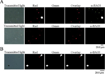
To investigate whether DsRed Express and GFP were expressed stage specifically, PLK/DUAL zoites were stained with anti-SAG1 antibody or anti-BAG1 antiserum. Under pH 7·0 conditions, before the antibody staining procedure, almost all parasites expressed Ds Red Express but no zoite expressed GFP (Fig. 1B, C). When the parasites cultured under pH 7·0 conditions, all zoites were stained by anti-SAG1 antibody but specific staining was not revealed by anti-BAG1 antiserum (Fig. 2A). SAG1-/DsRed Express+zoite was not observed. Although some parasites looked like SAG1+/DsRed Express – (Fig. 2A, arrows), it must be a result of artificial reduction of fluorescent intensity by the staining procedure because almost all zoites showed strong red fluorescence before the staining procedure (Fig. 1B). These results indicate that the DsRed Express-positive zoites, cultured under pH 7·0 conditions, were in the tachyzoite stage.
Stage-specific expression of DsRed Express and GFP by PLK/DUAL. (A) Purified free zoites from PLK/DUAL-infected Vero cells cultured in pH 7·0 medium. The purified free zoites were stained with anti-SAG1 antibody (upper panels) or anti-BAG1 antiserum (lower panels). Goat anti-mouse IgG (H+L)-Alexa 350, secondary antibodies were used for both experiments. Transmitted light images, red fluorescence images, green fluorescence images, overlay images of red and green fluorescence, and blue fluorescence images (α-SAG1 or α-BAG1) are shown. The blue fluorescence images were converted into grey scale. Arrow in the upper panels indicates zoites in which the red fluorescence had been reduced by the staining procedure. (B) Purified free zoites from PLK/DUAL-infected Vero cells cultured in medium containing SB202190. The purified free zoites were stained with anti-BAG1 antiserum. Goat anti-mouse IgG (H+L)-Alexa 350 secondary antibody was used. Transmitted light images, red fluorescence images, green fluorescence images, overlay images of red and green fluorescence, and blue fluorescence images (α-BAG1) are shown. Arrowhead and arrow indicate red and green fluorescent zoites, respectively. The blue fluorescence images were converted into grey scale.

When PLK/DUAL zoites were cultured under stage conversion-inducing conditions, either high pH or the presence of a p38 MAPK inhibitor, a number of zoites expressed not only DsRed Express, but also GFP (Fig. 1B, C, D, and Fig. 2B). The intensity of GFP expression varied between individual zoites (Fig. 2B). Zoites cultured in medium containing a p38 MAPK inhibitor were stained with anti-BAG1 antiserum. Fig. 2B demonstrates a zoite that was strongly expressing GFP and was BAG1 positive, as well as a zoite showing weak GFP expression that was weakly BAG1 positive. Although only a few zoites are shown in Fig. 2B, all observed zoites in this culture demonstrated a similar trend (data not shown). These results indicate that BAG1 and GFP expression patterns were synchronized; in other words, GFP expression indicated bradyzoite stage-specific expression.
DsRed Express and GFP in vivo expression in the PLK/DUAL strain
To confirm in vivo, stage-specific expression of DsRed Express and GFP by the PLK/DUAL strain, C57BL/6J mice were infected with PLK/DUAL tachyzoites. The brain samples were observed at acute, latent, and reactivating phases. In the acute phase, disseminated red fluorescence-positive zoites were observed in the brain samples (Fig. 3A and D, left panel). However, GFP expression was not detected (Fig. 3A). During the latent phase, green fluorescence-positive cyst and green fluorescence-positive bradyzoites within the cyst were observed in the brain samples (Fig. 3B and D, centre panel). As shown in Fig. 3B, red fluorescence was not detected in these cysts by short-time exposure (1/3·0 sec). Whole brain tissues from 1 latently infected mouse were examined by short-time exposure. Analysis resulted in neither red fluorescence-positive cysts nor tachyzoites. The expression of fluorescent proteins was detected in the parasites but secretion into the parasitophorous vacuole was not observed (Fig. 3D). To investigate whether reactivated tachyzoites from latent cysts also expressed DsRed Express, latent cysts were reactivated by administration of dexamethasone to the latently infected mice. After 28 days, red fluorescence-positive zoites were observed in the brain; however, green fluorescence-positive zoites were not detected (Fig. 3C). These results indicate that stage specificity could be identified in the PLK/DUAL living zoites by fluorescence.
Red and green in vivo fluorescence in the PLK/DUAL strain. (A-C) PLK/DUAL fluorescence in the brains of infected C57BL/6J mice during the acute phase (A), latent phase (B) and reactivating phase (C). Red fluorescence was detected by short exposure (1/3·0 sec). Green fluorescence was detected by long exposure (1·5 sec). Transmitted light images, red fluorescent images, green fluorescent images, and overlay images of transmitted, red fluorescent, and green fluorescent images are shown. (D) Area shown by the dotted frames in (A) (left panel), (B) (centre panel) and (C) (right panel) were observed by confocal laser microscopy. Note that images in (D) are shown as mirrored images of (A), (B) and (C) because of the difference in structure of the fluorescent and confocal laser microscopes.

Low-level DsRed Express expression in PLK/DUAL cysts
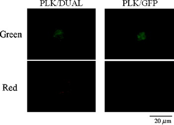
As described above, red fluorescence-positive PLK/DUAL latent cysts were not detected by short-time exposure (Fig. 3B). To determine whether PLK/DUAL bradyzoites in the latent cysts express a trace amount of DsRed Express or none at all, PLK/DUAL latent cysts were observed using a long-time exposure. The results demonstrated that a portion of the bradyzoites in latent cysts showed weak red fluorescence (Fig. 4 left panel). Bradyzoites in latent PLK/GFP cysts did not show red fluorescence (Fig. 4 right panel). These results indicate that the weak red fluorescence in the PLK/DUAL bradyzoites was not because of GFP bleed-through into the red fluorescent channel, but rather was caused by the presence of DsRed Express molecules in the bradyzoites.
Expression of DsRed Express in latent cysts of the PLK/DUAL strain. Latent cysts of the PLK/DUAL (left panels) and PLK/GFP (right panels) strains in the brains of infected C57BL/6J mice are shown. Red fluorescence was detected by long exposure (1·5 sec). Green fluorescence was detected under the conditions described in Fig. 3.

DISCUSSION
The present study demonstrated the use of a transgenic T. gondii strain, PLK/DUAL, as a tool to investigate stage conversion in T. gondii. The transgenic parasites express DsRed Express during the tachyzoite stage and GFP during the bradyzoite stage. Several transgenic parasite-expressing fluorescent proteins have been reported as tools for investigation for stage conversion in T. gondii and/or visualization of parasites in host tissue (Vanchinathan et al. Reference Vanchinathan, Brewer, Harb, Boothroyd and Singh2005; Takashima et al. Reference Takashima, Suzuki, Xuan, Nishikawa, Unno and Kitoh2008; Nishikawa et al. Reference Nishikawa, Zhang, Ibrahim, Ui, Ogiso and Xuan2008). This method is a powerful tool with which to observe one-way stage conversion, from tachyzoite to bradyzoite, or from bradyzoite to tachyzoite. However, none of these methods have allowed for the observation of stage conversion in both directions. Vanchinathan et al. (Reference Vanchinathan, Brewer, Harb, Boothroyd and Singh2005) constructed a transgenic parasite that fluoresced only during the bradyzoite stage. Using this transgenic parasite, stage conversion from tachyzoites into bradyzoites could easily be detected as an emission of fluorescence. However, it was impossible to detect reactivation of the parasite (stage conversion from bradyzoite to tachyzoite) using fluorescence. We have also reported a transgenic parasite that fluoresced only during the tachyzoite stage (Takashima et al. Reference Takashima, Suzuki, Xuan, Nishikawa, Unno and Kitoh2008), and have visualized T. gondii reactivation in brain tissue. However, it is difficult to observe entrance into a latent state (stage conversion from tachyzoite to bradyzoite), because bradyzoites in brain cysts do not show bright fluorescence. To better understand the life cycle of T. gondii in the intermediate host, it is important to study the mechanisms of stage conversion between tachyzoites and bradyzoites in both directions. Although much knowledge regarding stage conversion has been independently provided by several research groups (Vanchinathan et al. Reference Vanchinathan, Brewer, Harb, Boothroyd and Singh2005; Takashima et al. Reference Takashima, Suzuki, Xuan, Nishikawa, Unno and Kitoh2008), various strains of T. gondii were used for these studies. Because of the diverse properties among parasite strains (Gross et al. Reference Gross, Kempf, Seeber, Lüder, Lugert and Bohne1997; Grigg et al. Reference Grigg, Bonnefoy, Hehl, Suzuki and Boothroyd2001), an assortment of independent reports does not directly result in an overall understanding of the T. gondii life cycle. The PLK/DUAL strain, in which stage conversion is visualized in both directions by dual fluorescence, could provide a greater understanding of the T. gondii life cycle in the intermediate host. T. gondii cysts are primarily formed in brain and muscle tissue of the host (Turner, Reference Turner1978). Latent cysts have also been reported in the placenta (Dubey, Reference Dubey1987). This indicates that T. gondii tachyzoites can convert to bradyzoites in the placenta. In addition, it was reported that there is lag-time between T. gondii infection in the placenta and the fetus in some pregnant mouse models (Shiono et al. Reference Shiono, Mun, He, Nakazaki, Fang, Furuya, Aosai and Yano2007). There is a possibility that the bradyzoites existing in the cysts in the placenta could reactivate and reach the fetus later. Thus, to understand the behaviour of T. gondii in the placenta, observation of stage conversion in both directions is necessary. Therefore, the PLK/DUAL strain could contribute to further understanding of the mechanisms of T. gondii vertical infections.
Stage conversion between tachyzoite and bradyzoite is associated with morphological and molecular biological changes, including stage-specific antigen expression, many of which have been identified (Lyons et al. Reference Lyons, McLeod and Roberts2002). To construct transgenic T. gondii stage-specifically expressing fluorescent proteins, the promoters of the SAG1 and BAG1 genes were utilized, thereby specifically targeting expression in tachyzoites and bradyzoites, respectively (Ferguson, Reference Ferguson, Hutchison and Pettersen2004). In addition, it has been reported that stage-specific expression of SAG1 and BAG1 is controlled at the transcriptional level (Cleary et al. Reference Cleary, Singh, Blader, Brewer and Boothroyd2002; Bohne et al. Reference Bohne, Wirsing and Gross1997). SAG1 gene transcription occurs during the tachyzoite stage, and decreases drastically during stage conversion from tachyzoite to bradyzoite (Cleary et al. Reference Cleary, Singh, Blader, Brewer and Boothroyd2002). By contrast, the upregulation of BAG1 mRNA occurs early during differentiation from tachyzoite to bradyzoite (Bohne et al. Reference Bohne, Wirsing and Gross1997). To confirm stage-specific expression of fluorescent proteins by PLK/DUAL zoites, immunostaining was performed. Although zoites showing weak and no red fluorescence were observed after SAG-1 staining, this loss of red fluorescence of zoite may be artifact of the staining procedure because almost all PLK/DUAL showed strong red fluorescence before staining. DsRed Express positive and GFP negative zoites exhibited SAG1-expression, but no BAG1-expression. When PLK/DUAL zoites were cultured in medium containing SB202190, a number of zoites expressed BAG1, as well as GFP. Strongly BAG1-positive zoites exhibited brighter GFP expression compared with BAG1-negative or BAG1-slightly positive zoites. These results indicate that DsRed Express and GFP expression patterns coincided with SAG1 and BAG1, respectively; in other words, tachyzoites were DsRed Express and SAG1 positive, and bradyzoites were GFP and BAG1 positive. In vivo analysis demonstrated that zoites in the acute and reactivating phases, most likely tachyzoites, exhibited DsRed Express expression; however, GFP expression was undetectable. By contrast, zoites within latent cysts, most likely bradyzoites, exhibited GFP expression, and DsRed Express expression was undetectable. Therefore, in vivo PLK/DUAL zoite stages could also be identified by fluorescence. Although it is necessary to consider the effect of expressed fluorescent proteins to immune reactions and parasites' behaviours, the PLK/DUAL strain could be a tool for investigation of in vivo stage-conversion of T. gondii. The intensity of the DsRed Express expressed by PLK tachyzoites was strong enough to detect zoites in the brain tissue from the outside (Takashima et al. Reference Takashima, Suzuki, Xuan, Nishikawa, Unno and Kitoh2008). However, despite the usage of a strong bradyzoite-specific BAG1 promoter (Bohne et al. Reference Bohne, Wirsing and Gross1997), the intensity of green fluorescence of GFP expressed by bradyzoites was not enough to detect latent cysts in that manner. Trying out other fluorescent proteins and bradyzoite-specific promoters, it might be possible to construct more useful recombinant parasites, of which latent cysts can be identified by strong fluorescence without either squashing tissue or making thin sections.
Bohne et al. (Reference Bohne, Heesemann and Gross1993) observed a parasitophorous vacuole containing both tachyzoites and bradyzoites during the in vitro stage-converting phase. It was also reported that there is a point when parasites are expressing both SAG1 and BAG1 (Ferguson, Reference Ferguson2004). We also observed a PLK/DUAL cyst containing both red fluorescence +/green fluorescence – and red fluorescence +/green fluorescence+zoites in the brain at 28 days after PLK/DUAL infection (data not shown). It suggested that PLK/DUAL also has an SAG1/BAG1 double-positive phase during stage-conversion. Although SAG1 is regarded as a tachyzoite-specific antigen, latent cysts of the T. gondii ME-49 strain have been shown to contain some slightly positive SAG1 zoites (Silva et al. Reference Silva, Gazzinelli, Silva, Ferro, Kasper and Mineo1998). The PLK strain is a clonal derivative of ME-49 (Bohne et al. Reference Bohne, Hunter, White, Ferguson, Gross and Roos1998). Therefore, we attempted to detect low levels of red fluorescence in the PLK/DUAL latent cysts using a long-time exposure. As shown in Fig. 4, weak red fluorescence was detected in PLK/DUAL latent cysts. Considering that weak expression was detected not only in the cyst shown in Fig. 4, but also in all observed PLK/DUAL cysts (data not shown), it is unlikely that the weak red fluorescence indicates reactivation. We have previously reported that latent cysts of the PLK/RED strain exhibit much brighter red fluorescence at the point of inception of reactivation (Takashima et al. Reference Takashima, Suzuki, Xuan, Nishikawa, Unno and Kitoh2008). Our present results suggest low-level SAG1 promoter activity during the latent phase in the PLK strain.
It has been reported that T. gondii modifies many signalling cascades within the host cell (Seaij et al. Reference Saeij, Coller, Boyle, Jerome, White and Boothroyd2007; Fouts and Boothroyd, Reference Fouts and Boothroyd2007). It has also been demonstrated that tachyzoites and bradyzoites can induce host transcriptional changes (Fouts and Boothroyd, Reference Fouts and Boothroyd2007). However, it is not yet known what biological activity exists between tachyzoite- and bradyzoite-infected cells to induce various host transcriptional changes in different manners. To elucidate the difference in the mechanisms of biological activity, that is, their reactions to exogenous stimuli, it is necessary to identify the stages of the infected parasites without killing them. As well, it is necessary to observe the parasite and/or host cells over a period of time. The PLK/DUAL strain could be an effective tool for investigating the biological activities of tachyzoite- and/or bradyzoite-infected host cells.
The following reagents were obtained through the AIDS Research and Reference Reagent Program, Division of AIDS, NIAID, NIH from Dr David Roos: Toxoplasma gondii Host Strain P(LK) HXGPRT and Toxoplasma gondii Selection Plasmid (pminiHXGPRT). The plasmid Bag/cat was provided by Dr Wolfgang Bohne. We thank Dr David Roos and Dr Wolfgang Bohne for supplying these materials. We thank Dr Fumie Aosai for discussion. This work was supported by the Health Labor Sciences Research Grant, Research on HIV/AIDS from the Ministry of Health, Labor and Welfare of Japan.