Behavioral inhibition (BI), a temperament identified in infancy, involves fear of novelty and vigilance to threat (Fox, Henderson, Rubin, Calkins, & Schmidt, Reference Fox, Henderson, Rubin, Calkins and Schmidt2001; Kagan, Reznick, & Snidman, Reference Kagan, Reznick and Snidman1987). Although BI increases risk for anxiety disorders (Clauss & Blackford, Reference Clauss and Blackford2012), many children with this temperament do not develop psychopathology and hence demonstrate resilience (Degnan & Fox, Reference Degnan and Fox2007). Research examining the neural correlates of BI has largely focused on anxiety-related outcomes rather than the processes that promote resilience. While impairments in threat/safety discrimination during extinction recall are associated with anxiety disorders, the ability to correctly distinguish threat from safety cues may be one pathway involved in moderating the development of anxiety. Hence, research on extinction recall following conditioned fear learning provides a particularly promising avenue to map aspects of resilience related to anxiety disorders in this temperamentally at-risk population. The current study compares brain regions engaged during extinction recall, in healthy adults with and without a childhood history of BI.
While BI confers risk for anxiety disorders (Chronis-Tuscano et al., Reference Chronis-Tuscano, Degnan, Pine, Perez-Edgar, Henderson, Diaz and Fox2009; Corcoran, Desmond, Frey, & Maren, Reference Corcoran, Desmond, Frey and Maren2005), only about half of children with BI manifest anxiety later in life (Clauss & Blackford, Reference Clauss and Blackford2012). Thus, while BI is a risk factor for anxiety disorders, it is phenomenologically distinct from this class of mental illness. Studies focusing on the function of the amygdala (Guyer et al., Reference Guyer, Benson, Choate, Bar-Haim, Perez-Edgar, Jarcho and Nelson2014; Perez-Edgar et al., Reference Perez-Edgar, Roberson-Nay, Hardin, Poeth, Guyer, Nelson and Ernst2007), striatum (Bar-Haim et al., Reference Bar-Haim, Fox, Benson, Guyer, Williams, Nelson and Ernst2009; Guyer et al., Reference Guyer, Benson, Choate, Bar-Haim, Perez-Edgar, Jarcho and Nelson2014; Helfinstein et al., Reference Helfinstein, Benson, Perez-Edgar, Bar-Haim, Detloff, Pine and Ernst2011; Jarcho et al., Reference Jarcho, Fox, Pine, Etkin, Leibenluft, Shechner and Ernst2013, Reference Jarcho, Fox, Pine, Leibenluft, Shechner, Degnan and Ernst2014) and prefrontal cortex (PFC); (Guyer et al., Reference Guyer, Nelson, Perez-Edgar, Hardin, Roberson-Nay, Monk and Ernst2006, Reference Guyer, Benson, Choate, Bar-Haim, Perez-Edgar, Jarcho and Nelson2014; Jarcho et al., Reference Jarcho, Fox, Pine, Etkin, Leibenluft, Shechner and Ernst2013, Reference Jarcho, Fox, Pine, Leibenluft, Shechner, Degnan and Ernst2014) have established long-term associations between BI in early life and brain function well into adolescence and young adulthood. These studies recognize BI as a risk factor and examine the neural correlates of BI predicting the development of anxiety. However, brain imaging research to date has not focused on the unique characteristics of those with BI in childhood who do not develop anxiety disorders later in life.
Research on fear conditioning and extinction has generated insights on risk and resilience for anxiety disorders (for a review, see Shechner, Hong, Britton, Pine, & Fox, Reference Shechner, Hong, Britton, Pine and Fox2014). During fear conditioning, a neutral stimulus (CS+) is paired with an aversive stimulus (UCS) until a CS+-threat association is formed. In extinction, the CS+ is presented in the absence of the aversive UCS, resulting in a new CS+-safe association. Extinction recall occurs when the extinguished CS+ is presented again at a later time. Preliminary research in BI suggest that when at-risk children are exposed to mildly anxiety provoking situations during childhood, they are less likely to develop anxiety disorders compared to BI children who are not (Lewis-Morrarty et al., Reference Lewis-Morrarty, Degnan, Chronis-Tuscano, Rubin, Cheah, Pine and Fox2012). Similar associations manifest in adolescents with a history of BI, whereby naturally events may mirror forms of exposure associated with diminution of anxiety (Frenkel et al., Reference Frenkel, Fox, Pine, Walker, Degnan and Chronis-Tuscano2015). Both sets of findings have been interpreted in the context of research on extinction. The findings may suggest that fear extinction through exposure to natural stressors represents an important moderating factor for risk and resilience in children with BI (Pine & Fox, Reference Pine and Fox2015).
While the amygdala is important for both fear conditioning and extinction, the ventromedial PFC (vmPFC) has a special role in extinction and its recall (Quirk & Mueller, Reference Quirk and Mueller2008). When processing previously extinguished threats, vmPFC engagement may facilitate the ability to distinguish dangerous from safe stimuli. Anxious relative to nonanxious adults and adolescents show less engagement in the subgenual anterior cingulate (sgACC) and vmPFC during extinction recall (Britton et al., Reference Britton, Grillon, Lissek, Norcross, Szuhany, Chen and Pine2013; Indovina, Robbins, Nunez-Elizalde, Dunn, & Bishop, Reference Indovina, Robbins, Nunez-Elizalde, Dunn and Bishop2011; Milad et al., Reference Milad, Pitman, Ellis, Gold, Shin, Lasko and Rauch2009). Engagement of the vmPFC when processing previously extinguished threats may facilitate the ability to distinguish dangerous from safe stimuli, particularly during CS–/CS+ discrimination in an extinction recall task.
Unlike animal research, extinction recall tasks in humans allow the interrogation of self-reported feeling states evoked by the stimuli (e.g., threat appraisal and explicit memory of the CS-UCS contingency). Moreover, assessing such feeling states during scanning may draw attention to these states and moderate engagement of brain regions. Consistent with this possibility, prior imaging studies manipulate task context by asking subjects to rate feeling states (Beesdo, Knappe, & Pine, Reference Beesdo, Knappe and Pine2009; McClure et al., Reference McClure, Parrish, Nelson, Easter, Thorne, Rilling and Pine2007). During a study of extinction recall in adolescent anxiety, the most consistent differences between anxious and nonanxious participants in the amygdala and the ventral PFC emerge during threat appraisal, when participants are asked to assess their internal reaction to fearful cues (Britton et al., 2011; Gold et al., Reference Gold, Shechner, Farber, Spiro, Leibenluft, Pine and Britton2016). Although anxiety-related differences emerge in the sgACC during threat appraisal, both anxious and healthy adults exhibited quadratic patterns of vmPFC activation across morphed images varying from the CS– to the CS+ in the explicit memory condition (Britton et al., Reference Britton, Grillon, Lissek, Norcross, Szuhany, Chen and Pine2013). Thus, examining threat appraisal and memory separately is particularly important when studying the underlying brain circuitry involved in extinction recall.
The current study used similar procedures as in these prior imaging studies. This allowed us to compare neural responding during extinction recall in longitudinally followed adults who were characterized as BI or no-BI in early childhood. We used an extinction recall paradigm that requires individuals to appraise and remember conditioned stimuli approximately 2–4 weeks after undergoing fear acquisition and extinction (Britton et al., Reference Britton, Grillon, Lissek, Norcross, Szuhany, Chen and Pine2013). We extended prior research by testing the hypothesis that young adults with childhood BI, but without anxiety disorders, exhibit increased vmPFC activation during extinction recall task relative to noninhibited (non-BI) young adults.
Methods and Materials
Participants
Sixty-seven young adults participated as paid volunteers. Participants were a subsample of 153 individuals who were selected at 4 months of age and assessed for BI at ages 14 months and 24 months, and for social reticence at 4 and 7 years of age. At each time point, maternal ratings of shyness were also collected. A composite BI score was computed from maternal report and observations at each time point. Higher composite scores correspond to higher levels of BI (full cohort M = 0.019, SD = 0.60, Cronbach α = 0.83; Hardee et al., Reference Hardee, Benson, Bar-Haim, Mogg, Bradley, Chen and Perez-Edgar2013). Individuals taking psychotropic medications, reporting acute psychopathology in need of immediate treatment, taking recreational drugs, or having any contraindications to MRI (e.g., permanent retainer) were excluded from the current study. All other individuals from the longitudinal study were asked to participate if they were physically healthy based on medical examination and history and had an IQ of >70. All participants received a comprehensive psychiatric assessment, the Structured Clinical Interview for DSM-IV-TR for adults (First, Spitzer, Gibbon, & Williams, Reference First, Spitzer, Gibbon and Williams2002; Kaufman et al., Reference Kaufman, Birmaher, Brent, Rao, Flynn, Moreci and Ryan1997).
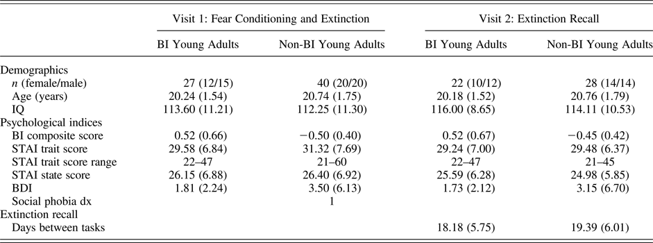
After describing the study fully, written informed consent was obtained. Procedures were approved by the University of Maryland, College Park and National Institute of Mental Health Institutional Review Boards. All 67 participants attended a first visit, during which they completed a fear conditioning and extinction task in the clinic. The second visit included an extinction recall task during fMRI scanning. A number of participants were unable to return for the second visit due to scheduling conflicts (4 individuals), equipment failure (1 individual), contraindications to MRI (2 individuals), initiation of psychotropic medication (4 individuals), and recent use of recreational drugs (6 individuals). Thus, the final sample consisted of 50 participants completing both Visits 1 and 2 of the study. None of the participants in the final sample were diagnosed with anxiety disorder. This sample included 22 participants who were classified as behaviorally inhibited as children (BI) and 28 who were classified as non-behaviorally inhibited (non-BI). Sample demographics are presented in Table 1.
Sample demographics
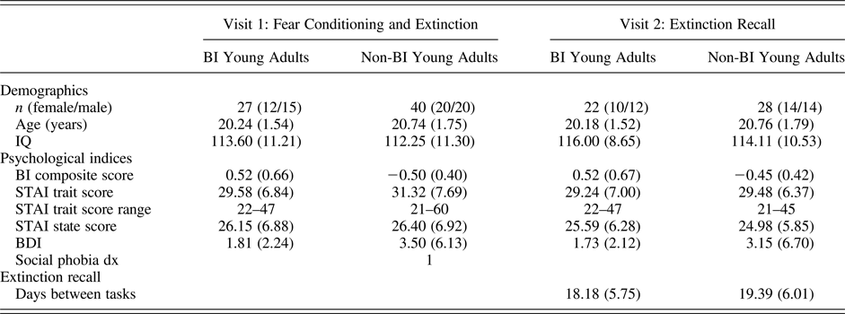
Note: BI, behavioral inhibition; STAI, State Trait Anxiety Inventory; BDI, Beck Depression Inventory; dx, diagnosis. No other anxiety diagnoses were present.
Procedures
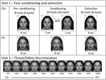
This study adapted the “screaming lady” differential fear conditioning and extinction paradigm used in earlier work (Britton et al., Reference Britton, Grillon, Lissek, Norcross, Szuhany, Chen and Pine2013; Lau et al., Reference Lau, Lissek, Nelson, Lee, Roberson-Nay, Poeth and Pine2008). The paradigm is composed of three phases. During preacquisition, participants passively viewed neutral faces of two women, the conditioned stimuli (CSs). During fear acquisition, one woman, the CS+, predicted the UCS, a fearful face coterminating with an aversive scream, while the other woman, the CS–, did not. Participants were instructed that they could learn to predict when the UCS would occur, but were not informed of the CS/UCS contingency. During extinction, the CS+ and CS– were presented repeatedly in the absence of the UCS. A gray screen presented for 8–21s (average 15 s) was used as the intertrial interval.
Stimulus presentation and recording of psychophysiological indices were controlled by PsyLab psychophysiological recording system (PsyLab SAM System Contact Precision Instruments, London). Skin conductance response (SCR) and self-reported anxiety were used to assess fear acquisition and extinction. SCR was collected continuously during fear conditioning and extinction using two Ag/AgCl electrodes filled with nonsaline gel attached to the medial phalanx of the middle and ring fingers of the left hand. Prior to and immediately after fear conditioning, and immediately after fear extinction, participants rated their level of fear to the CS+ and the CS– using a 10-point Likert scale (1 = none to 10 = extreme).
Approximately 3 weeks after fear conditioning and extinction (mean = 18.8 ± 5.8 days), participants underwent MRI scanning during an extinction recall task. Neuroimaging data were collected on a 3T General Electric 750 scanner, using a 32-channel head coil. During 3 runs, 272 functional image volumes, with 47 contiguous interleaved axial slices (in-plane resolution 3 mm) were obtained with a T2*-weighted echo-planar sequence (repetition time = 2300 ms, echo time = 25 ms, flip angle = 50°, field of view = 240 mm, matrix = 96 × 96). Functional data were anatomically localized and coregistered to a high-resolution T1-weighted volumetric scan of the whole brain, using a magnetization prepared gradient echo sequence (echo time = min full, inversion time = 425 ms, flip angle = 7°, field of view = 256 mm, matrix = 256 × 256, in-plane resolution = 1.0 mm).
The extinction recall task was designed to capture the transitional boundary between threat and safety using morphed images (Greenberg, Carlson, Cha, Hajcak, & Mujica-Parodi, 2012; Lissek et al., Reference Lissek, Rabin, McDowell, Dvir, Bradford, Geraci and Grillon2009, Reference Lissek, Rabin, Heller, Lukenbaugh, Geraci, Pine and Grillon2010; Figure 1). Participants viewed the CS+ and CS– and nine morphed images consisting of different blends of the CS– and CS+ (CS–, 10%CS+, 20%CS+, 30%CS+, 40%CS+, 50%CS+, 60%CS+, 70%CS+, 80%CS+, 90%CS+, and 100%CS+). This stimulus array was used to measure behavioral and neural response gradients (e.g., linear or quadratic trends) along a continuum.
(Color online) The fear conditioning, extinction, and extinction recall tasks.
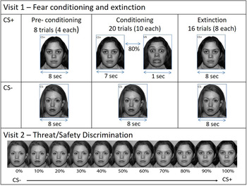
Participants used a slider to answer one of two questions on a scale from 0 to 6 in response to each face: how afraid are you when you look at these pictures now? (threat appraisal); and how likely was the woman to scream in the past? (explicit memory). In three runs, four blocks of each task instruction (threat appraisal and explicit memory) were presented in random order. These reiterations resulted in 12 presentations of each morph for each task instruction. Blocks were composed of 11 morphs and 3 blank images presented randomly for 4000 ms with a 500-ms interstimulus interval. The task was programed in E-prime (PST Inc., Pittsburgh, PA) and presented using a front projector.
Data processing and analysis
Fear conditioning and extinction: Visit 1
SCR was measured as the maximal deflection occurring within 1–5 s after the CS onset. For each participant, SCR scores were square root transformed. The effects of stimulus were averaged across all trials excluding the first CS+ and CS– presentations during conditioning and extinction.
Repeated measures analyses of variance (ANOVAs) with phase (preconditioning, conditioning, and extinction) and stimulus type (CS+, CS–) as within-subject factors and BI group (BI, non-BI) as between-subject factor, was used to examine main effects and interactions of SCR. Similar analyses were used to examine self-report measures obtained prior to and immediately after fear conditioning, and immediately after fear extinction. All statistical analyses were performed using SPSS software, and statistical significance was set to α < 0.05.
Extinction recall: Visit 2
Imaging data were analyzed using Analysis of Functional NeuroImages (AFNI; http://afni.nimh.nih.gov/afni/). Individual preprocessing of echo-planar data included slice-time correction, motion correction, spatial normalization to the Talaraich template, and spatial smoothing with a 6-mm full width at half maximum kernel. Blood oxygen level dependent data were scaled at the voxelwise time series by their temporal means so that the effect estimates could be interpreted as percent signal change relative to the mean. For each subject, a general linear model included 22 regressors of interest (11 regressors for each morph in each of the task instructions); 6 regressors of no interest were included to control for residual motion effects. Estimated betas, one for each regressor, were generated at the individual level and were then included in the group-level analysis.
Group-level analyses were conducted in AFNI, using linear mixed-effects analysis. Statistical analysis tested interactions between BI groups (BI, non-BI), task instructions (threat appraisal, explicit memory) and linear (morph-level) and quadratic (morph2-level) trends across morphed images. The linear and quadratic trend regressors consisted of the morphed level for each image and its square, respectively. Two additional regressors of no interest were modeled to control for internalizing symptoms, and days between fear conditioning/extinction and extinction recall. We corrected for the family-wise error of multiple comparisons at the cluster level through Monte Carlo simulations with the cluster threshold probability set at α = 0.05 and a voxelwise peak p value of .005. Statistical significance was defined as 83 voxels (1406 mm3) for whole brain. These thresholds were determined using AFNI's AlphaSim program using methods described previously (Britton et al., Reference Britton, Grillon, Lissek, Norcross, Szuhany, Chen and Pine2013).
Post hoc tests were conducted within each significant cluster identified by higher level interactions. This post hoc analysis was done to facilitate interpretation of complex results. Percentage signal change values, relative to baseline, were averaged across voxels within each significant cluster for each of the 22 effects of interest for all individual subjects. Using these extracted values in SPSS, linear mixed model analysis and post hoc tests with Bonferroni correction decomposed group differences that had emerged from the omnibus analysis. Post hoc tests determined group differences in activation levels and patterns of response. Behavioral data were analyzed similarly using mixed-effects model with bootstrapped confidence intervals.
Results
Fear conditioning and extinction: Visit 1
SCR
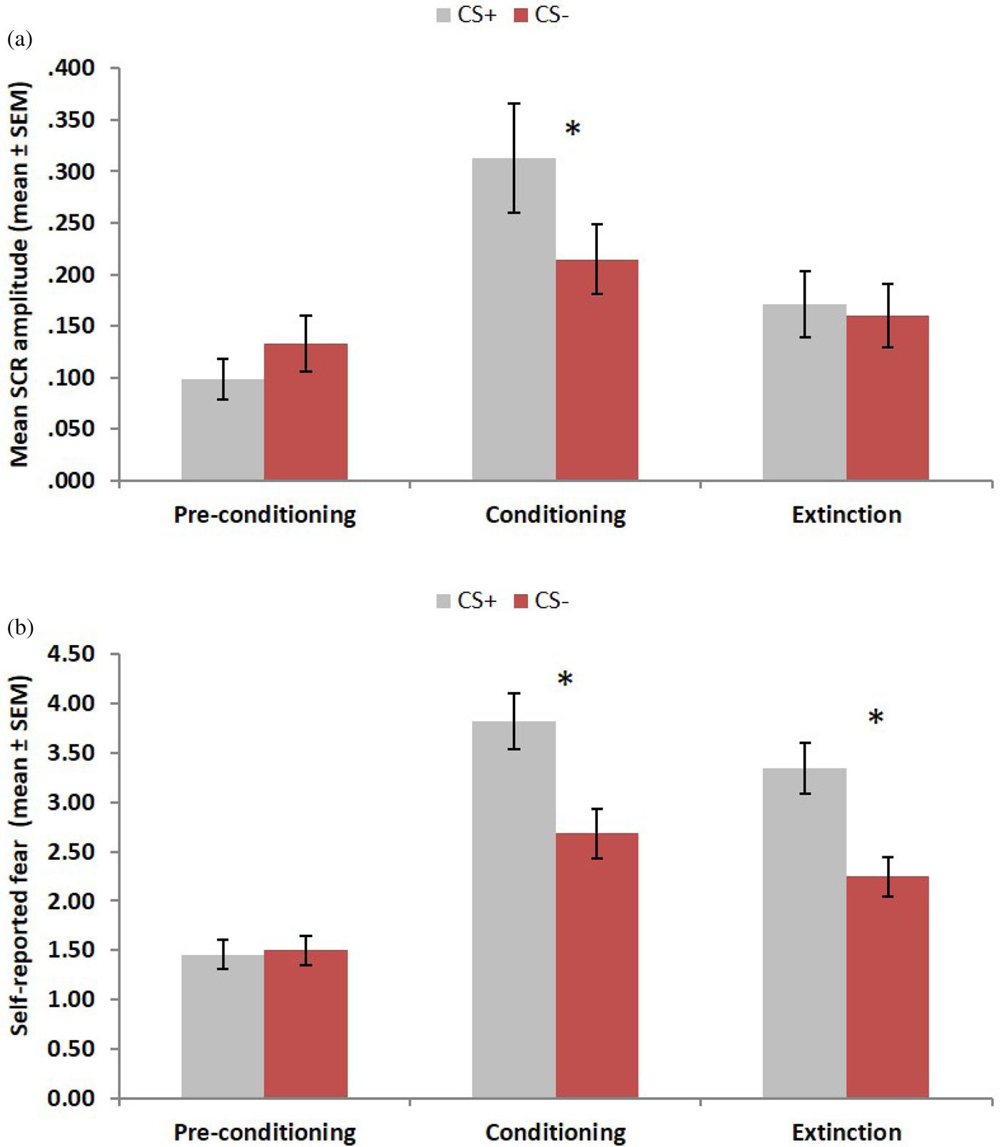
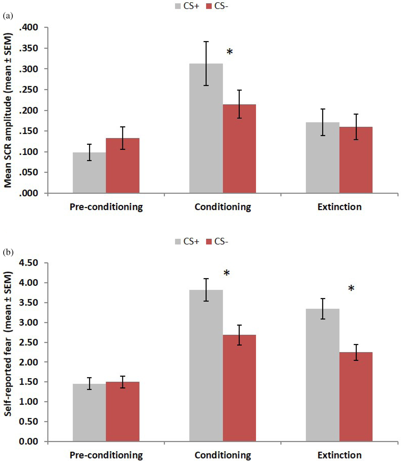
The ANOVA for the Phase × Stimulus × BI group on SCR resulted in a significant Phase × Stimulus interaction, F (2, 124) = 4.13, p = .018, partial η2 = 0.06 (Figure 2a). Paired-samples t tests revealed greater response to the CS+ relative to the CS– during conditioning, t (63) = 2.26, p = .028, but not during preconditioning or extinction (ps > .10). These results indicate successful differential conditioning and extinction for the entire sample, as indexed by SCR. In addition, the main effect of phase was significant, F (2, 124) = 11.22, p < .001, partial η2 = 0.15, with elevated SCR responses during conditioning relative to the other two phases (all ps < .021). There were no main or interaction effects with BI (ps > .11).
(Color online) (a) Skin conductance response (SCR) and (b) self-reported fear during fear conditioning and extinction. Error bars indicate standard errors. CS, conditioned stimulus.
Self-reported fear
The ANOVA for the Phase × Stimuli × BI group on self-reported fear yielded a significant Phase × Stimulus interaction, F (2, 130) = 14.06, p < .001, partial η2 = 0.18 (Figure 2b). While similar in preconditioning (p = .356), paired-samples t tests revealed greater levels of reported fear to the CS+ relative to CS– after conditioning and extinction (ps < .001). Like SCR, these results indicate successful self-reported differential conditioning, for the entire sample; however, evidence of successful extinction based on subjective report was lacking. In addition, the main effect of stimulus was significant, F (1, 65) = 22.85, p < .001, partial η2 = 0.26. As expected, participants reported higher levels of fear to the CS+ than the CS–. Finally, the main effect of phase was also significant, F (2, 130) = 46.47, p < .001, partial η2 = 0.42. Self-reported fear was higher during conditioning relative to the other two phases (all ps < .001) and during extinction relative to preconditioning (p < .001). There were no main or interaction effects with BI (ps > .34).
Extinction recall: Visit 2
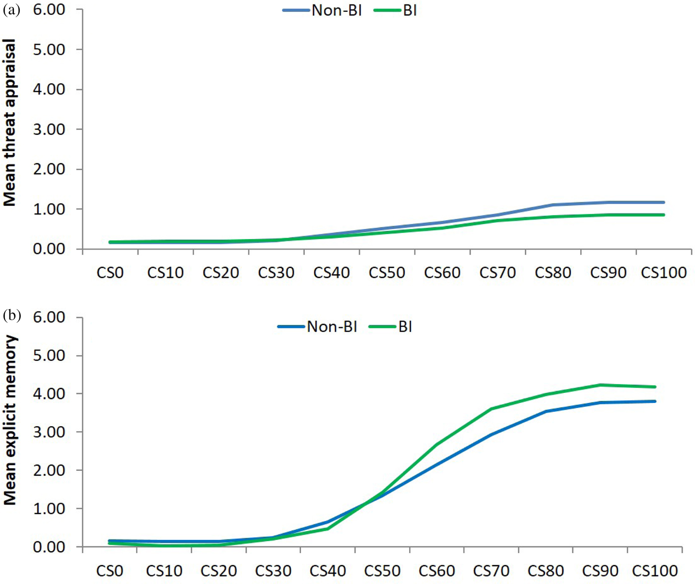
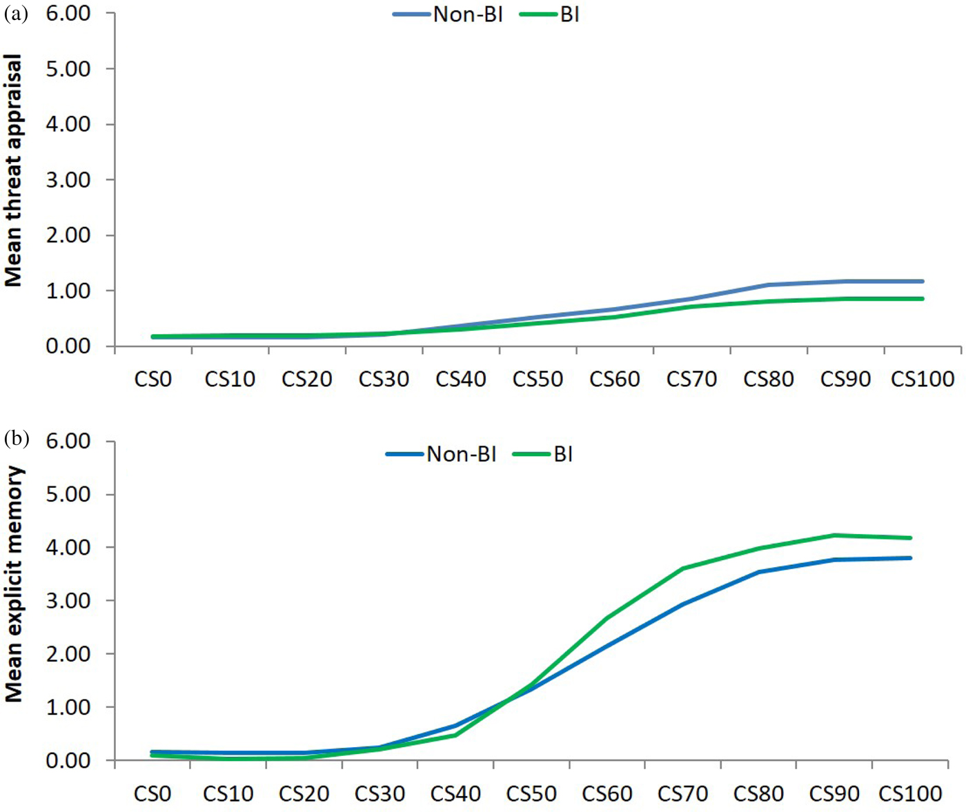
Averaged trends for threat appraisal and explicit memory are depicted in Figure 3.
(Color online) Behavioral response to task instruction during extinction recall. BI, behavioral inhibition; CS, conditioned stimulus.
Threat appraisal
Participant behavioral responses for threat appraisal (Figure 3a) followed linear and quadratic patterns across morphed images (linear: B = 0.105, SE = 0.005, p = .001, quadratic: B = 0.005, SE = 0.00, p = .001). There was a significant interaction between BI group and linear trend (BI × Morph: B = –0.059, SE = 0.006, p = .001), as well as the quadratic trend across morph (BI × Morph2: B = –0.003, SE = 0.002, p = .047). The negative coefficient of this interaction indicates less fear in the BI group.
Explicit memory
Participant behavioral responses for explicit memory (Figure 3b) also followed linear and quadratic patterns (linear: B = 0.488, SE = 0.01, p = .001, quadratic: B = 0.024, SE = 0.004, p = .001). Only the interaction between BI group and morph linear was significant (B = 0.038, SE = 0.017, p = .021). This interaction was derived from group differences in memory of the CS+-UCS contingency. Specifically, for morphs with higher percentages of the original CS+ (>60% CS+), the BI group remembered the CS+-UCS contingency better than the non-BI group.
fMRI results
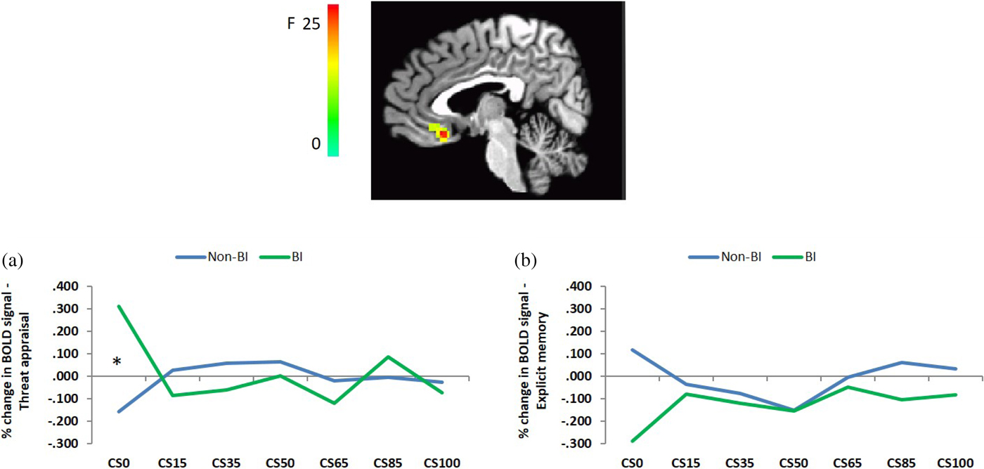
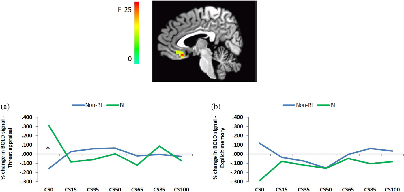
In line with our hypothesis, whole-brain random effects analyses indicated significant BI × Task Instruction × Morph2 interaction in the left subgenual anterior cingulate cortex (LPI coordinates, 4, 24, 11, 84 voxels), F (1, 1,030) = 25.38, p < .005 (Figure 4). This interaction was decomposed according to task instructions. As shown in Figure 4a, a BI × Morph interaction emerged only for threat appraisal, F (10, 480) = 3.30, p < .001, and not for the memory condition, F (10, 480) = 1.16, p = .314 (Figure 4b). Post hoc tests with Bonferroni correction indicated greater sgACC activation in the BI group than in the non-BI only to the CS– (the safe signal), t (48) = 3.31, p = .002. In addition, one-sample t tests indicated that activation during the CS– presentation in the BI group was significantly different from zero, t (21) = 2.479, p = .022; a trend in the opposite direction emerged in the non-BI group, t (27) = –2.025, p = .053.
(Color online) Behavioral inhibition (BI) × Task Instruction × Morph2 interaction in the left subgenual anterior cingulate. BOLD, blood oxygen level dependent; CS, conditioned stimulus.
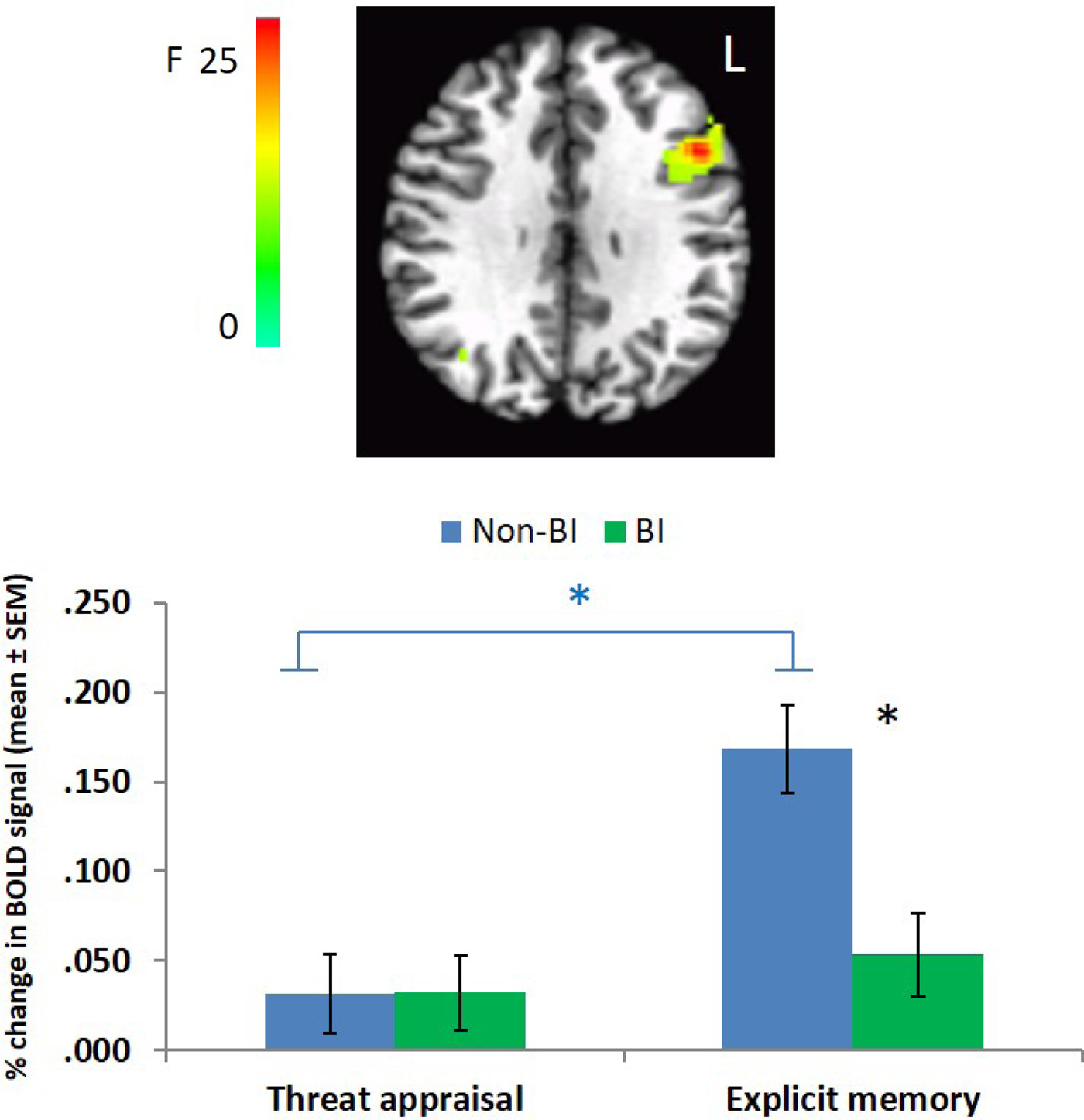
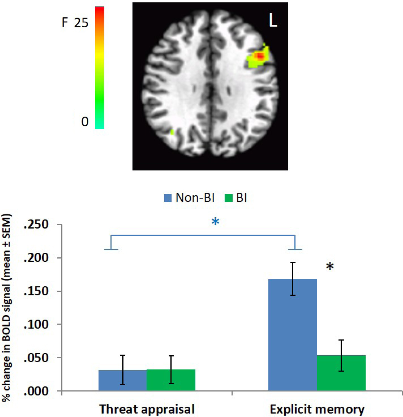
A BI Group × Task Instructions interaction emerged in the left dorsolateral PFC (dlPFC; LPI coordinates: –46, 14, 29, 159 voxels), F (1, 1,030) = 29.46, p < .005 (Figure 5). This interaction derives from greater activation in the memory compared to the threat appraisal task only in the non-BI group, t (27) = –6.095, p < .001, but not in the BI group, t (21) = –0.766, p = .452. In addition, greater dlPFC activation was observed in the non-BI compared to the BI group during the memory task only, t (48) = 3.68, p = .001.
(Color online) Behavioral inhibition (BI) Group × Task Instruction interaction in the left dorsal lateral prefrontal cortex. Error bars indicate standard errors. BOLD, blood oxygen level dependent.
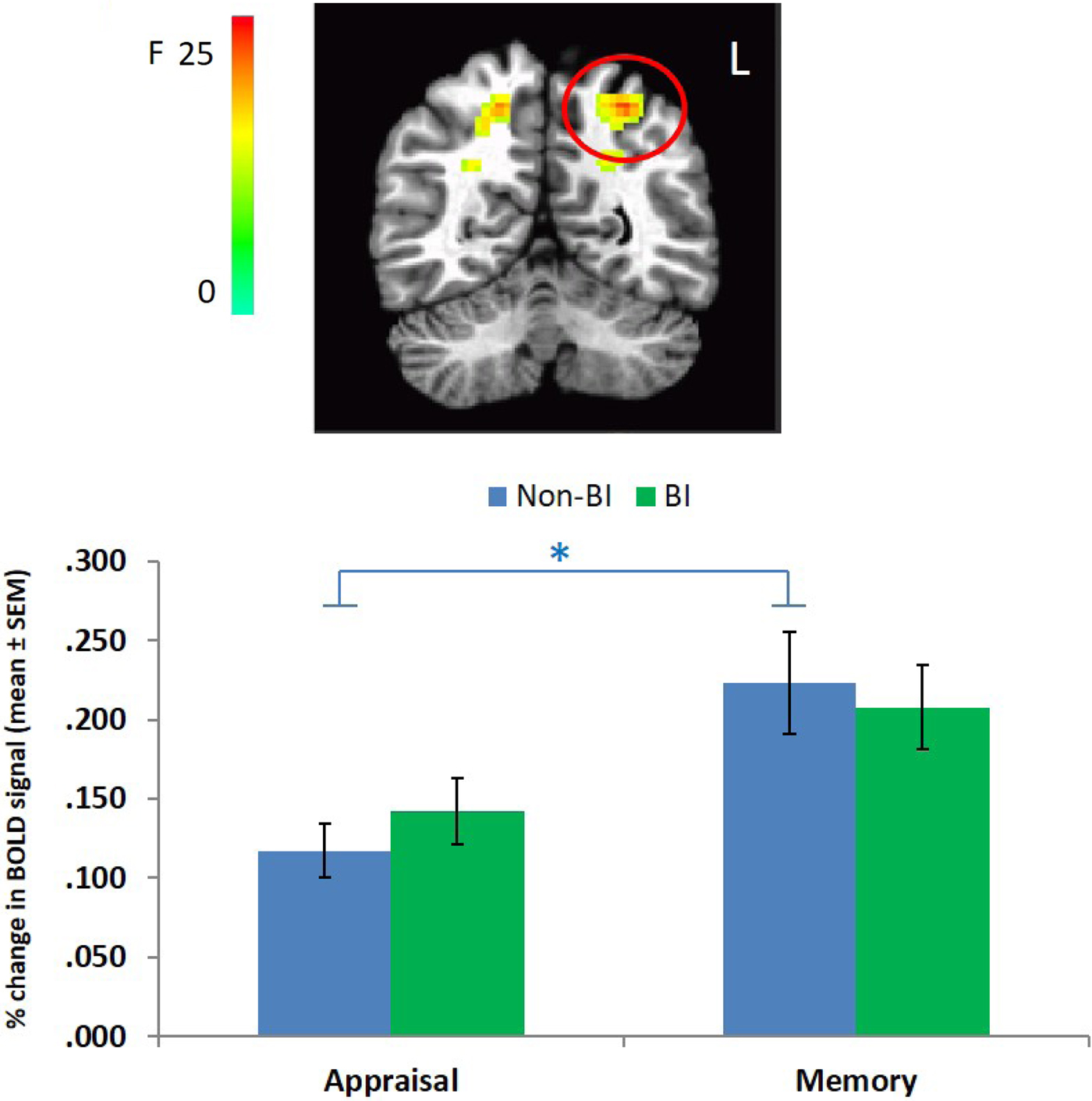
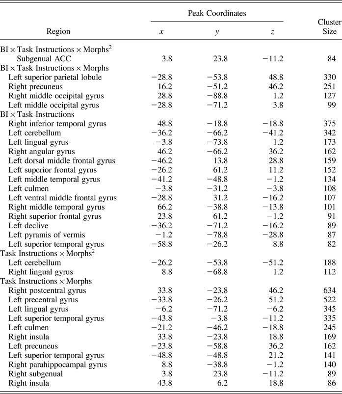
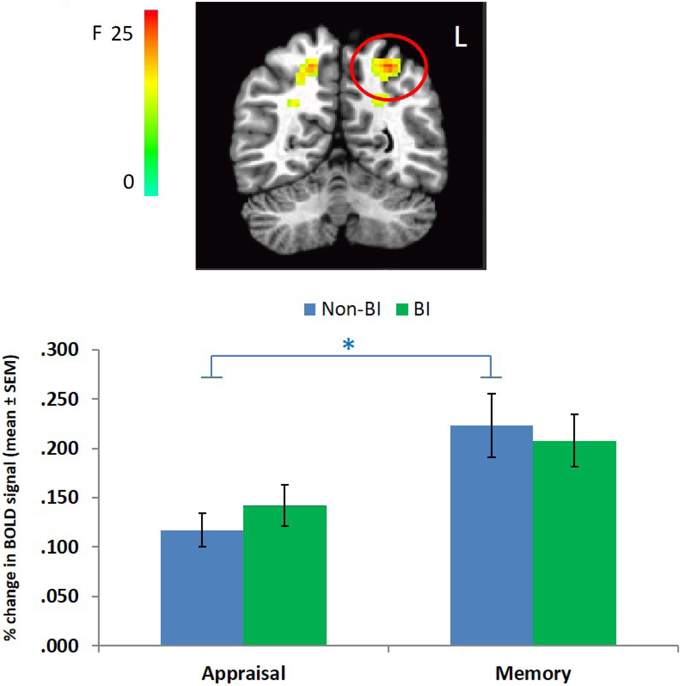
A significant BI Group × Task Instruction × Morph linear interaction emerged in the left superior parietal lobule (LPI coordinates: –29, –54, 49, 330 voxels), F (1, 1,030) = 23.86, p < .005 (Figure 6). When decomposed according to task instructions, a main effect of morph emerged for each type of task instructions (ps < .017). However, a significant difference between the two tasks, collapsed across morphs, emerged only for the non-BI group, F (1, 27) = 37.15, p < .001, not for the BI group, F (1, 21) = 3.65, p > .05. For completeness, other brain regions that showed significant interactions in activation during the task are reported in Table 2.
(Color online) Behavioral inhibition (BI) Group × Task Instruction × Morph linear interaction in the left superior parietal lobule. Error bars indicate standard errors. BOLD, blood oxygen level dependent.
Areas that survived whole brain correction analysis
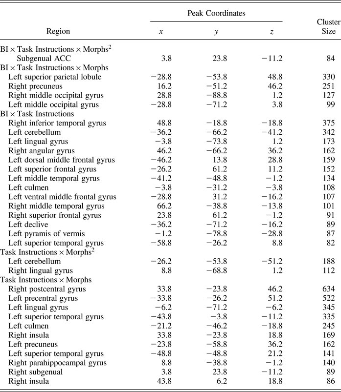
Note: ACC, anterior cingulate cortex.
Discussion
This study compared a group of young adults who are free of anxiety disorder diagnoses, characterized in childhood with BI to those with non-BI on measures of fear conditioning, extinction, and extinction recall. Three major findings emerged. First, no group differences were detected during fear conditioning or extinction. Both objective (SCR) and subjective (self-reported) measures of fear indicated robust differential conditioning (CS+ > CS–); however, SCR but not self-report provided evidence of extinction. Second, groups differed in their memory of the conditioning and extinction procedure during extinction recall (measured during fMRI) 3 weeks later. Namely, while self-reported responses to questions about threat appraisal and threat memory during extinction recall followed a quadratic trend in both groups, the young adults characterized with BI reported better CS+-UCS contingency awareness (i.e., explicit memory) and less fear of the previous CS+ relative to the non-BI group. Third, the BI group showed greater sgACC activation during threat appraisal than the non-BI group, particularly when appraising the CS–.
Groups performed similarly during fear conditioning and extinction. Prior meta-analyses have consistently shown similar performance during differential fear learning for healthy and anxious adults (Duits et al., Reference Duits, Cath, Lissek, Hox, Hamm, Engelhard and Baas2015; Lissek et al., Reference Lissek, Powers, McClure, Phelps, Woldehawariat, Grillon and Pine2005). Because differences fail to emerge across groups that would be most likely to diverge in the context of fear learning, it is not surprising that our sample of healthy BI and non-BI adults also show similar patterns of responding. The sample as a whole demonstrated robust fear conditioning as indicated by SCR and subjective self-report of fear; however, extinction was observed only through SCR. Again, such findings were expected. Discrepancies between observable fear extinction as measured objectively (SCR) and subjectively (self-report) have been reported in previous studies that used similar differential conditioning paradigms in both clinical and nonclinical samples of youth and adults (Britton et al., Reference Britton, Grillon, Lissek, Norcross, Szuhany, Chen and Pine2013; Shechner et al., Reference Shechner, Britton, Ronkin, Jarcho, Mash, Michalska and Pine2015; Waters, Henry, & Neumann, Reference Waters, Henry and Neumann2009).
Despite their similarities during fear conditioning and extinction, group differences emerged in extinction recall via threat appraisal and explicit memory of the CS+-UCS association. As expected, in both groups, participants’ fear responses increased in a quadratic fashion with increased CS+ features, suggesting that fear levels are greater with increased similarity to the CS+. Similar response patterns, including quadratic trends in threat appraisal and explicit memory as well as low levels of fear (threat appraisal), were previously reported using a comparable extinction recall paradigm (Britton et al., Reference Britton, Grillon, Lissek, Norcross, Szuhany, Chen and Pine2013). In the current study, both groups were free of anxiety disorders; therefore, it seems reasonable that the overall fear levels would be low while still maintaining a similar gradient with maximal fear of stimuli containing more CS+ features. The interaction between BI and the quadratic pattern across morphs was statistically significant for threat appraisal, with the BI group reporting less fear of the CS+. However, low fear levels overall (range = 0–1 on a 0–6 scale in both groups) indicate that caution is warranted in interpretation.
Compared to threat appraisal, participants’ responses followed a stronger quadratic pattern during the explicit memory condition, suggesting that subjects successfully differentiated between the CS+ and the CS–. Yet, as evidenced by a steeper linear slope, the BI group exhibited stronger memory for morphs that were similar to the CS+ (>80%). Previous research has implicated memory of CS-UCS contingency as one of the most robust conditioning markers of pathological anxiety (Lissek et al., Reference Lissek, Kaczkurkin, Rabin, Geraci, Pine and Grillon2014). Namely, anxious adults with less accurate memory of CS-UCS contingency exhibited greater fear responses (as indicated by self-report and fear potentiated startle) than anxious adults who more accurately remembered the CS-UCS contingency (Grillon, Reference Grillon2002). Taken together, the BI group in the current study demonstrated even lower levels of one common conditioning marker of pathological anxiety (reduced memory of CS-UCS) than the non-BI group did, and accordingly reported less fear to the CS+.
Greater left sgACC activation during threat appraisal was detected in the BI group as compared to the non-BI group only when the CS– was presented. Previous studies implicate this brain region in affective regulation and threat/safety discrimination particularly during threat appraisal (Dunsmoor, Prince, Murty, Kragel, & LaBar, Reference Dunsmoor, Prince, Murty, Kragel and LaBar2011; Kalisch et al., Reference Kalisch, Korenfeld, Stephan, Weiskopf, Seymour and Dolan2006; Milad et al., Reference Milad, Wright, Orr, Pitman, Quirk and Rauch2007). Compared to nonanxious individuals, prior research suggests that anxious individuals exhibit less activation in sgACC/vmPFC regions during extinction recall (Britton et al., Reference Britton, Grillon, Lissek, Norcross, Szuhany, Chen and Pine2013; Graham & Milad, Reference Graham and Milad2011). As previously noted, the temperament of BI is a risk factor for anxiety later in life, but only about half of children with BI develop clinical anxiety. Hence, the greater activation within this region in the nonanxious BI group relative to the non-BI group seen in the current study could reflect a compensatory mechanism promoting threat/safety discrimination. Significant differences emerged only when a pure safe signal (0% of CS+) was presented.
The sgACC plays a role in emotion–cognition interactions and may function as part of a circuit encompassing the amygdala and various components of the PFC. Engagement of this circuit may lead the organism to manifest relatively high or low levels of reactivity to stimuli (Cromheeke & Mueller, Reference Cromheeke and Mueller2014). The current sample represented a resilient group of individuals who have a history of BI temperament. While BI is a risk factor for anxiety in the current sample, no subjects studied here manifested psychopathology. As a result, these BI subjects may possess greater top-down processing skills, and these skills may sustain the observed enhanced activation of the sgACC in the current study to the safety signal and greater memory of the CS contingency. This idea is further supported by the lower levels of reported fear in BI as compared to non-BI individuals to morphs that were perceptually similar to the CS+. Finally, greater PFC activation in the resilient BI group is in line with recent reviews emphasizing the important role of adequate recruitment of PFC in mental health (Cole, Repovs, & Anticevic, Reference Cole, Repovs and Anticevic2014).
Previous research has uncovered similar neurocognitive processes in individuals with a history of BI and in anxious individuals, with some studies demonstrating that impairments in information processing and underlying neural circuitries predict risk for anxiety (McDermott et al., Reference McDermott, Perez-Edgar, Henderson, Chronis-Tuscano, Pine and Fox2009; Shechner et al., Reference Shechner, Britton, Perez-Edgar, Bar-Haim, Ernst, Fox and Pine2012). Greater vmPFC activation in the resilient BI group observed in the current study, together with evidence of reduced activation of the vmPFC among anxious individuals during extinction recall task, implicates the vmPFC in the expression of anxiety.
Finally, group differences were found in other brain areas that may be relevant for this task but for which we had no a priori hypotheses. Greater left dlPFC and left superior parietal lobule activation were detected in explicit memory compared to threat appraisal only in the non-BI and not in the BI group. The dlPFC is involved in monitoring and evaluating affective information (Etkin, Reference Etkin2010; Etkin, Egner, & Kalisch, Reference Etkin, Egner and Kalisch2011) and in discrimination between stimuli (Lau et al., Reference Lau, Lissek, Nelson, Lee, Roberson-Nay, Poeth and Pine2008). The superior parietal lobule is implicated in manipulation of information in working memory (Kornigs, Barbey, Postle, & Grafman, Reference Koenigs, Barbey, Postle and Grafman2009) and in motor response. This result may indicate that, for the non-BI group, greater effort was required to complete the memory task compared to the threat appraisal task. This was not the case in the BI group. This interpretation is consistent with the behavioral findings from that same task in which the BI group remembered better than the non-BI group the contingency between the morphs and the UCS. These findings could be interpreted as reflecting another marker of resilience in which the BI group demonstrated better memory while using fewer resources manifested in lower dlPFC activation. In addition, several other brain regions for which group differences were found are reported and should be further examined in future studies.
Results from the current study should be considered in light of some notable limitations. First, given the cross-sectional design of the study (e.g., fear learning processes were only tested at one time point), the causality of relationships is unknown. Namely, greater sgACC activation during threat/safety discrimination may lead to resilience for anxiety, or vice versa. Future studies should target brain activation during extinction recall in earlier developmental stage and then examine predictors of longitudinal outcomes. Second, the unique sample of young adults characterized with BI in childhood and absent of anxiety was obtained partially as a result of stringent exclusion criteria not allowing individuals who were treated with psychotropic medications to participate in the current study. Hence, generalization of our results is limited to this very specific BI subgroup. Future studies should include all three groups (affected BI, resilient-BI, and non-BI groups) to more wholly understand these relationships.
Taken together, our findings are in line with other recent studies that show promise in implementing extinction recall tasks to detect behavioral and neural differences associated with risk for and resilience to anxiety. This task could be applied to individuals at risk for anxiety at different stages of development and pathology to better understand and manage potential extinction-processing deficits throughout the life span and course of illness.