Introduction
Bovine ovaries are commonly collected from abattoirs as a source of oocytes for experiments involving reproductive science and in vitro fertilization. Bovine oocytes are usually recovered from ovaries that have been transported from an abattoir within 2–4 h at 30–35°C (Blondin et al., Reference Blondin, Coenen, Guilbault and Sirard1997; Yao et al., Reference Yao, Wan, Hao, Gao, Yang, Cui, Wu, Liu, Liu, Chen and Zeng2009) or 4–7 h at 20–25°C (Flood et al., Reference Flood, Gage and Bunch1993; Nagao et al., Reference Nagao, Saeki, Hoshi, Takahashi and Kanagawa1995). Ovaries need to be collected and returned to the laboratory immediately after slaughter in order to make effective use of the oocytes contained within. In clinical medicine, many different types of preservative have been developed for organ transplantation effectively prolonging the preservation period for intra-abdominal organs by up to 24 h (Jamieson, Reference Jamieson, Sundberg, Lindell, Claesson, Moen, Vreugdenhil, Wight, Southard and Belzer1988; Collins et al., Reference Collins and Wicomb1992; Roberts et al., Reference Roberts, Nishanian, Carey, Sakamaki, Starnes and Barr1999). Prolonged preservations periods are highly useful as they permit patients to be transported to the hospital for transplantation from distant locations, as well as improving the chances of matching organs to recipients. Recently, research has focused upon the storage life of the bovine ovary (Matsushita et al., Reference Matsushita, Tani, Kato and Tsunoda2004; Iwata et al., Reference Iwata, Hayashi, Sato, Kimura, Kuwayama and Monji2005). Successful preservation of bovine ovaries for longer time periods without reducing the developmental potential of the oocytes contained within may enhance the effective application of such oocytes in assisted reproductive technologies for animal reproduction. Screening tests for BSE carried out at abattoirs can also cause delays of up to 8–24 h in Japan until ovaries become available for scientific use. However, little is currently known of the effect of extended preservation upon bovine ovaries, particularly the quality of the oocytes contained within. Moreover, we currently do not yet have an optimal protocol for the preservation of bovine ovaries. During the preservation of human organs for transplantation, organ injury has been shown to cause cellular swelling, cell membrane destruction and oxygen free-radical injury (Jamieson et al., Reference Jamieson, Sundberg, Lindell, Claesson, Moen, Vreugdenhil, Wight, Southard and Belzer1988). Oxygen free radicals, in particular, are major contributors to organ damage during preservation (Koyama et al., Reference Koyama, Bulkley, Williams and Im1985). To suppress the detrimental effects of oxygen free radicals, reduced glutathione (GSH) has been used as a supplement in preservation solutions under cold conditions (Jamieson, Reference Jamieson, Sundberg, Lindell, Claesson, Moen, Vreugdenhil, Wight, Southard and Belzer1988; Collins et al., Reference Collins and Wicomb1992; Roberts, et al., Reference Roberts, Nishanian, Carey, Sakamaki, Starnes and Barr1999). Natural occurring anti-oxidants such as GSH and polyphenols may also play an important role for preventing oxygen radical injury during the preservation of bovine ovaries. GSH (Matos et al., Reference Matos, Furnus, Moses and Baldassarre1995), polyphenols including EGCG (Guang et al., Reference Guang, Yu and Xu2007) other antioxidants (Ali et al., Reference Ali, Bilodeau and Sirard2003) have all been shown previously to promote embryo development in bovine oocytes. To obvious optimal conditions for the overnight preservation of bovine ovaries for subsequent reproductive use, we examined the effects of temperature, preservation solution and the use of antioxidant supplementation upon subsequent embryonic development.
Materials and Methods
Ovary preservation
Bovine ovaries were obtained all year round from culled Holstein at an abattoir. Paired ovaries derived from individual females were held collectively in the same sample containing saline/0.1% antibiotic–antimycotic (Invitrogen Corp: AB) (saline). Ovaries were then washed to remove blood contamination by gently massaging in saline for 2–5 min (5–7 times). Ovaries were transported to the laboratory within 1–3 h in saline. Upon arrival at the laboratory, paired ovaries were divided; one ovary remained in the saline as a control and the other was transferred to an experimental bottle containing different solutions as per the experimental protocol.
Oocyte collection and in vitro maturation
Oocyte collection and in vitro maturation (IVM) of oocytes were carried out as previously described (Saeki et al., Reference Saeki, Hoshi, Leibfried and First1990), with some modifications. In short, cumulus oocyte–complexes (COCs) were aspirated from follicles 2–6 mm in diameter using 20-gauge needle attached to 5 ml syringe. Oocytes with intact cumulus cells and evenly granulated cytoplasm were selected for further study. Selected oocytes were washed and cultured in 50 μl drops of modified TCM-199 (m-TCM199) covered with mineral oil (M8410, Sigma-Aldrich Corp.) for 22–25 h at 39°C under 5% CO2 in air with high humidity. The m-TCM199 consisted of HEPES-buffered medium 199 (no. 12340, Invitrogen Corp.) supplemented with 0.1% (w/v) polyvinyl alcohol (P8136, Sigma-Aldrich Corp.; PVA), 0.5 mM sodium pyruvate (Nacalai Tesque Inc.), 1% AB, 0.02 AU/ml FSH (Antrin, Denka) and 1 μg/ml estradiol-17β (E2758, Sigma-Aldrich Corp.).
In vitro fertilization of bovine oocytes
In vitro fertilization (IVF) was carried out as previously described (Nagao et al., Reference Nagao, Iijima and Saeki2008), with some modifications. Briefly, frozen–thawed semen from one bull was washed with a discontinuous Percoll solution. Matured oocytes were then inseminated with the washed spermatozoa (1–2 × 10⁶ cells/ml) in BO supplemented with 10 μg/ml heparin (H0777, Sigma-Aldrich Corp.) and 3 mg/ml bovine serum albumin (A4378, Sigma-Aldrich Corp.: BSA). Oocytes were then incubated with spermatozoa in 100 μl drops of the medium covered with mineral oil (M8410, Sigma-Aldrich Corp.) for 18–20 h after insemination. All incubations were performed at 39°C under 5% CO2 in air at high humidity.
In vitro culture of bovine oocytes
In vitro culture (IVC) of embryos was carried out as previously described (Nagao et al., Reference Nagao, Iijima and Saeki2008). In short, 18–20 h after insemination, presumptive zygotes were removed from the fertilization medium, stripped of cumulus cells by vortexing and washed twice in modified-SOF supplemented with 0.05% (w/v) PVA and 1% AB. Then, 15–50 of these embryos were placed in 50 μl drops of modified-SOF supplemented with 3 mg/ml BSA and 1% AB covered with mineral oil (M8410, Sigma-Aldrich Corp.) and cultured at 39°C under 5% CO2, 5 % O2 and 90% N2 at high humidity for 7–8 days.
Evaluation of oocytes after IVM, IVF and IVC
To evaluate nuclear stage after maturation, a sample of oocytes were fixed after IVM on a slide with 3:1 acetic acid: ethanol and stained with 1% orcein. Oocytes found to be undergoing germinal vesicle breakdown (GVBD) and maturation phases to the metaphase II (M II) stage were assessed. To evaluate pronuclear stage after fertilization, a sample of oocytes post-IVF was processed in the same way as described above. Oocytes with a sperm tail in the cytoplasm were considered to be fertilized and were classified into one of three groups: ‘2PN’ (female pronucleus, male pronucleus and sperm tail; normal fertilization), ‘polyspermy’ (two or more sperm tails in the cytoplasm with condensed heads or two or more decondensed heads in the cytoplasm) and ‘asynchrony’ with a condensed sperm head (female pronucleus and a condensed sperm head). A stereomicroscope (×60) was used to evaluate the developmental stage after IVC, cleavage and development to the blastocyst stage.
Experimental design
Experiment 1
In Experiment 1-1, the effect of ovary preservation temperature upon the maturation and fertilization rate was examined for 24 h. After ovaries were transported to the laboratory, they were preserved in saline for 24 h at 5, 15, 25 (control) and 35°C; IVM and IVF were carried out as described above. In Experiment 1–2, the effect of ovary preservation temperature upon the cleavage and blastocyst formation rate was examined for 24 h. After ovaries were transported to the laboratory, they were preserved in saline for 24 h at 5, 15, 25 (control) and 35°C, IVM, IVF and IVC were carried out as described above.
Experiment 2
In Experiment 2-1, the effect of the solution used to preserve ovaries and its effects the maturation and fertilization rate were examined over a period of 24 h. After ovaries were transported to the laboratory, they were preserved in saline, D-PBS (no. 14287, Invitrogen Corp.) containing 0.1% antibiotic–antimycotic (PBS) or University of Wisconsin solution (ViaSpan, Du Pont Pharmaceuticals) for 24 h at 15°C. IVM and IVF were carried out as described above. In Experiment 2-2, the effect of the solution used to preserve ovaries and its effects cleavage and blastocyst formation rate were examined over a period of 24 h. After ovaries were transported to the laboratory, they were preserved in saline, PBS or UW for 24 h at 15°C. IVM, IVF and IVC were carried out as described above.
Experiment 3
The effect of supplementing preservatives with reduced glutathione (GSH) during ovary preservation and its effect upon cleavage and blastocyst formation rate was examined. After ovaries were transported to the laboratory, they were preserved in saline supplemented with 0 (control group), 0.3 or 3 mM GSH (G6013, Sigma-Aldrich Corp.) for 24 h at 15°C. IVF and IVC were carried out as described above. GSH was supplemented into saline just before use in order to prevent oxidization.
Experiment 4
The effect of supplementing preservatives with epigallocatechin gallate (EGCG) during ovary preservation and its effect upon cleavage and blastocyst formation rate was also examined. After ovaries were transported to the laboratory, they were preserved in saline supplemented with 0, 10 or 100 μM EGCG (E4143, Sigma-Aldrich Corp.) for 24 h at 15°C. IVF and IVC were carried out as described above. EGCG was supplemented into saline just before use in order to prevent oxidization.
Data analysis
Cleavage and blastocyst formation rate rates were analysed using the chi-squared test. A p-value of less than 0.05 was considered to be statistically significant.
Results
Experiment 1
In Experiment 1-1, ovaries derived from the 35°C group were rotton and could not be used for subsequent experiments. The effect of preservation temperature upon in vitro maturation and fertilization in the other groups are shown in Table 1-1. The maturation rates of oocytes recovered from ovaries stored at 15 and 25°C was higher than that of the control group (p < 0.05). The fertilization of oocytes stored at 5, 15 and 25°C were not different.
Effect of temperature for 24 h preservation of ovaries on the maturation and fertilization of oocytes.

a, bValues with different superscripts within each column differ (p < 0.05).
The experiments were repeated five times.
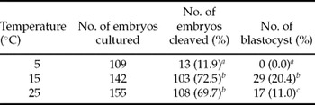
The effects of preservation temperature upon in vitro development in the other groups are shown in Table 1-2 (Experiment 1-2). Cleavage rate of oocytes recovered from ovaries stored at 15 and 25°C was higher than that of the control group (p < 0.05). Developmental rate to the blastocyst stage of oocytes derived from ovaries stored at 15°C was significantly higher than that of the other groups.
Effect of temperature for 24 h preservation of ovaries on the cleavage and blastocyst rate of the embryos.

a–cValues with different superscripts within each column differ (p < 0.05).
The experiments were repeated six times.
Experiment 2
The effect of preservation temperature upon in vitro maturation and fertilization in the other groups are shown in Table 2-1 (Experiment 2-1). Maturation and fertilization of the oocytes stored in saline, PBS and UW were not different among the experimental groups.
Effect of medium for 24 h preservation of ovaries on the maturation and fertilization rate of the embryos.

The experiments were repeated five times.
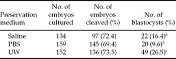
The effects of preservation solution upon in vitro development are shown in Table 2-2 (Experiment 2-2). Developmental rate to the blastocyst stage of oocytes derived from ovaries stored in UW solution was higher than that from PBS and saline groups (p < 0.05).
Effect of medium for 24 h preservation of ovaries on the cleavage and blastocyst rate of the embryos.

a–cValues with different superscripts within each column differ (p < 0.05).
The experiments were repeated six times.
Experiment 3
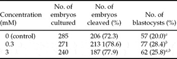
The effects of supplementing preservation fluid with GSH upon in vitro development are shown in Table 3. There was no significant difference in the rates of cleavage among the groups. However, the development rates to the blastocyst stage of oocytes in the 0.3 mM GSH group were higher than that of the oocytes in the 0 mM GSH groups (p < 0.05).
Effect of GSH supplementation for 24 h preservation of ovaries on the cleavage and blastocyst rate of the embryos.

a, bValues with different superscripts within each column differ (p < 0.05).
The experiments were repeated eight times.
Experiment 4
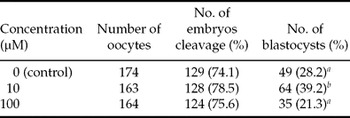
The effect of supplementing preservation fluid with EGCG upon in vitro development is shown in Table 4. Oocyte cleavage did not differ among the groups. However, developmental rate to the blastocyst stage of the oocytes from the 10 μM EGCG group was higher than that of the control group (p < 0.05).
Effect of EGCG supplementation for 24 h preservation of ovaries on the cleavage and blastocyst rate of the embryos.

a, bValues with different superscripts within each column differ (p < 0.05).
The experiments were repeated six times.
Discussion
The purpose of the present study was to develop optimal conditions to preserve bovine ovaries overnight whilst maintaining the developmental potential of the oocytes. We examined the effect of temperature, ischemic condition, type of preservation fluid and the supplementation of preservation fluids with antioxidant agent upon the fertilization and developmental capacity of oocytes derived from the preserved ovaries.
In Experiments 1-1 and 1-2, we examined the optimal temperature for preservation of ovaries. A preservation temperature of 15°C increased the rates of cleavage and development to the blastocyst stage significantly when compared with the other groups (Table 1-2). Previous studies describe protocols in which bovine ovaries have been preserved and transported at 30–35°C for 3–4 h (Blondin et al., Reference Blondin, Coenen, Guilbault and Sirard1997; Yao et al., Reference Yao, Wan, Hao, Gao, Yang, Cui, Wu, Liu, Liu, Chen and Zeng2009) or at 20–25°C 4–7 h (Flood et al., Reference Flood, Gage and Bunch1993; Nagao et al., Reference Nagao, Saeki, Hoshi, Takahashi and Kanagawa1995) for in vitro production and reproductive research. Preservation of bovine ovaries at 37–39°C for 5–8 h has been shown to reduce the maturation rate of oocytes and the developmental ability to blastocysts after in vitro fertilization (Abe et al., Reference Abe and Shioya1996). We also demonstrated that oocytes derived from ovaries preserved at 35°C for 24 h died and could not maintain developmental potential (data not shown). Furthermore, developmental potential to the blastocyst stage of oocytes derived from ovaries preserved at 25°C was significantly lower than that of oocytes derived from ovaries preserved at 15°C. These results were similar to some previous reports (Abe et al., Reference Abe and Shioya1996; Matsushita et al., Reference Matsushita, Tani, Kato and Tsunoda2004). At preservation temperatures of 25 and 35°C, it is possible that certain proteins which are necessary for in vitro development to the blastocyst stage might be destroyed or damaged by enzymes in the oocytes as the metabolism and enzymes of warm-blooded animals work most efficiently at body temperature. Preservation of ovaries at 15°C may not be deleterious for protein synthesis in nuclear and cytoplasmic maturation. In a previous report, livers for transplantation were preserved at 4°C (Jamieson et al., Reference Jamieson, Sundberg, Lindell, Claesson, Moen, Vreugdenhil, Wight, Southard and Belzer1988) in order to reduce intracellular oxygen consumption and enzymatic activity. However, in this experiment, most oocytes from the ovaries preserved at 4°C did not have the ability to fertilize. In ovaries, the pre-requisites for protein synthesis and effective gap junction connections between cumulus cells and the oocyte itself in order to allow transition from the MI–MII stages may be interrupted by the prolonged exposure to low temperature. Therefore, the preservation of ovaries at 15°C may represent a better option for improving developmental competence of the oocyte.
In Experiment 2-1 and 2-2, we examined the type of preservation solution used during ovary preservation. The three types of preservation solution tested (saline, UW and PBS) did not affect the rates of maturation, fertilization and cleavage, although UW increased the rate of development to the blastocyst stage when compared with the other groups. In humans, UW prolongs the period of preservation for intra-abdominal organs by up to 24–48 h (Jamieson et al., Reference Jamieson, Sundberg, Lindell, Claesson, Moen, Vreugdenhil, Wight, Southard and Belzer1988), thus enabling patients to reach hospital from distant locations for transplantation operations and help improve recipient matching for organs such as the liver, pancreas and kidney. Many components have been formulated in order to minimize organ damage, including cellular swelling, cell membrane destruction and oxygen free-radical injury (Jamieson, Reference Jamieson, Sundberg, Lindell, Claesson, Moen, Vreugdenhil, Wight, Southard and Belzer1988). In particular, oxygen free radicals are a major contributor to organ damage during preservation (Koyama et al., Reference Koyama, Bulkley, Williams and Im1985). To suppress the detrimental effects of oxygen free radicals, reduced glutathione (GSH) was supplemented into UW. Natural occurring anti-oxidants, such as GSH and polyphenols, may play an important role in the prevention of oxygen radical injury in bovine ovaries. In the present study, we found that UW was optimal for preserving ovaries in cold conditions, whilst UW is too expensive for routine use in reproductive studies. Thus, in Experiments 3 and 4, we examined the effect of supplementing preservation fluids with antioxidants such as GSH, one of the chief components of UW and EGCG, a polyphenol group.
In Experiment 3, ovaries preserved in saline containing 0.3 mM GSH exhibited enhanced embryo development to the blastocyst stage when compared to the other groups (Table 3). GSH has been shown to be soluble in water and permeate organs during preservation in cold condition and can be an important factor in the suppression of oxygen free-radical injury (Vreugdenhil et al., Reference Vreugdenhil, Belzer and Southard1991; Delcorso et al., Reference Delcorso, Cappiello and Mura1994; Lafleur et al., Reference Lafleur, Hoorweg, Joenje, Westmijze and Retel1994). This molecule might efficiently protect bovine oocytes against oxidative stress (Matos et al., Reference Matos, Furnus, Moses and Baldassarre1995). These earlier studies, combined with the results of the present study, indicate that GSH protects the developmental potential of oocytes from oxygen free radicals in preserved ovaries.
In Experiment 4, the supplementation of saline preservation fluid with 10 μM EGCG significantly enhanced embryo development to the blastocyst stage compared with the other groups (Table 4). EGCG is one of the strongest antioxidants of the catechin family and is a scavenger of oxygen free radical (Vinson et al., Reference Vinson, Dabbagh, Serry and Jang1995; Guo et al., Reference Guo, Zhao, Li, Shen and Xin1996; Schroeder et al., Reference Schroeder, Klotz and Sies2003), is easily soluble in water and is useful as well as GSH. Supplementation of IVM media with green tea polyphenols (GTP), including EGCG, has been shown to convey beneficial effects upon subsequent embryo development (Guang et al., Reference Guang, Yu and Xu2007). These earlier reports demonstrated that the supplementation of GTP during IVM significantly increases intracellular GSH concentration in the oocytes, indicating that the improvement of fertilization and developmental competence in oocytes. Therefore, EGCG may have the effect of increasing the level of GSH in oocytes during preservation and thus improving early development, as also seen in Experiment 3.
In additional experiment, we demonstrated that blastocysts derived from oocytes preserved in saline supplemented with 0.3 mM GSH at 15°C exhibited normal chromosomal integrity (72.7%, 16/22) and normal developmental ability to term (pregnancy rate: 50.0%, 3/6). These results indicate a useful method of preserving ovaries in a manner, which can maintain the developmental ability of oocytes to reach term successfully.
In conclusion, bovine ovaries can be preserved in saline supplemented with GSH or EGCG at 15°C for 24 h and still represent a quality biological resource for reproductive study. Oocytes preserved using this method maintain developmental ability to the blastocyst stage and reach term efficiently.
Acknowledgements
We thank Dr Eiji Kobayashi, Jichi Medical University, for providing University of Wisconsin solution, Miyagi Prefectural Livestock Experiment Station for providing the frozen semen and Chikusei Meat Center and Nakao Chikusan Co., Ltd. for providing the ovaries, used for this study.