Introduction
Laryngopharyngeal reflux (LPR) is common: between 4 and 10 per cent of patients presenting to ENT out-patient clinics have reflux-related symptoms.Reference Koufman1 The condition commonly presents with symptoms of hoarseness, persistent throat-clearing, dysphagia, cough and sore throat, which are often intermittent and recurring.Reference Koufman, Aviv, Casiano and Shaw2 In addition, there are several long term sequelae associated with LPR, including subglottic stenosis, laryngospasm, paradoxical vocal fold movement and laryngeal carcinoma.Reference Koufman, Aviv, Casiano and Shaw2, Reference Maronian, Azadeh, Waugh and Hillel3
Given the functional impairment and possible serious complications of LPR, accurate diagnosis and effective management is important. Several diagnostic tools are available to the clinician. The ‘gold standard’ is thought to be 24 hour pH monitoring using a double lumen (oesophageal and pharyngeal) probe.Reference Postma4 However, this is seldom conducted in routine clinical practice as it is invasive and time-consuming. Other diagnostic tools include the reflux symptom index (a validated patient questionnaire) and the reflux finding score (based on pathological features of LPR seen on direct visualisation of the larynx and supraglottis). In the UK, these tools are commonly used in conjunction to diagnose LPR.
The treatment of LPR remains controversial. Many centres treat suspected LPR empirically with proton pump inhibitors (PPIs). However, this approach offers the clinician no quantifiable means of disease surveillance. In addition, a recent systematic review has indicated that treatment of LPR with PPIs confers no benefit over placebo.Reference Karkos and Wilson5
Laryngeal sensory testing was first described by Aviv et al. in 1993 as a component of flexible endoscopic evaluation of swallowing with sensory testing.Reference Aviv, Martin, Keen, Debell and Blitzer6 The former procedure tests the integrity of the brainstem-mediated laryngeal adductor reflex, using administration of pulsed air to the laryngopharynx.Reference Aviv, Martin, Kim, Sacco, Thomson and Diamond7 Stimulation of the laryngeal mucosa in this way results in adduction of the vocal folds in the normal individual. Laryngeal sensory testing was originally used as a diagnostic adjunct in the assessment of swallowing disorders, but has more recently been applied to patients suffering from LPR. Patients with LPR have been shown to have sensory deficits of the posterior larynx, which lead to an impaired laryngeal adductor reflex on stimulation of the laryngeal mucosa.Reference Aviv, Liu, Parides, Kaplan and Close8
The use of laryngeal sensory testing in patients with LPR enables the clinician to quantify the degree of laryngeal sensory impairment, which has important implications for both the diagnosis and surveillance of the disease.
Laryngeal sensory testing
Materials
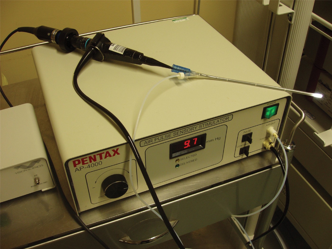
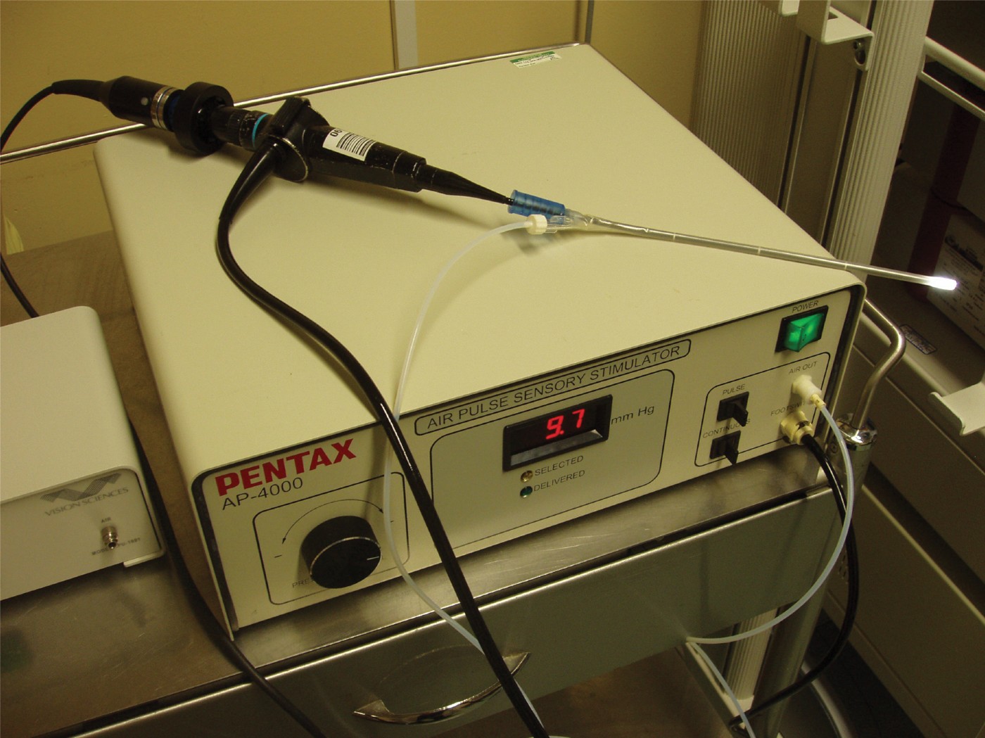
The following materials were used: a flexible nasolaryngoscope (25–30 cm length, 3.6 mm diameter; Olympus P4; Olympus, Southend-on-Sea, United Kingdom); disposable endoscope sheaths with air delivery port (Figure 1) (Vision Sciences, MedTronic, New York, United States); a videolaryngoscopy system with video-recording capacity (Xion; Sheffmed, Sheffield, United Kingdom); and an air pulse sensory stimulator (Figure 1) (Vision Sciences).
Flexible nasolaryngoscope attached to air pulse sensory stimulator.
Technique
Laryngeal sensory testing is carried out using a specially designed sensory stimulator (Figure 1). The device is calibrated to endoscopically administer an air pulse of varying pressure (2–10 mmHg) to the arytenoid mucosa, in an attempt to elicit the laryngeal adductor reflex. Each pulsed administration of air lasts 50 milliseconds. If no response to the air pulse is achieved at any pressure, continuous air stimulation can be used. The air pulse sensory stimulator must be calibrated every time it is used.
The air pulse threshold is initially set to 10 mmHg. The tip of the endoscope is brought to within 2 mm of the arytenoid mucosa and the air pulse delivered. As the air is delivered, it is possible to see the resulting indentation on the mucosa. Once the pulse has been delivered, the endoscope must be slightly withdrawn to preserve the endoscopist's field of vision as the laryngeal adductor reflex occurs. If the laryngeal adductor reflex is absent on pulse stimulation at maximum strength, continuous air flow is used. If this fails to elicit adduction of the vocal folds, the patient is assumed to have an absent laryngeal adductor reflex. Laryngeal sensory testing is performed on both the left and right arytenoid mucosa to determine a sensory threshold for each.
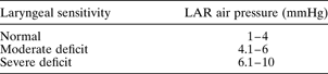
If the laryngeal adductor reflex is elicited at an air pressure of 10 mmHg, sequentially lower air pressures (at intervals of 2 mmHg) are used to determine the sensory threshold for each side. When the laryngeal adductor reflex becomes absent during this process, the air pressure is increased in smaller increments until a precise sensory threshold for each side of the laryngeal mucosal is determined. The laryngeal sensory thresholds for deficits of varying severity are shown in Table I.
Sensory capacity of laryngopharynx as determined by laryngeal sensory testing
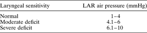
If no effect is seen at 10 mmHg air pressure with continuous flow, the laryngeal adductor reflex is assumed to be absent. LAR air pressure = air pressure at which laryngeal adductor reflex is elicited
Discussion
Loss of sensory discrimination in the laryngopharynx as a result of LPR has been well documented.Reference Aviv, Liu, Parides, Kaplan and Close8 Despite this, laryngeal sensory testing is not commonly used in the assessment of this condition in the UK. The authors have found that laryngeal sensory testing provides a useful adjunct to the assessment and monitoring of patients with LPR, when used in combination with the reflux finding score and the reflux symptom index. This finding is supported by work conducted by Aviv et al., who demonstrated that the laryngopharyngeal sensory deficits of patients with LPR improve with PPI therapy, and that both were correlated with an improvement in patient symptoms and laryngopharyngeal oedema.Reference Aviv, Liu, Parides, Kaplan and Close8 In addition to providing a quantitative assessment of laryngeal sensitivity, laryngeal sensory testing has been shown to be reproducible, with good intra-observer reliability, thus allowing the clinician to monitor the response to treatment.Reference Cunningham, Halum, Butler and Postma9, Reference Aviv, Kim, Thomson, Sunshine, Kaplan and Close10
Laryngeal sensory testing has been shown to be a safe procedure, with no significant adverse side effects. In a series of 1340 patients, epistaxis occurred in only one, and there were no instances of airway compromise.Reference Aviv, Murry, Zschommler, Cohen and Gartner11 In another study of 500 consecutive examinations, the most frequently occurring side effect of laryngeal sensory testing was mild discomfort, which occurred in 71 per cent of patients.Reference Aviv, Kaplan, Thomson, Spitzer, Diamond and Close12 In the same study, 99 per cent of the patients stated they would undergo the procedure again if clinically indicated.
Conclusion
Given the prevalence of LPR and the workload burden this places on ENT departments, a quick, easy, cost-effective assessment tool is desirable. Laryngeal sensory testing fulfils these criteria and is well tolerated by patients, making it a useful adjunct to the assessment and monitoring of patients with LPR.