Introduction
First described in 1871 by Joseph Jones,Reference Jones1 a Confederate Army surgeon in the American Civil War, necrotising fasciitis is a rapidly spreading soft tissue infection characterised by progressive destruction of fascia and subcutaneous tissue.Reference Jones1 In 1952, Wilson found this infection to be primarily caused by staphylococcal species.Reference Wilson2 However, recent literature has revealed a polymicrobial aetiology including streptococci and anaerobes. Necrotising fasciitis is most common in the extremities, perineum and abdominal wall, and predominantly occurs in elderly and immunocompromised patients.
Necrotising fasciitis of the head and neck (cervicofacial necrotising fasciitis) carries high rates of morbidity and mortality, the latter ranging from 20 to 73 per cent.Reference Lin, Yeh, Lin, Ma, Hwang and Shen3 Symptoms usually develop quickly, and timely diagnosis is critical to optimising outcome. Misdiagnosis and delayed treatment can result in death from sepsis, mediastinitis, carotid artery erosion, jugular vein thrombophlebitis or aspiration pneumonia. Diagnosis is based on a combination of clinical history (including predisposing factors to infection), Gram staining and culture, imaging, and, ultimately, surgical exploration.
Cervicofacial necrotising fasciitis is uncommon, and the extant literature includes fewer than 200 reported cases in a number of case reports and small series.Reference Lin, Yeh, Lin, Ma, Hwang and Shen3 Any deep space neck infection can potentially lead to necrotising fasciitis. It is most often a mixed bacterial synergistic infection involving both aerobes and obligate anaerobes.Reference DeBacker, Bossuyt and Schoenaers4 Early and aggressive surgical management is critical to optimising outcome. Definitive and timely diagnosis can be challenging due to protean variability in this disease's symptoms, signs and bacteriology.Reference Krenk, Nielson and Christensen5
More so than in the extremities or perineum, cervicofacial necrotising fasciitis is rarely suspected initially, for three reasons: the disease is uncommon; presenting signs and symptoms are often benign; and there is frequently a dental infection or pharyngitis which is thought to explain all findings.
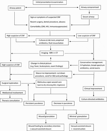
We propose an algorithm for the diagnosis and management of suspected cervicofacial necrotising fasciitis (see below), with the aim of facilitating early diagnosis and timely management.
Methods
This study was deemed exempt from review by the institutional review board of the New York University School of Medicine.
We retrospectively reviewed the medical records of seven consecutive patients ultimately diagnosed with cervicofacial necrotising fasciitis at two affiliated academic hospitals in New York City between July 2005 and August 2008. We noted the following parameters: age, sex, comorbidity, initial presentation, radiological and bacteriological findings, type of surgical intervention, presence of mediastinal involvement, airway management, complications, and patient survival.
Results
Patient characteristics
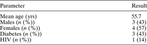
Seven consecutive patients ultimately diagnosed with cervicofacial necrotising fasciitis were included. Demographic and medical comorbidity data are summarised in Table I. The patients' mean age at presentation was 55.7 years (range 35–68). Three patients were men and four were women. Three patients had type two diabetes mellitus. One patient had human immunodeficiency virus (HIV) infection and was receiving highly active antiretroviral therapy.
Demographic and medical data of seven patients with cervicofacial necrotising fasciitis
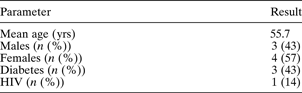
Yrs = years; HIV = human immunodeficiency virus infection
Presentation
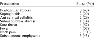
Patterns of presentation are summarised in Table II. Initial signs and symptoms were universally mild. Most patients experienced sore throat, fever and neck pain, with neck pain present in all cases. Each of the seven patients appeared well, comfortable, fully alert and in minimal or no distress. No patient exhibited shortness of breath, dysphagia, altered mental status or unstable vital signs. No patient had been subjected to recent trauma or surgery. There were no antecedent dental infections or dental procedures. Importantly, most patients were diagnosed with a localised infectious process, to which all signs and symptoms were initially attributed. At presentation, three patients had a peritonsillar abscess, while two presented with isolated anterior cervical cellulitis, two with supraglottitis and one with a submandibular abscess.
Initial presentation of seven patients with cervicofacial necrotising fasciitis

Pts = patients; ant = anterior
Imaging
Computed tomography (CT) imaging of the head and neck was obtained at the time of initial evaluation for all seven patients. In five patients, the initial radiological findings gave clear indications for urgent surgical exploration. These patients all had deep space neck abscesses, but only three had findings (i.e. subcutaneous emphysema) which prompted an unequivocal radiological diagnosis of cervicofacial necrotising fasciitis. Fascial plane blunting or dissection was seen in five patients, involving the platysma, sternocleidomastoid and strap muscles. Six patients had areas of low attenuation consistent with fluid accumulation. In each of these six cases, the low density regions involved multiple neck spaces and were not contained within cervical fascial compartments. Four patients had mediastinitis on initial imaging, indicated by streaky enhancement of mediastinal fat and fluid in the mediastinum. One patient had only peritonsillar phlegmon noted on initial CT scanning.
Diagnosis via cut-down procedure
Cervicofacial necrotising fasciitis was diagnosed on the day of presentation in six patients, and on hospital day six in one patient. In six patients, it was diagnosed either at the time of presentation (based on CT findings and clinical suspicion) or at the time of surgery.
A single patient had a delayed diagnosis. This diabetic patient was treated initially with intravenous antibiotics for a peritonsillar phlegmon and anterior neck cellulitis, and exhibited slow improvement in symptoms and physical signs over hospital days one to six. Because of an increase in leukocytosis and fever on day six, despite apparently improving clinical signs, a second CT scan was obtained, revealing only nonspecific low attenuation signal in various cervical compartments. Although there were no definitive signs of cervicofacial necrotising fasciitis, the nonspecific radiological findings and clinical events prompted a bedside cut-down procedure along the anterior border of the sternocleidomastoid muscle. Loose tissue planes and mildly friable muscle were encountered, and a decision made to undertake formal exploration in the operating theatre. This patient was ultimately determined to have extensive, bilateral cervical necrotising fasciitis with mediastinal extension, and required wide debridement in the neck and mediastinum.
Bacteriology
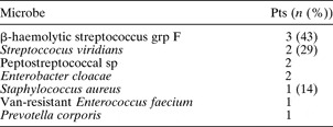
The causative bacterial species, identified on culture (Table III), were mixed in all but one case, and commonly involved aerobic and anaerobic oral flora. Specimens for bacterial culture included wound tissue, pus, blood and soft tissue biopsy material. The organism most often cultured was β-haemolytic group F streptococcus (n = 3), followed by Strepto-coccus viridians (n = 2), peptostreptococcal species (n = 2) and Enterobacter cloacae (n = 2). Vancomycin-resistant Enterococcus faecium and Prevotella corporis were cultured from one patient each as part of a mixed presentation. Isolated Staphylococcus aureus infection was seen in one patient.
Bacteriology of seven patients with cervicofacial necrotising fasciitis

Pts = patients; grp = group; sp = species; van = vancomycin
Management
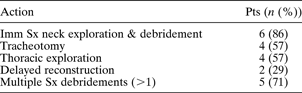
Management of the infection varied among the seven cases, as shown in Table IV. All seven patients were commenced immediately on fluid resuscitation and broad spectrum intravenous antibiotics, due to clinical suspicion of bacterial infection. Airway patency was evaluated in all patients on initial presentation, with flexible fibre-optic laryngoscopy. One patient was electively endotracheally intubated prior to obtaining any imaging, due to worsening supraglottitis; ultimately, this patient required a tracheotomy after 10 days of intubation. However, the remaining six patients did not develop respiratory embarrassment prior to surgery. The airway was secured by endotracheal intubation in these six patients at the time of primary surgery.
Management of seven patients presenting with cervicofacial necrotising fasciitis
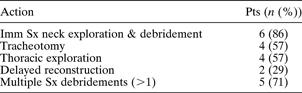
Pts = patients; imm Sx = immediate surgical
Six patients underwent immediate surgical exploration and debridement of necrotic tissue within 12 hours of suspicion of necrotising fasciitis. In four patients, initial surgery also included mediastinal and thoracic exploration via thoracotomy, wash-out and chest tube placement. Five patients required an additional procedure (four required multiple additional procedures) for serial debridements and wash-out of the neck and mediastinum. A delayed tracheotomy was performed in four cases within 7 to 14 days after the initial debridement, due to prolonged intubation.
Outcomes
Three of the four patients who underwent tracheotomy were successfully decannulated prior to hospital discharge. A single patient, who developed hypoxic brain injury due to mucus plugging of the tracheotomy tube, remained ventilator-dependent for the remainder of the hospital course.
A single patient with extensive, bilateral cervical necrotising fasciitis refused all surgical intervention, and was treated only with empirical, intravenous, broad-spectrum antibiotics. On hospital day three, areas of overlying skin broke down, permitting egress of copious purulent debris and ‘dishwater fluid’. The patient ultimately died on hospital day six due to progressive sepsis.
In each of the six patients who were aggressively debrided, meticulous wound care was continued, including twice-daily dressing changes. All six patients had open wounds that healed by secondary intention. A delayed reconstruction with a pectoralis major flap was performed in one patient. In this patient, progressive septic shock and residual local infection eventually resulted in flap failure.
In total, there were two deaths: the patient who refused surgery (who died on hospital day six), and another who suffered progressive septic shock, a massive post-operative myocardial infarction and ultimately multi-organ failure. One patient experienced major morbidity (hypoxic brain injury).
Discussion
While cervicofacial necrotising fasciitis is not rare, single institutions generally do not accrue sufficient cases to permit reporting of patterns of presentation and management, and the existing literature consists nearly entirely of individual case reports and several small case series.Reference Krenk, Nielson and Christensen5–Reference Skitarelic, Mladina and Morovic16 To date, we have not identified any systematic studies of the diagnosis or management of cervicofacial necrotising fasciitis.
In addition to providing further validation of the deceptively benign early appearance of cervicofacial necrotising fasciitis, our review emphasises two key points. First, cervicofacial necrotising fasciitis patients' initial CT will often appear to be normal or to involve deceptively minor pathology. Second, a cut-down procedure under local anaesthesia at the bedside – the use of which has not previously been reported in cervicofacial necrotising fasciitis cases – may be crucial to timely diagnosis in difficult cases.
The severity of cervicofacial necrotising fasciitis and its rapid spread necessitate early diagnosis. In many – but not all – cases, the diagnosis is straightforward. Because any delay in diagnosis or treatment significantly increases the risk of morbidity and mortality, it is imperative that challenging diagnoses also be made without delay. Figure 1 shows an algorithm for the contemporary management of cervical necrotising fasciitis, based on the available literature and our experience.
Algorithm for management of suspected cervicofacial necrotising fasciitis (CNF). Exam = examination; DM = diabetes mellitus; HIV = human immunodeficiency virus; lab = laboratory; MRI = magnetic resonance imaging; CT = computed tomography; IV = intravenous; C&S = culture and sensitivities; BID = twice daily

Cervicofacial necrotising fasciitis has widely varying presentations, ranging from mild, localised cellulitis to deep neck infections with subcutaneous emphysema and mediastinitis. While two of our patients presented with isolated anterior neck cellulitis, the remaining five all presented with a deep neck space infection, most commonly a peritonsillar abscess. There was no association between the location of the initial infection and the spread to mediastinitis or the presence of subcutaneous emphysema. Common presenting symptoms were fever, neck pain and sore throat. Additional symptoms or signs that should raise suspicion of necrotising fasciitis include cutaneous anaesthesia, ill-defined areas of erythema and tenderness, dusky or grey skin discoloration, and crepitus.
Our results are in agreement with the largest detailed review of cervicofacial necrotising fasciitis published to date, by Lin et al., which included 47 cases encountered over a 12-year period.Reference Lin, Yeh, Lin, Ma, Hwang and Shen3 In these patients, neck cellulitis was a universal sign, and pain was the only symptom present in the majority (85 per cent of patients). Otherwise, presentation was highly variable.
Diagnosis can be relatively straightforward in cases with extensive abscess formation and subcutaneous emphysema. Even if necrotising fasciitis is not immediately suspected, these patients generally undergo prompt surgical incision and drainage, leading to recognition of necrotising fasciitis intra-operatively. However, many cases are not this straightforward; in our series, two of seven patients did not have alerting CT findings at presentation.
Patients with a difficult diagnosis of cervicofacial necrotising fasciitis demonstrate a constellation of findings: diffuse neck cellulitis, neck tenderness or pain (generally mild) without a clear explanation, and comorbid diabetes or immunocompromise. This clinical picture should prompt a high index of clinical suspicion for cervicofacial necrotising fasciitis. The limited literature indicates a high prevalence of diabetes in these patients.Reference Lin, Yeh, Lin, Ma, Hwang and Shen3–Reference Skitarelic, Mladina and Morovic16 The initial infectious cause may be dental in origin, or related to tonsillitis or pharyngitis; these more commonplace diagnoses may be the focus of initial efforts, if cervicofacial necrotising fasciitis is not kept in mind. In the face of such a clinical picture, cervicofacial necrotising fasciitis should be strongly suspected, even if the patient is believed to simply have a straightforward otolaryngological infection. In our cases in which diagnosis was not straightforward, operative findings were far more extensive than suspected clinically.
A normal CT does not rule out necrotising fasciitis. Not every case will present with frank abscess formation or subcutaneous air, and in some cases initial findings are nonspecific. These early CT findings may be limited to small fluid collections, low density changes in soft tissue consistent with early necrosis, or fascial thickening. Minor or nonspecific radiological findings suggestive of early infection should be serially monitored if the patient fails to improve clinically. Progressive or persistent symptoms, elevation in leukocyte count, and fever are all symptoms that should prompt repeated imaging to assess disease progression. There is emerging evidence supporting a role for magnetic resonance imaging in early differentiation of non-necrotising cellulitis from necrotising fasciitis.Reference Edlich, Cross, Dahlstrom and Long17
Despite negative imaging, patients with diffuse cellulitis, persistent pain or fever, or symptoms and signs which are slow to improve should be considered for a cut-down procedure and incisional biopsy. At the bedside, the area of suspected involvement is locally anaesthetised, and a 1–2 cm incision is made through the skin and platysma to the level of the deep cervical fascia. Lack of bleeding or production of dishwater-coloured fluid are highly suspicious for necrotising fasciitis. A positive ‘finger test’ – i.e. easy separation of tissue planes under gentle finger dissection – is also highly suspicious.Reference Edlich, Cross, Dahlstrom and Long17 It should be stressed that, even if the fascia appears normal, specimens should be sent for culture and sensitivity, as well as frozen section histopathological analysis. Histological findings of obliterative vasculitis and soft tissue necrosis are characteristic of necrotising fasciitis.
A cut-down procedure with biopsy is sometimes performed in suspected extremity necrotising fasciitis, but we are not aware of any reports of its use in the head and neck.Reference Edlich, Cross, Dahlstrom and Long17 In many cases, appropriate use of this technique may be life-saving. In our series, it was utilised in one case. This patient had persistent neck cellulitis and tenderness, and exhibited unsatisfactorily slow clinical improvement. A repeated CT, prompted by leukocytosis and fever on hospital day six, was not definitive. Findings at cut-down prompted formal surgical exploration. In retrospect, this patient would have benefited from an earlier cut-down procedure.
Once necrotising fasciitis is confirmed intra-operatively, the surgeon must be prepared to widely debride the affected area, including all grossly necrotic skin and muscle. Often, apparently normal tissues may become involved soon after surgery, and repeated debridements are the rule rather than the exception. A delay in appropriately aggressive surgery has been associated with a high mortality rate.Reference Krenk, Nielson and Christensen5
Immediate airway control is essential when there is extensive cervical involvement. Our experience suggests that most patients with extensive cervicofacial necrotising fasciitis will inevitably require tracheotomy. Although confirmatory data from other authors is not available, we advise strong consideration of early tracheotomy in patients with extensive disease.
Meticulous daily wound care is essential after surgical debridements, to facilitate further removal of compromised tissue. A twice-daily regimen of wound irrigation and wet-to-dry dressing changes is recommended. The margins of the wound should be explored at each dressing change. Vacuum-assisted closure devices have been demonstrated to be a useful adjunct in wound-healing and closure.Reference Cainzos and Gonzales-Rodriguez18
Wound defects which are excessively large or which do not close by secondary intention can be managed with a reconstructive procedure. Reconstruction should be delayed until infection resolution is well established.
• Cervical necrotising fasciitis is uncommon but carries high rates of morbidity and mortality
• Presenting signs and symptoms are often deceptively benign
• A normal computed tomography scan does not rule out necrotising fasciitis
• A high index of clinical suspicion must be maintained in unexplained cases of cervical cellulitis and tenderness, especially in diabetic and otherwise immunocompromised patients
• A cut-down procedure with fascial biopsy is sometimes useful in expediting diagnosis
Prognostic factors for the outcome of cervicofacial necrotising fasciitis have not yet been successfully defined. As a larger number of cases accrue in the literature, this analysis may become feasible.
Conclusion
Cervical necrotising fasciitis is an uncommon, life-threatening disease which can present with deceptively innocuous symptoms and signs. Early diagnosis and aggressive management are critical to reducing the associated morbidity and mortality. We propose an algorithm which may be helpful in facilitating prompt diagnosis and management of this entity.
Acknowledgements
The authors thank Professors David Myssiorek, Joe Watts and Kelvin Lee at the New York University School of Medicine for their expertise, assistance in developing a theraupeutic strategy, and for helpful discussions.