Epithelial cells in the normal breast can be phenotypically and functionally subdivided into multiple types, with each subtype expressing unique genes and representing cells at different levels of differentiation (Refs Reference Visvader and Lindeman1, Reference Raouf2, Reference Villadsen3). Phenotypic classification has mostly relied on the expression pattern of select surface molecules; only a few key signalling molecules that bestow the phenotype have been characterised (Refs Reference Raouf2, Reference Villadsen3). These key molecules are likely to be involved in regulating a gene expression programme for maintaining the phenotype or triggering differentiation of cells. Characterisation of such molecules is therefore critical for understanding normal breast development as well as breast cancer. In this context, the forkhead family transcription factor FOXA1 has generated considerable interest because of its role as a critical component in the hormonal signalling network required for the growth and differentiation of a specific subtype of breast epithelial cells (Refs Reference Carroll4, Reference Laganiere5, Reference Eeckhoute6). In this review, we first describe general aspects of FOXA1 and then relate its function to a specific subtype of luminal epithelial cells of the breast and in breast cancer.
A brief history of FOXA1/HNF3α
Hepatocyte nuclear factor 3α (HNF3α) was initially identified as the transcription factor required for high-level expression of transthyretin and α1-antitrypsin in the liver (Ref. Reference Costa, Grayson and Darnell7). Response elements in the regulatory regions of these genes were used to generate DNA affinity chromatography columns for purification of HNF3α from rat liver and its subsequent cloning (Ref. Reference Lai8). Screening of cDNA libraries with HNF3α cDNA as a probe under low-stringency hybridisation conditions allowed cloning of two additional members of the family: HNF3β and HNF3γ (Ref. Reference Lai9). Based on the homology of these proteins to Drosophila forkhead proteins, HNF3α, HNF3β and HNF3γ were renamed as FOXA1, FOXA2 and FOXA3, respectively (Ref. Reference Kaestner10). Obviously, initial functional studies of these genes were focused on their role in the development of the liver (Ref. Reference Kaestner10). Binding sites for these transcription factors were found in hundreds of genes encoding gluconeogenic and glycolytic enzymes (Ref. Reference Kaestner10). Genetic analysis in mice revealed an important role for FOXA1 in proglucagon expression in the pancreas, FOXA2 in the development of foregut endoderm from which liver and pancreas arise, and FOXA3 in the induction of gluconeogenic enzymes during fasting (Ref. Reference Kaestner10). Subsequent studies revealed their role in differentiation of epithelial cells and longevity (Refs Reference Matusik11, Reference Panowski12). The unique ability of FOXA family transcription factors to bind to target sites in silent chromatin in a dominant manner and initiate regulatory events distinguishes them from the rest of the transcription factors (~2000) in the mammalian genome (Ref. Reference Cirillo13); therefore, these transcription factors are dubbed ‘pioneer factors’.
The FOXA1 regulatory circuit
DNA binding and transactivation
FOXA1 binds to target DNA sequences as a monomer, using a helix–turn–helix motif of ∼110 amino acids (Refs Reference Clark14, Reference Cirillo and Zaret15). This DNA-binding domain is flanked on either side by ‘wings’ of polypeptide chain that determine the stability of DNA binding (Ref. Reference Cirillo and Zaret15). The DNA-binding domain and the wings bind to the consensus sequence A(A/T)TRTT(G/T)RYTY. While the DNA-binding domain makes contact with the major groove (underlined sequence), the wings make contact with the minor groove of the DNA (sequences adjacent to the underlined sequences). The wings interact more tightly with high-affinity binding sites compared with low-affinity binding sites (Ref. Reference Cirillo and Zaret15). The genome-wide distribution patterns of low-affinity and high-affinity binding sites and the impact of differential binding on target gene expression are yet to be determined.
FOXA2 but not FOXA1 has been analysed extensively for transactivation domains; however, based on sequence conservation, the same regions in FOXA1 as in FOXA2 may also function as transactivation domains (Ref. Reference Qian and Costa16) The N-terminus and the C-terminus both contain transactivation domains (Fig. 1). The N-terminal transactivation domain of FOXA2 is phosphorylated by casein kinase 1 (CK1) and the phosphorylatable amino acid is conserved between FOXA1 and FOXA2 (Ref. Reference Qian and Costa16). Although this phosphorylation is not required for the transactivation function of FOXA2, similar evaluation of CK1 phosphorylation on FOXA1 function has not been performed. In fact, there are no reports on post-translational modifications of FOXA1 and the effect of such modifications on its ability to bind silent chromatin.
FOXA1 functional domains. The schematic of the FOXA1 polypeptide chain indicates the winged helix–turn–helix DNA-binding domain (‘winged helix DBD’) and two transactivation domains (TADs). SMAD family member 3 (SMAD3), NK2 homeobox transcription factor (NKX2.1) and androgen receptor (AR) interact with the DNA-binding domain of FOXA1 (Refs 19, 20), whereas TLE3/GRG3 (transducin-like enhancer of split 3/Groucho-related protein 3) associates with the C-terminal TAD (Ref. Reference Sekiya and Zaret22). The casein kinase 1 (CK1) phosphorylation site (at amino acid 18) and the nuclear localisation signal (NLS) have been characterised for FOXA2 and the corresponding amino acids are present in FOXA1 (Ref. Reference Qian and Costa16). Blue bars indicate highly conserved domains; numbers indicate amino acid positions.

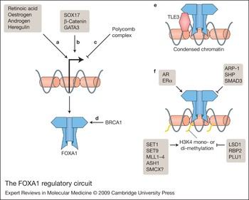
Two orphan nuclear receptors – apolipoprotein regulatory protein 1 (ARP-1; also called chicken ovalbumin upstream transcription factor II, COUP-TFII) and small heterodimer partner (SHP; also called NR0B2) – inhibit the transactivation function of FOXA1 (Refs Reference Achatz17, Reference Kim18). By contrast, androgen receptor (AR), another nuclear receptor, interacts with the DNA-binding domain of FOXA1 and this interaction leads to increased expression of target genes (Ref. Reference Gao19). FOXA1 also associates with the NK2 homeobox (NKX2.1) transcription factor, leading to repression of genes with the NKX2.1-binding site but lacking a FOXA1-binding site (Ref. Reference Minoo20). However, FOXA1 and NKX2.1 additively activate transcription of genes with both FOXA1- and NKX2.1-binding sites (Ref. Reference Minoo20). The effect of NKX2.1 on transcription of genes with only the FOXA1-binding site is unknown. SMAD family member 3 (SMAD3) transcription factor, which is activated by transforming growth factor β (TGF-β), associates with FOXA1 and prevents FOXA1 from interacting with NKX2.1 and subsequently with DNA (Ref. Reference Minoo21). The C-terminal region of FOXA1 associates with transducin-like enhancer of split 3 (TLE3; also called Groucho-related protein 3, GRG3) and recruits it to chromatin (Ref. Reference Sekiya and Zaret22) (Figs 1 and 2). The FOXA1–TLE3 association on chromatin results in the formation of locally closed chromatin and repression of FOXA1 target genes. Other coregulatory proteins that are essential for the transactivation function of FOXA1 are yet to be identified. It is likely that the ability of FOXA1 to open silent chromatin is dependent on the corecruited positive and negative coregulatory proteins. These coregulatory proteins may impart cell-type specificity to FOXA1 function.
The FOXA1 regulatory circuit. Regulation of FOXA1 expression and activity at multiple levels is shown. FOXA1 gene expression can be increased by (a) soluble factors such as oestrogen and (b) transcription factors such as GATA3. (c) Polycomb complex may repress expression by binding to the FOXA1 gene. (d) BRCA1 increases the stability of FOXA1. Several factors influence the activity of FOXA1. (e) Recruitment of TLE3 by FOXA1 to chromatin represses transcription. (f) Chromatin-modifying enzymes may indirectly control FOXA1 activity by redirecting FOXA1 to specific regions of the chromatin with histone H3K4 methylation (yellow tails), and additional trancription factors may regulate the function of DNA-bound FOXA1 at such sites. Abbreviations: AR, androgen receptor; ARP-1, apolipoprotein regulatory protein 1 (also known as COUP-TFII, chicken ovalbumin upstream-transcription factor II); ASH1, absent, small or homeotic; BRCA1, breast cancer 1; LSD1, histone demethylase lysine-specific demethylase 1 (also known as BHC100); MLL1–MLL4, mixed-lineage-leukaemia proteins; PLU-1, putative DNA/chromatin-binding motif and PHD/LAP domain 1 (also known as JARIB1B); RBP2, retinoblastoma binding protein 2; SET, set-domain-containing protein; SHP, small heterodimer partner (also known as NR0B2); SMCX, X-linked mental retardation gene (also known as JARID1C); SOX17, HMG box transcription factor; TLE3, transducin-like enhancer of split 3 (also called Groucho-related protein 3, GRG3).

Regulation of FOXA1 DNA binding through histone modification
The DNA recognition element for FOXA1 lacks a GC-rich sequence; thus, DNA methylation is not likely to play a role in restricting FOXA1 binding to DNA within chromatin. Epigenetic modifications of histones involving specific modifications of amino acids in histone tails is another mechanism that dictates binding affinity and/or accessibility of transcription factors to DNA within chromatin (Ref. Reference Kouzarides23). Accessibility to such DNA is not a regulatory mechanism for FOXA1 as it can bind to silent chromatin (Ref. Reference Cirillo13), but binding affinity may be regulated by specific chromatin modifications. This possibility has recently been addressed through genome-wide FOXA1-binding site analysis in MCF-7 breast cancer cells (Ref. Reference Lupien24). In these cells, 12 904 motifs bound FOXA1, representing only 3.7% of sites harbouring FOXA1-recognition motifs. FOXA1 recruitment correlated with methylation of histone H3 lysine 4 (H3K4) (Ref. Reference Lupien24). H3K4 undergoes mono-, di- or tri-methylation and these methylation events are associated with active transcription (Ref. Reference Li, Carey and Workman25). The majority of FOXA1-binding sites displayed H3K4 mono- or di-methylation (Ref. Reference Lupien24). It thus appears that FOXA1 binds to both silent and actively transcribed regions of chromatin; the effect of FOXA1 binding on transcription depends on corecruitment of TLE proteins to chromatin (silencing) or histone H3K4 methylation status around the binding site (activation) (Fig. 2).
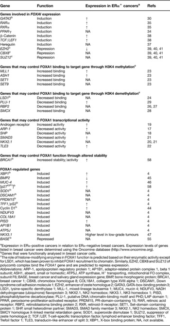
The binding of FOXA1 to chromatin regions with H3K4 mono- or di-methylation indicates that the expression levels of enzymes that control mono- or di-methylation of H3K4 will influence the binding affinity of FOXA1 to DNA within chromatin. FOXA1 is not required for these histone modifications (Ref Reference Lupien24). Mixed-lineage-leukaemia proteins MLL1–MLL4, set-domain-containing proteins SET1 and SET9, and ASH1 (‘absent, small or homeotic’) methylate H3K4 (Ref. Reference Kouzarides23). Several demethylases are known: histone demethylase lysine-specific demethylase 1 (LSD1; also called BHC100) demethylates mono- and di-methylated H3K4 (Ref. Reference Kouzarides23); retinoblastoma binding protein 2 (RBP2) demethylates tri- and di-methylated H3K4 (Refs Reference Klose26, Reference Christensen27); X-linked mental retardation gene SMCX/JARID1C demethylates tri-methylated H3K4 to di- or mono-methylated but not to unmethylated H3K4 (Ref. Reference Iwase28); and PLU-1 (also called Jumonji, AT-rich interaction domain 1B, JARIB1B) also demethylates tri- and di-methylated H3K4 (to unmethylated H3K4) but not mono-methylated H3K4 (Ref. Reference Yamane29) (Table 1). Overexpression of LSD1 leads to specific reduction in FOXA1 recruitment to chromatin, suggesting that the expression levels of LSD1 will impact FOXA1 function in cells (Ref. Reference Lupien24). Similar studies examining the effects of overexpression or reduction in the levels of other demethylases or methyltransferases on the recruitment of FOXA1 are yet to be reported. It is likely that SMCX increases FOXA1 binding as it demethylates tri-methylated H3K4 to the di- or mono-methylated form, which is preferred by FOXA1. Nonetheless, these recent advances have begun to elucidate specific control mechanisms for the recruitment of FOXA1 to chromatin.
Modulators of FOXA1 function and their expression in breast cancer in relation to ERα status

aExpression in ERα-positive cancers in relation to ERα-negative breast cancers. Expression levels of genes listed in breast cancer were determined using the Oncomine database (http://www.oncomine.org).
bGenes that were functionally analysed in breast cancer cells.
cThe role of histone-modifying enzymes in FOXA1 function is predicted based on their enzymatic activity except for LSD1, which has been proven to inhibit FOXA1 recruitment to chromatin. Similarly, EZH2, CBX8 and SUZ12 of polycomb complex bind the FOXA1 gene and are predicted to repress expression.
Abbreviations: ARP-1, apolipoprotein regulatory protein 1; AP1B1, adaptor-related protein complex 1, beta 1 subunit; ASH1, absent, small or homeotic; ATP5J, ATP synthase, H+ transporting, mitochondrial FO complex, subunit F6; BASE, breast cancer and salivary gland expression gene; BMP, bone morphogenic protein; BRCA1, breast cancer 1; CBX8, chromobox homologue 8; COL18A1, collagen type XVIII alpha 1; DSCAM1, Down syndrome cell adhesion molecule 1; EZH2, enhancer of zeste homologue 2; GATA3, GATA-box-binding protein 3; LSD1, lysine-specific demthylase 1; MLL-1, mixed-lineage leukaemia 1; MUC4, mucin 4; NDUFV3, NADH dehydrogenase (ubiquinone) flavoprotein 3; NKX2.1, NK2 homeobox; NKX3.1, NK3 homeobox 1; PISD, phosphatidylserine decarboxylase; PLU-1, putative DNA/chromatin-binding motif and PHD/LAP domain 1; PPAR, peroxisome-proliferator-activated receptor; PRDM15, PR-domain-containing 15; RAR, retinoic acid receptor; RBP2, retinoblastoma binding protein 2; RXR, retinoid-X-receptor; SET1, Set-domain containing protein 1; SHP, small heterodimer partner; SMAD3, SMAD family member 3; NKX2.1, NK2 homeobox; SMCX, SMCY homologue X-linked mental retardation gene; SOD1, superoxide dismutase 1; SUZ12, suppressor of zeste homologue 2; TCF/LEF, T-cell-specific transcription factor/lymphoid enhancer binding factor; TFF1, Trefoil factor 1; TLE3, transducin-like enhancer of split 3; XBP1, X-box binding protein; NA, not available.
Regulation of FOXA1 expression
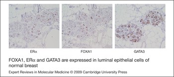
Transcription factors that control FOXA1 expression are just beginning to be identified (Fig. 2). In mammary epithelial cells, FOXA1 expression is positively regulated by GATA3 (Ref. Reference Kouros-Mehr30). GATA3 is a lineage-restricted transcription factor that is essential for the differentiation of mammary luminal progenitor cells (Refs Reference Kouros-Mehr30, Reference Asselin-Labat31). Oestrogen induces FOXA1, possibly through oestrogen (estrogen) receptor α (ERα) (Ref. Reference Laganiere5). Therefore, luminal epithelial cells that express ERα and GATA3 are expected to express the highest levels of FOXA1 in the mammary gland. Indeed, FOXA1, ERα and GATA3 are each expressed in a specific subpopulation of normal luminal epithelial cells (Refs Reference Kouros-Mehr30, Reference Badve32, Reference Clarke33) (Fig. 3), and are likely coexpressed.
FOXA1, ERα and GATA3 are expressed in luminal epithelial cells of normal breast. Immunohistochemistry of serial sections of normal breast reveals expression of each of these transcription factors is restricted to a subpopulation of luminal cells, as previously described (Refs 30, 32, 33).

FOXA1 is induced by the nuclear receptor peroxisome proliferator activated receptor γ (PPARγ) (Ref. Reference Varley34). Retinoic acid induces FOXA1, possibly through nuclear receptors retinoic acid receptors (RARs) and retinoid-X-receptors (RXRs) (Ref. Reference Jacob, Budhiraja and Reichel35). In a subset of ERα-negative breast cancer cells, AR induces the expression of FOXA1 (Refs Reference Doane36, Reference Naderi and Hughes-Davies37). Thus, several among the 49 known nuclear receptors of the human genome control either the expression (ERα, PPARγ, RARs, RXRs and AR) or the function (SHP and ARP-1) of FOXA1 (Fig. 2).
The HMG box transcription factor SOX17 and the WNT-regulated transcription factor β-catenin positively regulate the expression of FOXA1 (Ref. Reference Sinner38). In a subset of ERα-negative breast cancer cells, heregulin induces FOXA1 expression (Ref. Reference Naderi and Hughes-Davies37). Heregulin activates cell-surface receptors ERBB2 (v-erb-b2 erythroblastic leukaemia viral oncogene homologue 2; also called HER2) and ERBB3; the transcription factor(s) downstream of heregulin-activated signalling involved in FOXA1 expression is yet to be identified.
The expression of FOXA1 in only a specific subset of luminal epithelial cells suggests ‘epigenetic control’ of FOXA1 expression. Polycomb complexes are well-characterised repressor complexes involved in epigenetic negative regulation of gene expression (Ref. Reference Schuettengruber39). Recent genome-wide analysis revealed specific interaction of polycomb proteins suppressor of zeste homologue 12 (SUZ12) and chromobox homologue 8 (CBX8) with the FOXA1 gene (Refs Reference Bracken40, Reference Lee41). As polycomb proteins show variable expression in epithelial cells, the expression levels of these polycomb proteins along with the levels of positive regulatory factors described above may demarcate the subpopulation of epithelial cells that express FOXA1.
FOXA1 and ERα axis in breast cancer: from obscurity to sudden prominence
Although FOXA1 was in the forefront of research into liver and prostate development and into metabolic disease for several years, its prominence in breast cancer was established only in 2005 by pioneering work of Myles Brown and colleagues (Ref. Reference Carroll4). While studying the binding pattern of ERα to the genome, these investigators observed specific enrichment of FOXA1-binding sites in genes that bound to ERα. These observations and subsequent studies linking GATA3 and ERα to FOXA1 expression enabled identification of a cell-lineage-specific hormonal transcription factor network composed of ERα, FOXA1 and GATA3 (Refs Reference Eeckhoute6, Reference Kouros-Mehr30). In this network, GATA3 is upstream of FOXA1, whereas ERα functions both upstream of and parallel to FOXA1 (Fig. 2).
As discussed below, genes downstream of this network are just beginning to be identified, and use of expression signatures containing these downstream genes may help to establish the activity of this hormonal network in breast cancer. Recently, a ten-gene expression signature was reported for the diagnosis of ERα-positive invasive ductal carcinoma (Ref. Reference Schneider42). Subsequent meta-analysis of ERα- and GATA3-coregulated genes revealed a four-gene set that overlapped with the ten-gene signature (Ref. Reference Wilson and Giguere43). This four-gene set includes FOXA1 and SLC39A6 [solute carrier family 39 (zinc transporter), member 6; also known as LIV-1] in addition to ERα and GATA3. The expression levels of SLC39A6 may serve as a marker of a functional ERα–FOXA1–GATA3 network.
Downstream targets of FOXA1
Induction of expression
The transcription factor XBP1 (X-box binding protein 1) is a direct target of ERα–FOXA1–GATA3 as it has binding sites for all three of these transcription factors (Ref. Reference Carroll4). Reduction of FOXA1 in MCF-7 breast cancer cells through short interfering (si) RNA caused loss of oestrogen-inducible expression of XBP1. Oestrogen-regulated expression of cyclin D1 and resveratrol-mediated ERα-dependent expression of bone morphogenic protein 2 (BMP2) require FOXA1 (Refs Reference Eeckhoute44, Reference Su45). MUC-4, a transmembrane protein involved in ERBB receptor signalling, requires FOXA1 for expression (Ref. Reference Jonckheere46); however, the roles of GATA3 and ERα in MUC-4 expression are unknown. Several oestrogen-inducible genes on chromosome 21 and 22 require FOXA1 for their expression (Ref. Reference Carroll4).
In prostate epithelial cells, FOXA1 increases the expression of BMP7 and NKX3.1 (Ref. Reference Gao47). Whether FOXA1 similarly regulates the expression of these genes in breast epithelial and cancer cells remains to be determined. In this respect, oestrogen positively and negatively regulates NKX3.1 and BMP7 expression, respectively (Refs Reference Korkmaz48, Reference Kusumegi49). NKX3.1 expression is of particular interest because of its role in differentiation and its well-characterised tumour suppressor function (Ref. Reference Dong50). Additionally, in MCF-7 breast cancer cells, NKX3.1-binding sites are enriched around ERα-binding sites, and NKX3.1 prevents ERα binding to certain regions of chromatin and modulates oestrogen-regulated expression of such genes (Ref. Reference Holmes51). Therefore, FOXA1-regulated expression of NKX3.1 may be critical for attenuation of oestrogen-inducible expression of select genes. NKX3.1 expression in breast cancer is mostly observed in the lobular histological type compared with ductal carcinomas (26% versus 9%) (Ref. Reference Gelmann, Bowen and Bubendorf52). Lobular carcinomas are usually ERα-positive and better differentiated, a characteristic shared with FOXA1-positive breast cancers (Ref. Reference Pestalozzi53). In this regard, FOXA1 expression is more frequent in lobular and tubular carcinoma and lower in medullary carcinoma of the breast (Ref. Reference Habashy54). Future studies may have to focus on elucidating the biological and prognostic relevance of the ERα–FOXA1–NKX3.1 differentiation network specific for lobular carcinomas.
Repression of expression
Although most of the research focus is on the ability of ERα and FOXA1 to induce gene expression, these factors together may also repress gene expression. For example, oestrogen-mediated repression of the BASE (‘breast cancer and salivary gland expression’) gene requires recruitment of both ERα and FOXA1 to the chromatin (Ref. Reference Bretschneider55) Therefore, the histone modifications that occur after recruitment of both factors to the BASE gene may be distinct from the modifications that may occur on inducible genes such as XBP1. Alternatively, FOXA1 may promote the recruitment of TLE3, which interferes with chromatin association of positively acting transcription factors (Ref. Reference Sekiya and Zaret22). Since the mechanism of oestrogen-mediated repression of gene expression is relatively poorly understood, BASE may serve as a model to study the ERα–FOXA1-mediated chromatin modifications in oestrogen-mediated repression of genes. Studies related to oestrogen-mediated repression of genes are as relevant as those of inducible genes because the majority of oestrogen-regulated genes are repressed by oestrogen in MCF-7 cells (Ref. Reference Frasor56).
In prostate epithelial cells, FOXA1 suppresses the expression of several stemness-associated genes (Ref. Reference Gao47). These include the Hedgehog signalling pathway molecules SHH, GLI1 and GLI2, the Notch pathway molecule NOTCH1 and the WNT pathway molecule β-catenin. Whether these genes are similarly regulated by FOXA1 in breast epithelial and cancer cells is unknown. In breast cancer, stemness-associated gene expression is usually observed in ERα-negative breast cancers (Ref. Reference Ben-Porath57). Therefore, it is likely that FOXA1 negatively regulates the expression of these genes in ERα-positive breast cancers. Additional genes negatively regulated by FOXA1 in prostate epithelial cells that may be relevant to breast cancer include fibroblast growth factor (FGF)7, FGF10 and BMP4 (Ref. Reference Gao47).
A recent study described FOXA1-inducible genes that negatively regulate oestrogen-stimulated cell proliferation. For example, FOXA1 in co-operation with BRCA1 (‘breast cancer 1’) increases the expression of the cell cycle kinase inhibitor p27KIP1 (CDKN1B) (Ref. Reference Williamson58). Oestrogen, in contrast, represses p27KIP1 function, mostly at the post-transcriptional level (Ref. Reference Prall59). Interestingly, FOXA1 is coexpressed with BRCA1 in primary breast cancers (Ref. Reference Habashy54). Thus, it appears that the ERα–FOXA1 network establishes ‘one-step forward’ (through cyclin D1 induction) and ‘one-step backward’ (through p27KIP1 induction) modes to check cell cycle progression. These checks and balances may depend on the function of the tumour suppressor BRCA1.
Breast cancer classification and prognostic relevance of FOXA1
The traditional immunohistochemistry (IHC)-based classification scheme for breast cancer is being increasingly supplanted by molecular classification models. The ‘intrinsic’ molecular classification, which is based on cDNA microarray expression data, classifies breast cancer into five groups: luminal A, luminal B, HER2-positive, basal and normal subtypes (Ref. Reference Perou60). Expression of ERα is seen in the luminal groups but not in other subtypes. These molecular classification models will dramatically impact clinical practice as individualised treatments are likely to become standard care in the near future. However, surrogate histopathological markers that mimic the molecular classification are being sought for clinical use (Ref. Reference Badve and Nakshatri61). The most commonly used definition categorises tumours that express both ERα and progesterone receptor (PR) as luminal A and the other ERα-positive tumours including those that express HER2 as luminal B (Ref. Reference Carey62). FOXA1 also appears to be a marker that can effectively bridge the two types of classification tools.
When we analysed FOXA1 protein expression in breast cancers by IHC in relation to intrinsic subtypes, FOXA1high (score greater than 3 by IHC) was associated with ERα positivity, GATA3 positivity and luminal A subtype (Ref. Reference Badve32). Most significantly, improved event-free survival was seen in these patients compared with patients in the FOXA1low group, and this difference was significant even at 20 years. Additionally, FOXA1 expression was found to be a better predictor of survival than PR. When the analysis was stratified based on lymph-node positivity, FOXA1-positive patients in both node-positive and unknown nodal status groups had a better prognosis than FOXA1-negative patients. It is important to note that a specific subpopulation of normal luminal epithelial cells express FOXA1 at levels similar to those in FOXA1high group of patients. Therefore, FOXA1 expression in breast cancers may indicate cell type origin and differentiation status of breast cancer rather than cancer-specific overexpression.
Analysis of FOXA1 expression in another tissue microarray of ∼250 patients treated with standard adjuvant therapy also demonstrated the prognostic value of FOXA1 (Ref. Reference Thorat63). A third study analysed the expression of FOXA1 in a series of ∼850 patients (Ref. Reference Habashy54). This study differed significantly from the previous two studies with respect to methodologies; different reagents were used for IHC (mouse monoclonal rather than goat polyclonal antibody in the other two studies) as well as a different cut-off point for positivity (H-score of 10 rather than 30). Despite these differences, the study demonstrated almost identical results in terms of association with luminal A subtype. FOXA1 expression was also associated with disease-free survival and overall survival; however, this association did not retain significance in multivariate analysis. One of the findings of this study is intriguing as it relates to hormonal therapy. In the group of patients that did not receive hormonal therapy, FOXA1 expression was found to be associated with favourable breast-cancer-specific survival. However, no significant differences in breast-cancer-specific survival or disease-free interval were observed between FOXA1high and FOXA1low patients treated with the hormonal therapy tamoxifen. This suggests that response to tamoxifen therapy depends more on luminal type A or B phenotype rather than FOXA1 expression alone.
Expression of FOXA1 in ERα-negative tumours has been correlated with the expression of AR (Ref. Reference Doane36). FOXA1 expression in these cells is suggested to be due to crosstalk between AR and ERBB2 signalling and to represent part of an AR-mediated hormone-responsive programme in ERα-negative/PR-negative breast cancers (Ref. Reference Naderi and Hughes-Davies37). Although three IHC-based studies and several gene expression analyses showed an association of FOXA1 with ERα-positive/luminal type A breast cancer, a cell-line-based study found an association between FOXA1 mRNA expression and ERBB2/HER2-positive phenotype (Ref. Reference Yamaguchi64).
FOXA1 expression and current clinical practice
Oncotype DX is one of the major commercially available, clinically applicable diagnostic tools that has emerged from gene expression analysis (Ref. Reference Conlin and Seidman65). This diagnostic tool is being used to decide adjuvant therapy for patients with ERα-positive and node-negative breast cancers. This reverse transcription–polymerase chain reaction (RT-PCR) assay examines the expression of 16 genes (Ki67, STK15, survivin, CCNB1, MyBL2, MMP11, CTSL2, GRB7, HER2/ERBB2, GSTM1, CD68, BAG1, ERα, PR, BCL2 and SCUBE2). The expression pattern of each of these genes is tabulated using an equation, and a recurrence score (RS) is assigned to each case. Among these genes, higher expression levels of ER, PGR, BCL2, SCUBE2, GSTM1 and BAG1 reduce RS (negative values), whereas higher expression levels of remaining genes increase RS. ERα-positive patients with higher RS benefit from chemotherapy in addition to hormonal therapy (Ref. Reference Paik66).
Chromatin immunoprecipitation (ChIP)-coupled microarray (ChIP-on-chip) studies in MCF-7 breast cancer cells revealed oestrogen-dependent binding of ERα to seven of the above the 16 genes (survivin, MyBL2, HER2/ERBB2, BAG1, ER, PR and SCUBE2) (Ref. Reference Bhat-Nakshatri67). Currently, the influence of oestrogen on the expression of only five of the 16 genes is known: oestrogen increases PR, CCNB1 and BCL2 expression but decreases HER2/ERBB2 expression, and derepresses survivin from p53-mediated repression (Ref. Reference Sayeed68). Additionally, GRB7 confers oestrogen independence to breast cancer cells (Ref. Reference van Agthoven69). The requirement of FOXA1 in ERα binding and oestrogen-regulated expression of the above genes is unclear at present. We propose that FOXA1 and its downstream targets such as NKX3.1 influence RS by modulating ERα-mediated induction or repression of some of these genes.
MapQuant DxTM (previously called genomic grade index) is a newly commercialised diagnostic tool that combines the expression levels of 97 genes to help discriminate between histological grades 1 to 3 (Ref. Reference Sotiriou70). It can be used to decide whether ERα-positive and node-negative patients should receive chemotherapy (Ref. Reference Loi71). Among these genes, 42 are regulated by oestrogen in MCF-7 breast cancer cells (Ref. Reference Bhat-Nakshatri67). Interestingly, FOXA1 is not one of the 97 genes. Elucidating the role of FOXA1 in oestrogen-dependent and oestrogen-independent expression of these genes may reveal the mechanism of histological-grade-dependent increase in the expression of some of these genes in ERα-positive breast cancers. If indeed reduction in FOXA1 expression in an experimental setting leads to elevated expression of the above genes, one could replace the expensive MapQuant DxTM with simple IHC for FOXA1 to determine additional benefits of chemotherapy for ERα-positive breast cancer.
Do modulators of FOXA1 expression and/or activity add prognostic value to FOXA1 in breast cancer?
Cell-culture-based studies have clearly shown a role for multiple proteins in regulating the function of FOXA1. The fact that some patients in the FOXA1high group still succumb to disease raises the possibility that certain functions of FOXA1 are impaired in these patients. Also, the expression levels of FOXA1 in normal breast epithelial cells and FOXA1high cancer cells are similar. This observation suggests that FOXA1 function but not the expression is impaired in cancer cells compared with normal cells. There are plenty of unanswered questions regarding cancers with an ERα-positive and FOXA1low phenotype. Is this phenotype due to a progressive loss of FOXA1 expression in cancer cells due to elevated expression of polycomb proteins? Alternatively, do these cancers originate from ERα-positive cells that naturally express low levels of FOXA1? This can be partially addressed through data mining of already available breast cancer gene expression studies looking specifically at the expression of genes that regulate FOXA1 function. Several genes that positively regulate FOXA1 function are overexpressed in ERα-positive breast cancers compared with ERα-negative breast cancers (Table 1). For example, MLL, which mediates H3K4 methylation, is overexpressed in node-negative and ERα-positive breast cancers compared with ERα-negative breast cancers. Similarly, ASH1, which functions similarly to MLL, is overexpressed in ERα-positive breast cancers. PLU-1, which demethylates tri- and di-methylated H3K4, is overexpressed in ERα-positive breast cancers. PLU-1 functions as a transcription repressor in breast cancer cells by repressing the expression of BRCA1 and is required for breast cancer proliferation (Ref. Reference Yamane29). BRCA1 has been shown to interact with FOXA1 and increase its stability (Ref. Reference Williamson58). Thus, in ERα-positive breast cancers that overexpress PLU-1, FOXA1 expression and/or activity may be attenuated. RAR, a positive regulator of FOXA1 function, and ARP-1, a negative regulator of FOXA1 function, are overexpressed in ERα-positive breast cancers. Similarly, TLE3, which represses FOXA1 target gene expression (Ref. Reference Sekiya and Zaret22), is overexpressed in ERα-positive breast cancers. Therefore, the ability of FOXA1 to direct ERα to specific genes as well as to regulate the expression of differentiation- or proliferation-associated genes may depend on the expression pattern of genes that influence FOXA1 activity.
Is FOXA1 a target for differentiation therapy?
All four studies that compared FOXA1 expression in primary breast cancer have established FOXA1 as a good prognostic marker for ERα-positive breast cancers (Refs Reference Badve32, Reference Habashy54, Reference Thorat63, Reference Wolf72). In vitro studies have clearly shown that FOXA1 and ERα are functionally linked (Refs Reference Carroll4, Reference Laganiere5). The functional link between these transcription factors may be associated with a higher level of differentiation and lower histological grade. If that is the case, can differentiation of ERα-positive breast cancers be enhanced by therapeutic increase in FOXA1 expression? Ligands for nuclear receptors have been reported to induce FOXA1. One is the ligand for PPARγ (Ref. Reference Williamson58), which is already in clinical use as antidiabetic therapy. The second is retinoic acid or its synthetic derivatives (Ref. Reference Jacob, Budhiraja and Reichel35). Retinoic acids have previously been shown to induce growth arrest or apoptosis of ERα-positive breast cancer cells (Ref. Reference Roman73). Although synthetic retinoids have been tested in clinical trials (Ref. Reference Veronesi74), targeting these retinoids to only ERα-positive breast cancers with low FOXA1 expression may enhance their therapeutic value. The acute-phase response, inflammation and polycomb repressor complexes are likely to decrease FOXA1 expression (Refs Reference Bracken40, Reference Lee41, Reference Qian75). Therefore, drugs with anti-inflammatory properties or that reduce polycomb complex may increase FOXA1 expression and induce differentiation of ERα-positive breast cancers.
Future directions
Significant progress has been made in understanding the role of FOXA1 in development and differentiation. Its function in cancer is mostly studied in the context of prostate cancer. Some of the knowledge gained from these studies is beginning to be applied to breast cancer. However, significant gaps exist in our knowledge of the normal luminal cell type that expresses FOXA1 and of the relationship between FOXA1, ERα and GATA3 expression. There is no evidence to indicate a cancer-specific increase in FOXA1 expression due to amplification or mutations in breast cancer. However, FOXA1 expression may decrease as the cancer progresses due to an increase in polycomb complex activity. Therefore, future efforts need to focus on testing whether a lack of FOXA1 expression in cancer is indicative of cell type origin or a progressive increase in the activity of polycomb complexes leading to the epigenetic silencing of FOXA1 expression. Cancers with active silencing of FOXA1 expression may benefit from epigenetic/differentiation therapies.
Only a subset of normal luminal epithelial cells express ERα, FOXA1 and GATA3 (Fig. 3). Methods that can simultaneously analyse the expression of ERα, FOXA1 and GATA3 in a single cell in a laboratory and/or clinical setting may help to dissect the luminal-cell-type-specific oestrogen response and the susceptibility of different cell types to cancer, and to assess response to hormonal therapy.
Acknowledgements and funding
We thank Indiana University Simon Cancer Center Translational Genomic and Immunohistochemistry cores for analysis. We also thank the peer reviewers for suggestions and bringing a few important publications on FOXA1 to our attention, and Dr Jay Patel for critical reading of the manuscript. Work in H.N.'s laboratory is supported by grants from National Institutes of Health (CA89153 and CA123561). H.N. is Marian J. Morrison Professor of Breast Cancer Research.