Coronary arterial anomalies, either in isolation or in association with other lesions, are relatively common in children, although the majority may not come to clinical attention. This paper will focus on anomalous aortic origin of a coronary artery from the wrong sinus of Valsalva.
Background
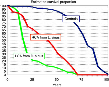
The most commonly reported coronary arterial malformation, in accounts of sudden deaths, is anomalous aortic origin of a coronary artery, with or without a proximal course between the aorta and the pulmonary artery. Anomalous coronary arteries may arise from the left, right, or non-coronary sinuses of Valsalva. Importantly, although the left coronary artery from the right sinus has the worst prognosis, sudden death has been reported in all variants of origin from the various sinuses of Valsalva (Fig 1).
Estimated survival proportion for patients known to have anomalous aortic origin of a coronary artery from the wrong sinus of Valsalva. All variants are at risk, as compared to the normal population. Patients with left coronary artery from the right aortic sinus of Valsalva have the worst prognosis. (This figure is modified from Hoffman JIE. Abnormal origins of the coronary arteries from the aortic root. In: Hoffman JIE. The natural and unnatural history of congenital heart disease. London: Wiley-Blackwell 2009:191, used with permission.)

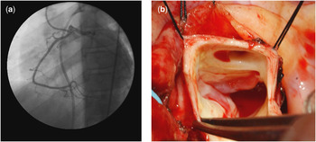
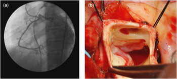
Cheitlin et al in 1974, described the clinical features and findings at autopsy in 13 patients in whom the left coronary artery arose from the wrong aortic sinus.Reference Cheitlin, De Castro and McAllister1 In a prospective study of 1950 patients undergoing coronary angiography, Angelini et al noted anomalous aortic origin of the right coronary from the left sinus in 0.92% of the cases, and anomalous aortic origin of the left coronary from the right sinus in 0.15%Reference Angelini2 (Fig 2). In a study of 2388 patients, who had echocardiographic imaging as part of an investigation for innocent murmur, and evaluation of performance status, four patients (0.2%) were found to have anomalous aortic origin of a coronary artery.Reference Davis, Cecchin, Jones and Portman3 Despite the fact that there would be referral bias in these two series, the suggestion is that several million people are probably affected by this anomaly in the United States of America alone.Reference Angelini2
(a) This coronary angiogram shows the origin of the left coronary artery in the right sinus of Valsalva. (b) This intraoperative view demonstrates an anomalous left coronary artery from the right aortic sinus of Valsalva, with a proximal intramural segment and interarterial epicardial course.
Sudden death is frequently the first presentation of this lesion, with only about one third of patients reporting some type of prodromal syndrome relating to the ischaemia.Reference Liberthson4, Reference Maron, Shirani, Poliac, Mathenge, Roberts and Mueller5 Presentations can include dyspnoea or syncope, with non-specific cardiovascular symptoms occurring in 18–30%.Reference Davis, Cecchin, Jones and Portman3–Reference Maron, Shirani, Poliac, Mathenge, Roberts and Mueller5 The important diagnostic differential is primary ventricular arrhythmia. Stress testing, even in individuals known to be affected, has a high incidence of yielding results that are falsely positive and falsely negative.Reference Liberthson4, Reference Basso, Maron, Corrado and Thiene6
Fatal ischaemic episodes occur during or shortly after strenuous exercise; and therefore, most victims are children who are old enough to play competitive sport.Reference Liberthson4, Reference Basso, Maron, Corrado and Thiene6, Reference Basso, Corrado and Thiene7 However, eight cases of sudden death have been reported in children below 1 year of age, and therefore the classical presentation is not universal.Reference Basso, Maron, Corrado and Thiene6
Several mechanisms are proposed to explain the occurrence of acute ischaemia in this potentially lethal anomaly.Reference Angelini2, Reference Davis, Cecchin, Jones and Portman3, Reference Basso, Corrado and Thiene7
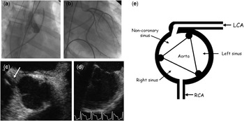
• Intermittent “flap closure” of the functionally stenotic slit-like coronary arterial orifice (Fig 3).
Figure 3(a) This aortic root angiogram demonstrates a patient with anomalous left coronary artery from the non-coronary sinus of Valsalva. An injection in the left sinus fails to demonstrate a coronary arterial orifice. (b) Following the engagement of the left coronary artery with a catheter, a normal distal vessel is present, suggesting ostial obstruction, which in this case was due to a proximal intramural course and slit-like orifice. (c, d) Echocardiographic imaging confirms anomalous aortic origin of the left coronary artery and global left ventricular hypokinesis related to prolonged ventricular tachycardia and acute myocardial infarction in an 11-year-old patient. (e) The schematic view of this anatomy is shown in the insert.

• An acute, non-orthogonal angle of exit of the coronary artery causes intermittent kinking of the proximal segment of the coronary artery.
• Compression of the intramural segment of the proximal coronary artery by the aortic commissure or aortic wall.
• Interarterial compression of the coronary artery between the aorta and the pulmonary artery, accentuated by exercise-related expansion of the pulmonary artery.
• Spasm of the coronary artery as a result of endothelial injury.
• Cumulative damage leads to focal myocardial necrosis, creating a pro-arrhythmic state.

In the presence of a proximal intramural segment of the coronary artery, ultrasonography has shown a thinning of the common aorto-coronary wall. This thinning could explain the luminal compression that occurs in systole, especially in patients who have a degree of ostial obstruction with a differential of pressure between the aorta and the proximal coronary. This effect might be less pronounced in older patients who have undergone some normal loss of compliance of the aortic wall, and in those who are less susceptible to sudden death from anomalous aortic origin of the coronary artery.Reference Cheitlin, De Castro and McAllister1
Surgical technique
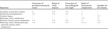
No effective medical treatment is available, but a number of surgical techniques are described to deal with the malformationReference Ono, Brown and Wolf8–Reference Rodefeld, Culbertson, Rosenfeld, Hanley and Thompson10 (Table 1). Even allowing for some uncertainty in the denominator, sudden death has been associated with all variants of anomalous aortic origin of the coronary artery. Therefore, in otherwise suitable candidates, it is probably justified to perform surgical correction whenever the diagnosis has been established. Our current technique was originally developed at the Royal Children’s Hospital, Melbourne, for congenital stenosis of a left coronary artery, and later applied to cases of Williams syndrome with ostial coronary arterial stenosis, and to patients with anomalous aortic origin of the coronary artery.Reference Kitayama, D’Orsogna and Karl11–Reference Guy, Tseng, Ratcliffe, Azakie and Karl14
Various operations designed to correct anomalous aortic origin of a coronary artery from the wrong sinus of Valsalva, with assessment of their potential to address the basic pathological features.

Repair is performed via median sternotomy using bicaval cannulation, at a temperature of 32 degrees centigrade. Once cardiac arrest has been achieved with anterograde and retrograde sanguineous cardioplegia, the aorta is transected just above the valve commissures. The pulmonary artery is transected close to the bifurcation.
An incision is made from the line of aortic transection into the ostium of the affected coronary artery, and then extended out onto the vessel wall for about 1 centimetre, either stopping short of the bifurcation or in some cases continuing on to the larger branch. The object is to open the coronary artery well beyond the intramural portion. A triangular patch of autologous pericardium treated with glutaraldehyde is advanced into the incision to create an ostium of 5 millimetres or larger.Reference Kitayama, D’Orsogna and Karl11, Reference Patel, Davidson and Karl12 The aortic segments are reconnected, incorporating the base of the pericardial triangle into the line of suture.
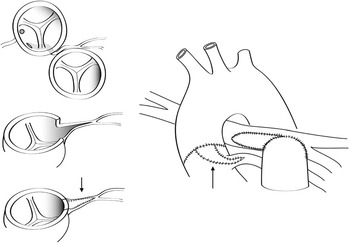
The left pulmonary artery is incised, stopping short of the bifurcation. The mobilised trunk of the pulmonary artery is moved leftwards and anastomosed to the left pulmonary artery, and the deficit in the main pulmonary artery is closed with a second pericardial patch (Fig 4). The goal of translocation of the pulmonary artery is to move the vessel away from the aorta, thereby reducing the chance of compression of the anomalous coronary artery between the great vessels.Reference Rodefeld, Culbertson, Rosenfeld, Hanley and Thompson10
This drawing demonstrates the technique for correction of an anomalous left coronary artery from the right aortic sinus of Valsalva, employing transection of the ascending aorta and pulmonary trunk, coronary arterial enlargement with a pericardial patch (arrows), and lateral translocation pulmonary trunk to the left pulmonary artery, with proximal right branch augmentation. (Please see the text for further details and also Alphonso et al13).

Results of surgery
Initial experience with this procedure at the University of California included seven patients, aged 2–52 years.Reference Alphonso, Anagnostopoulos and Nolke13, Reference Guy, Tseng, Ratcliffe, Azakie and Karl14 There was no operative mortality, although an 11-year-old patient with the left coronary artery arising from the non-coronary sinus required support with a left ventricular assist device for 72 hours after surgery. Her immediate pre-operative course was marked by extensive myocardial infarction and cardiogenic shock. All of the patients reported were free of symptoms at a median follow-up of 29 months. Echocardiographic imaging showed laminar flow through the neo-ostium in all cases. Of the patients who were examined, six had normal electrocardiograms and stress echocardiograms. To date, there have been no re-operations or late deaths and all patients are enjoying unrestricted activities.
Discussion
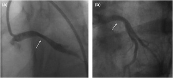
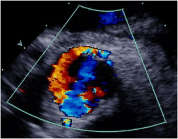
The technique described in this paper addresses all of the problems relating to anomalous aortic origin of a coronary artery. The ostial stenosis is corrected with the pericardial patch. The abnormal angle of exit created by the intramural portion of the coronary artery is overcome by the long incision into the coronary artery with subsequent enlargement with a patch. The plastic procedure also eliminates the possibility of compression by the aortic wall or a commissure during systole. The potential for compression of the coronary artery between the aorta and the pulmonary artery in systole is reduced by lateral translocation of the main pulmonary artery. Thus, both anatomic and physiological correction can be achieved (Figs 5 and 6).
(a) This coronary angiogram, in a patient with anomalous left coronary artery from the right aortic sinus of Valsalva, shows the left coronary artery compressed between the aorta and the pulmonary trunk in systole. (b) This coronary arteriogram shows the same patient, following repair. This image is taken at the same point in the cardiac cycle as panel (a) and suggests that the functional diameter of the vessel has increased.

This two-dimensional echocardiogram with interrogation with colour Doppler is performed on a patient with anomalous aortic origin of the left coronary artery from the non-coronary sinus of Valsalva, who has undergone enlargement of the proximal coronary artery with a patch plus translocation of the pulmonary artery. This study shows a supranormal diameter and relatively orthogonal angle of exit with laminar flow through the proximal vessel.

Sudden death in patients with anomalous aortic origin of a coronary artery from the wrong sinus of Valsalva may result from the cumulative effects of multiple episodes of myocardial ischaemia, which might be expected to predispose to ventricular tachyarrhythmia even after repair.Reference Liberthson4, Reference Basso, Corrado and Thiene7 However, this type of post-operative evolution is extremely rare, even for patients whose presentation was known to be with a ventricular arrhythmia.
The classical surgical technique for dealing with anomalous aortic origin of a coronary artery from the wrong sinus of Valsalva is ostial remodelling, in other words, excision of the common wall of the aorta and coronary artery.Reference Garcia-Rinaldi9 In some cases, it is necessary to detach a commissure of the aortic valve in order to accomplish the repair, and the potential for injury to the aortic valve is the main disadvantage of the technique. Despite being easy to perform, this operation may not be suitable when there is a single orifice to the right and left coronary arteries, or when there is a very eccentric location of a coronary arterial orifice within the aortic sinus. Furthermore, this unroofing technique also does not address the problem of compression of the coronary artery along the intra-arterial course.
The use of translocation of the pulmonary artery was described by Rodefeld.Reference Rodefeld, Culbertson, Rosenfeld, Hanley and Thompson10 As an isolated procedure, it has the advantage of simplicity, and avoids any surgery within the aortic root. This technique alone cannot address all of the abnormalities inherent in this syndrome, and therefore we regard it as a very useful adjunct to a direct coronary arterial procedure.
Coronary artery bypass grafting is described for use in patients with anomalous aortic origin of a coronary artery from the wrong sinus of Valsalva, although, with significant antegrade competitive flow, the patency of the graft will almost certainly be compromised in the long term.Reference Ono, Brown and Wolf8, Reference Garcia-Rinaldi9, Reference Mavroudis, Backer, Duffy, Pahl and Wax15 Reimplantation of the coronary artery, another strategy proposed to correct this problem,Reference Di Lello, Mnuk, Flemma and Mullen16 is technically demanding and can be problematic when there is a common origin of right and left coronary arteries. As in the simple unroofing operation, some potential exists for distortion of the aortic valve.
Despite the probable usefulness of all of these techniques in certain situations, we describe a universal solution to the problem that takes into account all of the potential mechanisms that can generate ischaemia. The technique can be used for any variant of anomalous aortic origin of a coronary artery, even when there is a common orifice for the right and left coronary arteries. Arterioplasty of the proximal coronary artery with an autologous pericardial patch has shown satisfactory long-term results in both children and adults.Reference Anagnostopoulos, Pigula, Myers, Beerman, Siewers and Gandhi17, Reference Meseguer, Hurle, Fernandez-Latorre, Alonso, Llamas and Casillas18
Conclusion
Anomalous aortic origin of a coronary artery from the wrong sinus of Valsalva is a potentially lethal cardiac anomaly that can be corrected in all cases using a simplified surgical technique that addresses the major anatomic and physiological problems.