Primary cardiac tumours are exceedingly rare in the paediatric population, with an estimated prevalence ranging from 0.027 to 0.49%.Reference Bédard, Becker and Gatzoulis 1 Their prevalence in pregnancies referred for fetal echocardiography was reported at 0.14%.Reference Holley, Martin and Brenner 2 Vascular tumours arising from the heart account for <5% of all primary paediatric cardiac tumours, with haemangiomas being most frequently encountered.Reference Mackie, Kozakewich, Geva, Perez-Atayde and Mulliken 3 , Reference Burke and Virmani 4 Although cardiac haemangiomas are histologically benign, and may even regress spontaneously, they can cause significant complications including large pericardial effusions, congestive heart failure, inflow or outflow tract obstructions, and disturbances in heart rhythm.Reference Laga, Gewillig, Van Schoubroeck and Daenen 5 – Reference Sharma, Hirata and Mosca 8
Rarely, cardiac tumours may also coexist with congenital heart lesions. As an example, a number of different structural cardiac defects have been found in patients with cardiac rhabdomyoma.Reference Burke and Virmani 4 , Reference Russell, Dhasmana, Berry and Gilbert-Barness 9
We present an extremely unusual coexistence of a cardiac haemangioma with a duct-dependent CHD in a newborn infant. To our knowledge, this is the first such case reported in the literature.
Case report
An apparently normal, full-term, male infant weighing 3400 g was born after an uneventful pregnancy and labour; however, the infant became severely cyanotic and dyspnoic in the first hour of birth. On physical examination, there was a single second heart sound, a faint systolic murmur at the left lower sternal border, and diffusely weak peripheral pulses. His transcutaneous oxygen saturation was about 60% on room air. The cyanosis was unresponsive to supplemental oxygen, and the child was intubated. Chest radiography showed mildly enlarged cardiac silhouette with diminished pulmonary vascular markings. Echocardiographic examination revealed a complex duct-dependent CHD. Intravenous prostaglandin E1 therapy was promptly initiated to maintain duct patency, and the patient was transferred to our hospital for further assessment and treatment.
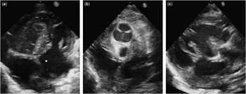
A repeat echocardiogram was performed on admission. It revealed usual atrial arrangement with normal systemic and pulmonary venous drainage. There was a restrictive atrial septal defect within the oval fossa with a right-to-left shunt across it. The atrioventricular connections were concordant with the tricuspid valve being atretic. The most prominent finding was a huge tissue mass filling the inlet and trabecular portion of the right ventricular cavity (Fig 1a). The mass was well-demarcated, measuring 25×18 mm in diameter, inhomogeneous, and poorly vascularised as demonstrated by color flow Doppler. There was ventriculoarterial discordance with pulmonary valve atresia (Fig 1b). The left ventricle emptied into the aorta through a large muscular ventricular septal defect (Fig 1c). The ascending aorta arising from the right ventricle continued into a normal left-sided aortic arch. The arterial duct was widely opened with left-to-right shunting of blood to the confluent pulmonary arteries. The coronary anatomy was usual for dextro-transposition of the great arteries. The left coronary artery, branching into the left anterior descending and the circumflex coronary artery, arose from sinus 1, whereas the right coronary artery arose from sinus 2. There was a small pericardial effusion.
( a ) The apical four-chamber view demonstrates a tumour mass filling much of the right ventricle. There is also a small pericardial effusion. ( b ) Parasternal short-axis view showing the antero-posterior relationship of the aortic valve and atretic pulmonary valve. ( c ) The parasternal long-axis view shows a large muscular ventricular septal defect opening into the right ventricular outflow tract.

The electrocardiogram displayed right-axis deviation with a pattern of right ventricular dominance, first-degree atrio-ventricular block, and non-specific ST and T wave abnormalities. The child was maintained on prostaglandin E1 and closely monitored in the ICU. Despite this, there was persistent hypoxia and metabolic acidosis.
The child’s parents did not consent to the proposed palliation consisting of balloon atrioseptostomy, and then placement of the systemic-to-pulmonary shunt. The following day, ventricular tachycardia followed by bradyarrhythmia developed, and the infant died in cardiogenic shock after unsuccessful resuscitation.
Findings on autopsy were consistent with the echocardiogram. Grossly, there was a 30×20-mm reddish mass occupying most of the right ventricular cavity. The tricuspid valve was imperforate, showing fusion of the leaflets.
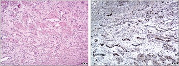
Histopathological examination revealed endothelium-lined vascular channels of various sizes and wall thicknesses (Fig 2a). There were also scattered adipocytes and foci of connective tissue. Surrounding cardiomyocytes showed signs of hypertrophy. Immunohistochemistry demonstrated negative staining for glucose transporter 1 (GLUT-1) and positive staining for cluster of differentiation 31 (CD31), cluster of differentiation 34 (CD34), factor VIII, α-smooth muscle actin (α-SMA), and Wilms’ tumor 1 (WT-1) protein (Fig 2b). Therefore, the diagnosis of a congenital haemangioma was established.
( a ) This low-power microscopic view of the tumour demonstrates numerous endothelium-lined vascular channels. The lesion is poorly demarcated from the surrounding myocardium (hematoxylin-eosin staining, ×40). ( b ) The vascular channels show positive immunohistochemical staining for endothelial marker CD31.

Discussion
Some cardiac tumours may be detected as early as 20 weeks of gestation, but the majority of them are observed later in pregnancy.Reference Uzun, Wilson, Vujanic, Parsons and De Giovanni 7 The location of haemangiomas within the heart appears to be related to the age of the patient. In fetuses and infants, haemangiomas predominantly originate from the right atrium, whereas in older children they can be found anywhere in the heart.Reference Mackie, Kozakewich, Geva, Perez-Atayde and Mulliken 3 , Reference Sharma, Hirata and Mosca 8 Therefore, it is very unusual for the haemangioma to arise from the right ventricle in utero, as in our case.
Presenting features mainly depend on the location and the size of the tumour.Reference Bédard, Becker and Gatzoulis 1 , Reference Mackie, Kozakewich, Geva, Perez-Atayde and Mulliken 3 In addition, a pericardial effusion might be the initial finding, as cardiac haemangiomas are secretory in nature.Reference Laga, Gewillig, Van Schoubroeck and Daenen 5 , Reference Sharma, Hirata and Mosca 8 Although it is known that cardiac tumours can mimic valvar stenoses or atresias by protruding into the chambers and causing obstruction to flow within the heart, their association with CHD is highly uncommon.Reference Bédard, Becker and Gatzoulis 1 , Reference Burke and Virmani 4 , Reference Russell, Dhasmana, Berry and Gilbert-Barness 9 It has been suggested that a space-occupying lesion may coexist with, or possibly induce, a malformation within the developing heart.Reference Russell, Dhasmana, Berry and Gilbert-Barness 9 In our case, the haemangioma was found in the presence of a complex transposition of the great arteries. Furthermore, the tumour caused the fusion of the tricuspid valve leaflets, thus producing functional tricuspid atresia.
It can be difficult to distinguish haemangiomas in this setting from other more common cardiac tumours, such as rhabdomyomas, fibromas, and teratomas. Moreover, the vascular nature of cardiac haemangiomas is not readily demonstrable with color flow Doppler, as the abnormal blood vessels are microscopic, although the main feeding vessel may occasionally be visualised within the tumour.Reference Tongsong, Sirichotiyakul, Sittiwangkul and Wanapirak 6
Surgical treatment for cardiac tumours is indicated in all symptomatic patients and in those with haemodynamic compromise.Reference Padalino, Vida and Boccuzzo 10 We believe that balloon atrioseptostomy followed by creation of the systemic-to-pulmonary shunt should be the treatment of choice in similar cases with functionally univentricular heart and duct-dependent pulmonary circulation. The occurrence of arrhythmias, however, might carry a poor prognosis.
This report shows that cardiac haemangiomas may not only coexist with congenital heart lesions but also cause severe haemodynamic impairment with deleterious effects on the developing heart. In addition, these findings could also suggest that cardiac haemangiomas may emerge earlier in pregnancy than previously reported.
Acknowledgements
The authors are greatly indebted to Prof. Ida Jovanović for her generous help and support as well as invaluable comments and suggestions.
Financial Support
This research received no specific grant from any funding agency or from commercial or not-for-profit sectors.
Conflicts of Interest
None.
Ethical Standards
The authors assert that all procedures contributing to this work comply with the ethical standards of the relevant national guidelines on human experimentation and with the Helsinki Declaration of 1975, as revised in 2008, and has been approved by the Ethics Committee of the University Children’s Hospital in Belgrade, Serbia.