Introduction
Pheochromocytoma and paraganglioma are tumours that originate from the adrenal medulla and paraganglia, respectively, and are characterised by catecholamine production. Compared with patients with noncyanotic CHD, those with cyanotic CHD have an approximately six-fold increased risk of pheochromocytoma and paraganglioma development. Reference Opotowsky, Moko and Ginns1 Pseudohypoxia-related pheochromocytoma and paraganglioma are characterised by the aberrant activation of hypoxia signalling pathways in chromaffin cells. Reference Opotowsky, Moko and Ginns1 In this context, chronic hypoxia in cyanotic CHD may further augment hypoxia signalling and promote tumorigenesis.
In the general population, pheochromocytoma and paraganglioma is rare in young children and is more commonly diagnosed in adults, often in the fourth to fifth decades of life. Reference Opotowsky, Moko and Ginns1,Reference Ogasawara, Fujii and Kakiuchi2 All published pheochromocytoma and paraganglioma cases in patients with CHD have developed during the second decade of life or later; the median age at the time of development is 31 years, and the youngest reported patient was 12 years old. Reference Opotowsky, Moko and Ginns1,Reference Ogasawara, Fujii and Kakiuchi2,Reference Arenillas, Celada and Ruiz-Cantador3 We present the case of an 8-year-old boy with pulmonary atresia with ventricular septal defect and major aortopulmonary collateral arteries who experienced persistent hypoxaemia without corrective surgery.
Case report
The patient was born in Indonesia and experienced cyanosis. Pulmonary atresia with ventricular septal defect and major aortopulmonary collateral arteries were diagnosed at age 1 month. Conservative management without corrective surgery was performed until age 6 years. At that time, he moved to Japan and was referred to our hospital because of cyanosis and oxygen saturation of 75% in room air. Because the major aortopulmonary collateral arteries were too narrow to be integrated and a confluent native pulmonary artery was present (Figure 1a, 1b), we elected to promote pulmonary artery growth. Palliative aortopulmonary shunts (4 mm at age 6 years and 5 mm at age 7 years) were placed, but oxygen saturation remained approximately 75% despite 1 L/min of supplemental oxygen.
(a) Angiography and (b) schematic images of major aortopulmonary collateral arteries (blue, orange, and green) and the native pulmonary artery (grey). (c) Contrast-enhanced abdominal computed tomography (CT) image showing a 3-cm enhancing tumour in the right adrenal gland (arrowhead). (d) 123I-Metaiodobenzylguanidine scintigraphy image demonstrating increased uptake at the tumour site (arrowhead) without metastasis or additional lesions.

Figure 1 Long description
The image consists of four separate elements. The first element is an angiography image showing the blood vessels around the heart. The second element is a schematic diagram illustrating major aortopulmonary collateral arteries in blue, orange, and green, along with the native pulmonary artery in grey. The third element is a contrast-enhanced abdominal computed tomography (CT) image displaying a 3-centimeter enhancing tumor in the right adrenal gland, indicated by an arrowhead. The fourth element is a 123I-Metaiodobenzylguanidine scintigraphy image demonstrating increased uptake at the tumor site, marked by an arrowhead, without showing any metastasis or additional lesions.
At age 8 years, the patient presented with dyspnoea, sweating, and hypertension (163/105 mmHg). Chest radiography showed increased pulmonary vascular markings. Transthoracic echocardiography revealed mildly reduced left ventricular systolic function (left ventricular ejection fraction, 45%); however, new valvular regurgitation and stenosis were not observed. Laboratory test results indicated marked elevation in B-type natriuretic peptide to 864.5 pg/mL; previously, however, this level was almost within the normal range (6.7–31.3 pg/mL). Cardiac troponin T, D-dimer, and inflammation marker (C-reactive protein) levels remained within the normal range. Haemoglobin and haematocrit levels were persistently elevated (range, 16–18 g/dL and 58–60%, respectively), consistent with long-standing hypoxia; however, erythropoietin data were not available.
Hypertensive heart failure exacerbation was diagnosed; therefore, the patient was urgently hospitalised. Urinary norepinephrine (2,660 μg/L; reference range: 29.2–118 μg/L) and normetanephrine (2.7 μg/L; reference range: 0.10–0.28 μg/L) levels were markedly elevated. Contrast-enhanced computed tomography (CT) revealed an enhancing tumour in the right adrenal gland (Figure 1c) and 123I-metaiodobenzylguanidine scintigraphy revealed increased uptake, thus confirming the diagnosis of pheochromocytoma (Figure 1d).
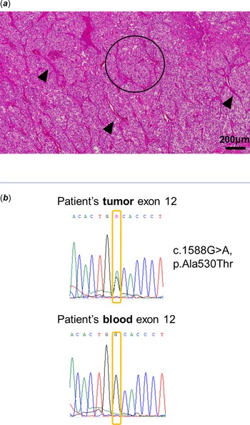
No metastases or additional lesions were observed. Surgical resection of the tumour resulted in spontaneous resolution of hypertension and related cardiac failure. The diagnosis of pheochromocytoma was confirmed by a pathological examination (Figure 2a).
(a) Histopathological examination of the resected tumour revealed a characteristic Zellballen pattern (indicated with the circle), consisting of tumour cell nests surrounded by a rich vascular stroma (indicated with the arrowheads) on haematoxylin and eosin staining (original magnification × 100). Scale bar = 200 μm. (b) Sanger sequencing identified an EPAS1 variant (NM_001430:c.1588G>A, p. Ala530Thr) in tumour deoxyribonucleic acid, but not in germline deoxyribonucleic acid extracted from the patient’s blood.

Figure 2 Long description
The image contains two elements: a histopathological examination and a genetic sequencing result. The first element is a histopathological image showing a resected tumor with a characteristic Zellballen pattern, indicated by a circle, and tumor cell nests surrounded by a rich vascular stroma, indicated by arrowheads. The scale bar is 200 micrometers. The second element consists of two genetic sequencing charts labeled 'Patient's tumor exon 12' and 'Patient's blood exon 12'. The tumor sequencing chart shows a variant c.1588G>A, p.Ala530Thr, which is not present in the blood sequencing chart.
Further investigation of underlying factors was prompted by the young age of the patient. Germline variants in genes that predispose individuals to pheochromocytoma and paraganglioma were analysed using a commercially available genetic testing panel. The pheochromocytoma and paraganglioma panel, including FH, MAX, NF1, RET, SDHA, SDHAF2, SDHB, SDHC, SDHD, TMEM127, and VHL, revealed no pathogenic variants. However, a Sanger sequence analysis of the tumour identified a somatic EPAS1 variant (c.1588G>A, p. Ala530Thr), which is recently recognised as a driver mutation for CHD-related pheochromocytoma (Figure 2b). Reference White, Nonaka and Chung4
Discussion
Hypoxia-associated pheochromocytoma and paraganglioma have been observed not only with systemic hypoxemic disorders such as cyanotic CHD Reference Opotowsky, Moko and Ginns1–Reference Arenillas, Celada and Ruiz-Cantador3 but also with exposure to chronic environmental hypoxia at high altitudes. Reference Kumar, Gupta, Thakur, Gupta and Bodh5 This association has been summarised in a recent review Reference Sanceau, Westbrook and Favier6 and supported by previous pathological and epidemiological studies.
Many previously identified genetic factors that result in pheochromocytoma and paraganglioma, such as SDHx and VHL, activate the hypoxia-inducible factor pathway, thus promoting tumorigenesis under pseudohypoxia. Reference López-Jiménez, Gómez-López and Leandro-García7 Chronic hypoxia in cyanotic CHD causes similar hypoxia-inducible factor activation, thereby promoting chromaffin cell proliferation and increasing the risks of pheochromocytoma and paraganglioma. Reference Ogasawara, Fujii and Kakiuchi2 EPAS1 encodes hypoxia-inducible factor 2α, and mutations in its oxygen-dependent degradation domain lead to hypoxia-inducible factor 2α stabilisation and transcriptional upregulation. Reference Ogasawara, Fujii and Kakiuchi2 Furthermore, the risks of pheochromocytoma and paraganglioma with the EPAS1 mutation are higher for patients with cyanotic CHD and severe hypoxaemia and those who have undergone palliative surgery. Reference Arenillas, Celada and Ruiz-Cantador3
To the best of our knowledge, pheochromocytoma associated with CHD in patients younger than the one described here has not been reported. Because the extent of cyanosis in our patient was relatively severe compared with that previously reported, Reference Opotowsky, Moko and Ginns1,Reference Ogasawara, Fujii and Kakiuchi2,Reference Arenillas, Celada and Ruiz-Cantador3 this case suggests that uncorrected cyanotic CHD and sustained severe hypoxaemia may contribute to the earlier onset of pheochromocytoma and paraganglioma, possibly through positive selection and clonal expansion of EPAS1-mutated cells as well as activation of the hypoxia-inducible factor pathway. Although erythropoietin data were not available, elevated haemoglobin and haematocrit levels may support a potential interaction between chronic hypoxia and EPAS1-related pseudohypoxia.
Previous studies have reported that recurrent CHD-associated pheochromocytoma and paraganglioma may occur as metachronous and/or multicentric tumours with independent clonal origins. Reference Ogasawara, Fujii and Kakiuchi2 In our patient, the persistence of residual adrenal medullary tissue indicated the possibility of an underlying EPAS1 mutation and the potential risk of pheochromocytoma and paraganglioma recurrence. Therefore, long-term follow-up comprising regular blood pressure monitoring and periodic assessments of urinary catecholamine levels is recommended. Identification of the somatic EPAS1 mutation supported and influenced our clinical management procedures, which included long-term surveillance because of the reported risk of metachronous or multicentric pheochromocytoma and paraganglioma associated with EPAS1-mutated clones and the possibility of independent development of additional EPAS1-mutated clones through positive selection in other chromaffin cell populations under chronic hypoxia.
Conclusion
Although chronic hypoxia and unrepaired cyanosis are recognised risk factors for EPAS1-mutated pheochromocytoma and paraganglioma, our case suggests that hypoxia-driven tumorigenesis may occur earlier than previously considered. Careful follow-up, including regular blood pressure monitoring and attention to related symptoms, may be warranted for such patients.
Acknowledgements
We would like to thank Editage (https://www.editage.com) for language editing. We thank Dr Masato Yonamine and Dr Kazuhiro Takekoshi for valuable insight regarding genetic testing for PPGL.
Financial support
This work was supported by the JSPS KAKENHI (grant numbers 22K17606 and 24K18874).
Competing interests
The authors declare none.
Ethical standard
The authors assert that all procedures contributing to this work comply with the ethical standards of the relevant national guidelines on human experimentation and with the Helsinki Declaration of 1975, as revised in 2008, and have been approved by the institutional committees (Ethics Committee of our institute, H23-128).