Neonates and young infants with isolated total anomalous pulmonary venous connection require surgical repair as soon as they are diagnosed. Previous reports have shown that surgical outcomes for total anomalous pulmonary venous connection are associated with not only preoperative obstruction of the vertical vein draining into the caval vein but also the small left ventricle owing to the overload of the right heart. Reference White, Ho and Faerber1–Reference Oliveira Lima, Valdes-Cruz and Allen4 We herein present the case of a newborn with supracardiac total anomalous pulmonary venous connection and mimicking hypoplastic left heart syndrome, including the small left ventricle, the ductus-dependent systemic circulation, and retrograde blood supply in the transverse arch. The patient successfully achieved biventricular circulation by a staged approach combined with surgical and transcatheter procedures.
Case report
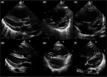
A newborn weighing 2,935 g at the 38 weeks’ gestational age was transferred to our hospital due to cyanosis and respiratory failure soon after birth. His systemic saturation was 80%. Chest X-ray showed the bilateral hazy lungs. Echocardiography showed that all pulmonary veins formed a common chamber and drained into the innominate vein via the vertical vein. There was no obstruction of pulmonary venous route. Further, echocardiography showed hypoplasia of the left heart; the left ventricular end-diastolic diameter: 7.6 mm (z-score: −7.5), the aortic annulus diameter of 5.6 mm (z-score: −1.8), the mitral annulus diameter of 8.0 mm (z-score: −1.8), the ductus-dependent systemic circulation, and retrograde blood supply in the transverse arch, which was mimicking hypoplastic left heart syndrome (Fig 1, Video S1). Doppler echocardiography showed diminished antegrade aortic blood flow with the velocity of 0.65 m/s but no accelerated trans-mitral blood flow. The interatrial communication was depicted as a foramen ovale with a right-to-left shunt. It was uncertain whether the patient could achieve biventricular circulation because the left ventricle was too small to maintain the systemic circulation as well as the aortic and mitral valves. Therefore, we performed the repair of the pulmonary venous return into the left atrium, creation of interatrial communication, and bilateral pulmonary arterial banding at the age of 2 days. The ductus was kept open on intravenous administration of prostaglandin E1. However, the left heart gradually developed after the first surgical intervention. Echocardiography at the age of 3 months showed that the diameters of left ventricular end-diastolic, the aortic, and mitral annulus were 14.3 mm (z-score: −4.56), 7.5 mm (z-score: −0.32), and 9.3 mm (z-score: −1.04), respectively (Fig 1, Video S2). The direction of blood flow in the transverse arch altered antegrade from retrograde. Doppler echocardiography showed increased antegrade aortic blood flow up to 1.10 m/s and the bidirectional interatrial shunt through a surgically enlarged atrial septal defect.
Echocardiograms after birth ( a , b , c ) and at the age of 3 months were shown. Echocardiograms after birth ( a , b , c ) show the left ventricular end-diastolic diameter of 7.6 mm (z-score: −7.5), the aortic annulus diameter of 5.6 mm (z-score: −1.8), and the mitral annulus diameter of 8.0 mm (z-score: −1.8). However, echocardiograms at the age of 3 months ( d , e , f ) show that the left ventricle grows up to 14.3 mm (z-score: −4.56).

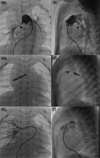
Thus, we performed the ductus occlusion test. A 5-French sheath was introduced from the right femoral vein, and a 5-Fr Berman angiographic catheterTM with a tip balloon (Gadelius Medical Japan, Tokyo, Japan) was placed into the main pulmonary artery through the right ventricle. Systemic blood pressure was monitored by the indwelling arterial line in the radial and the femoral arteries during the tip balloon was inflated to close the ductus. When the ductus was closed with a Berman angiographic catheterTM, the right radial arterial and the right femoral arterial pressures remained unchanged as 85/43 mmHg and 61/28 mmHg, respectively. Subsequently, we performed transcatheter de-banding of the bilateral pulmonary arteries. As angiograms showed that the diameters of left and right pulmonary arteries at the banding site were 2.8 mm and 2.0 mm, respectively, we successfully expanded the bilateral pulmonary arteries using a high-pressure non-compliant angioplasty balloon with a diameter of 5 mm (SHIDENTM, KANEKA Medix, Tokyo, Japan). After the procedure, the diameters of left and right pulmonary arteries were increased to 3.9 mm and 3.6 mm, respectively (Fig 2). We also attempt to close an atrial septal defect using Amplatzer Septal OccluderTM. However, the defect size of 14.0 mm was too large to close the device, so that the defect and the ductus were surgically closed after then. He was doing well in the outpatient clinic at the age of 3 years.
Transcatheter pulmonary artery de-banding is shown. The diameter of left (white arrowhead) and right (black arrowhead) pulmonary arteries at the banding site are 2.8 mm and 2.0 mm, respectively ( a , b ). The balloon angioplasty successfully dilates the pulmonary arteries ( c , d ); the diameters of left and right pulmonary arteries were increased to 3.9 mm and 3.6 mm, respectively ( e , f ).

Discussion
We present a newborn with total anomalous pulmonary venous connection mimicking hypoplastic left heart syndrome. Our staged approach could allow the patient to achieve biventricular circulation. Previous reports have shown that neonates with total anomalous pulmonary venous connection present the small left ventricle owing to compressed interventricular septum by the right ventricular overload. Reference Phoon and Silverman3–Reference Cohen and Rychik5 In this setting, the aortic and mitral valve annulus diameters are usually measured within the normal range. However, the present case showed not only the small left ventricle but also the small mitral and aortic annulus diameters. Moreover, the retrograde blood supply in the transverse arch was a characteristic feature of hypoplastic left heart syndrome. There are several predictive calculation formulas to identify the possibility of biventricular circulation in neonates with hypoplastic left heart syndrome. Reference Colan, McElhinney, Crawford, Keane and Lock6–Reference Mart and Eckhauser8 Colan et al. reported a predictive score: 10.98 × (body surface area) + 0.56 × (aortic annulus z-score) + 0.89 × (left ventricular to heart long-axis ratio)−0.79 × (grade 2 or 3 endocardial fibroelastosis), which a cut-off value of more than −0.65 predicted favorable outcome in neonates with critical aortic stenosis and hypoplastic left ventricle. Reference Colan, McElhinney, Crawford, Keane and Lock6 Mart et al. reported another scoring formula: [(the ratio of mitral annulus to aortic annulus)/(the ratio of left ventricular length to right ventricular length) + (pulmonary trunk diameter)]/(body surface area), in which a cut-off value less than 16.2 could predict to achieve biventricular circulation in neonates with borderline left heart hypoplasia, or hypoplastic left heart complex. Reference Mart and Eckhauser8 In the present case, Colan’s and Mart’s scores were −1.17 and 13.8, respectively, which suggested the possibility to achieve biventricular circulation in our present case. As the amount of pulmonary venous blood flow has been estimated to supply approximately 50% of the left ventricular output in a normal fetus, Reference Allan and Sharland9 the discrepancy between the aortic and mitral valve annulus sizes and the left ventricular diameter may suggest not maldevelopment of the left ventricular structures resulting in hypoplastic left heart syndrome but growth failure owing to a decreased pulmonary venous blood flow.
After the balloon occlusion test, we could not complete transcatheter procedures to close both an atrial septal defect and the ductus and dilate the pulmonary arteries. If we knew that the left ventricle might grow up enough to establish the biventricular circulation, we would make a small atrial septal defect at the first surgical procedure. Balloon angioplasty for the bilateral pulmonary arterial banding, or transcatheter de-banding, was a feasible procedure.
In conclusion, we supposed that a staged approach should be considered in neonates with total anomalous pulmonary venous connection and hypoplastic left ventricle.
Supplementary material
To view supplementary material for this article, please visit https://doi.org/10.1017/S1047951121001670
Acknowledgement
The authors thank the patient’s guardians for the written informed consent.
Financial support
This research received no specific grant from any funding agency, commercial, or not-for-profit sectors.
Conflicts of interest
The authors have no conflicts of interest to disclose.
Ethical standards
This work does not involve any human/animal experimentation.