INTRODUCTION
Holothuroids are echinoderms characterized by the reduction of the endoskeleton into small plates that are bound together by connective tissue (e.g. calcareous ring) and dermal ossicles widespread in the body. According to Miller et al. (Reference Miller, Kerr, Paulay, Reich, Wilson, Carvajal and Rouse2017), there are two major clades of holothuroids: the Apodida, characterized by the absence of tubefeet that were lost in the early evolution of the group, and the Actinopoda, characterized by the presence of shield-shaped tentacles. The apodids have some of the simple ossicles found in other holothuroids such as rods, hooks and miliary granules, but they have also developed elaborate wheel and anchor-shaped ossicles in the body wall, which are unique to this order (although they have been found in echinoids and ophiocistoids; Mortensen, Reference Mortensen1940; Reich & Kutscher, Reference Reich and Kutscher2014). The formation and function of most dermal ossicles are not well understood but studies have shown that anchors are important for locomotion (Berrill, Reference Berrill1966; Heffernan & Wainwright, Reference Heffernan and Wainwright1974). Apodids rely on peristaltic movements to explore the seafloor and in the absence of tubefeet, the synaptid apodids use their anchors to hold on to the substratum.
Some apodids have reduced their endoskeleton even further, giving up on anchors and wheels; three genera have completely lost their dermal ossicles. In this paper, we describe a new genus and a new species of ossicleless Apodida, based on shallow water specimens collected off the South-eastern Brazilian coast. The biodiversity of apodid holothuroids in Brazil is increased to eight species; an identification key with diagnostic traits is provided.
MATERIALS AND METHODS
The classification of holothuroids to lower taxonomic ranks relies mostly on the composition and shape of their dermal ossicles. However, since this species has lost ossicle formation, our classification relied only on the morphology of its calcareous ring and its internal morphology.
The methods used to examine the specimens followed Samyn et al. (Reference Samyn, VandenSpiegel and Massin2006). The internal morphology was analysed using the software Image-Pro Express (v. 6.0) linked to an Olympus CX31-RTSF optical microscope. We also performed scanning electron microscopy (SEM) (JEOL JSM-6390LV) to analyse if there were minute ossicles.
Specimens are deposited in the Museu de Zoologia at Universidade de São Paulo, Brazil (MZUSP).
RESULTS
SYSTEMATICS
Order APODIDA Brandt, Reference Brandt1835
Family CHIRIDOTIDAE Östergren, Reference Östergren1898
Subfamily CHIRIDOTINAE Östergren, Reference Östergren1898
DIAGNOSIS
Amended herein after Smirnov (Reference Smirnov2012): Chiridotidae with 12 or 18 tentacles. Ossicles are chiridotid wheels assembled in papillae and/or rods, or completely lacking. Radial segments of calcareous ring with perforation or deep notch on upper side. 1–30 Polian vesicles.
Genus Gymnopipina gen. nov. Souto & Martins
DIAGNOSIS
Chiridotinae with 12 tentacles and one Polian vesicle. Ciliated funnels lacking. Ossicles absent from body wall, tentacles and muscles.
TYPE SPECIES
Gymnopipina ikamiaba sp. nov. Souto & Martins, by monotypy.
ETYMOLOGY
Name derived from the Greek gymno (meaning ‘naked’) in reference to the absence of dermal ossicles in the body, and from the Portuguese pepino-do-mar (meaning ‘sea cucumber’). Gender feminine.
REMARKS
Gymnopipina gen. nov. was classified as a Chiridotidae because of its short anterior projections in the calcareous ring, and madreporite sitting at the end of the long stone canal. However, this genus lacks ciliated funnels, which are usually present (although sometimes reduced) in chiridotids.
There are three genera of apodid holothuroids without dermal ossicles: Gymnopipina gen. nov. differs from the Synaptidae Anapta by having one Polian vesicle (vs many) and no long terminal digit on tentacles; from Rhabdomolgus by having 12 tentacles (vs 10) with digits (vs without digits); and from the Myriotrochidae Achiridota by having short anterior processes in the calcareous ring (vs large processes). Among the Chiridotidae with reduced dermal ossicles, Gymnopipina gen. nov. differs from Paradota and Neotoxodora by the absence of ciliated funnels and ossicles (vs presence of minute rods in the tentacles and body wall, respectively); and from Kolostoneura by having 12 tentacles (vs 10) and perforations on the radial plates of the calcareous ring, and by lacking ciliated funnels and ossicles (vs presence of minute rods in the tentacles). Gymnopipina gen. nov. differs from all other chiridotid genera by lacking dermal ossicles.
Gymnopipina ikamiaba sp. nov. Souto & Martins
(Figures 1 & 2)
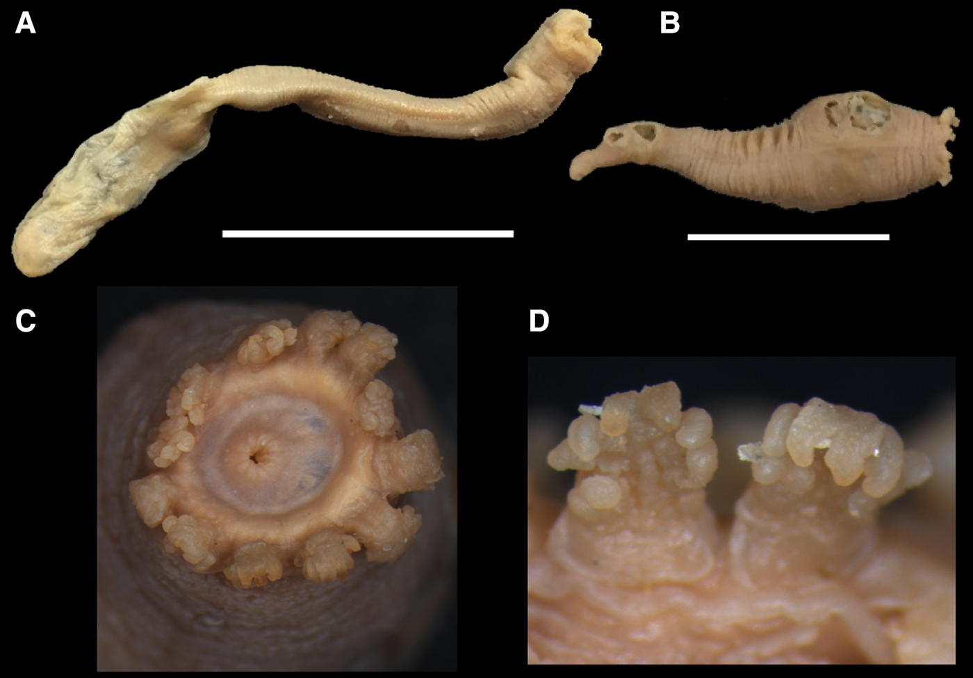
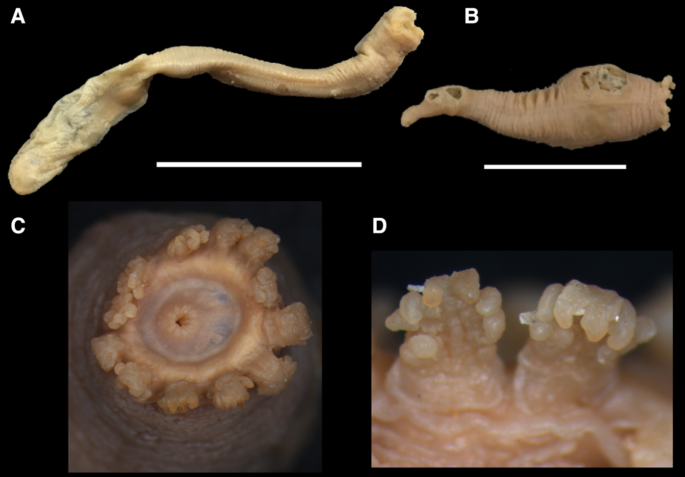
Gymnopipina ikamiaba gen. nov. et sp. nov.: (A) lateral view of holotype (MZUSP 1514) and (B) paratype (MZUSP 1515); and (C) apical view of mouth and peltate tentacles; and (D) detail of tentacle digits. Scale bars: A, 4 cm; B, 2 cm.
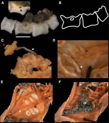
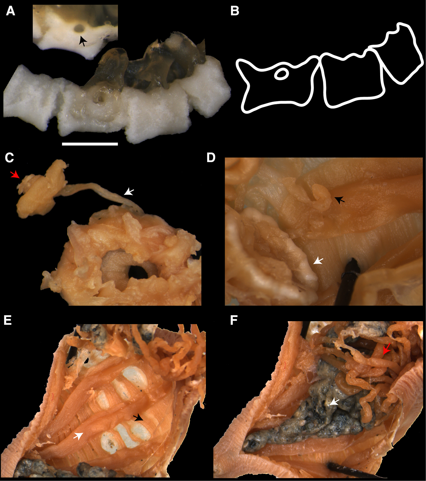
Gymnopipina ikamiaba gen. nov. et sp. nov. (MZUSP 1515): (A) calcareous ring after bleaching (arrow in upper image indicates the perforation for insertion of radial nerve); (B) drawing of calcareous ring; and detail of the internal morphology, including (C) madreporite (red arrow) and stone canal (white arrow); (D) posterior region of calcareous ring (white arrow) and Polian vesicle (black arrow); (E) longitudinal musculature (white arrow) and circular muscle bands (black arrow); and (F) gonads (red arrow) and digestive tract (white arrow). Scale bar: A, 1 mm.
TYPE MATERIAL
Holotype: São Sebastião, SP, Brazil, March 1956, 6.5 cm long (MZUSP 1514). Paratypes: same collection data as Holotype, five specimens 2–2.5 cm long (MZUSP 1515).
DIAGNOSIS
As for genus.
ETYMOLOGY
Named after the brave indigenous women who lived in the pre-colonized Amazon forest and were killed trying to protect their land.
DESCRIPTION
Body vermiform (Figure 1A, B), 6.5 cm long. 12 peltate tentacles (Figure 1C), each with 5 pairs of digits, distal pair larger than others (Figure 1D). Calcareous ring with 10 broad and delicate plates, posterior projections lacking (Figure 2A). Posterior region of plates flat to slightly concave, and anterior region W-shaped. Radial plates with anterior perforation for insertion of radial nerves (Figure 2A, B); interradial plates with a short anterior projection/tooth. Radials and interradials not necessarily intercalated, but complete arrangement of plates could not be observed because plates dissociated very quickly. Ciliated funnels absent, stone canal single and long, madreporite circular (Figure 2C). One Polian vesicle (Figure 2D). Longitudinal musculature in five bands along body; transversal muscles in multiple circular bands (Figure 2E). Gonad tufts with central trunk and simple branches, attached to body wall between middle region of body and calcareous ring (Figure 2F). Ossicles lacking from muscles, body wall and tentacles. Colour in ethanol brown.
TYPE LOCALITY
São Sebastião, São Paulo State, Brazil.
BATHYMETRY
Bathymetric data of the collecting site is not available but the maximum depth of the type locality is 40 m (Pires-Vanin et al., Reference Pires-Vanin, Muniz and Bromberg2014).
KEY TO APODID SPECIES FROM THE BRAZILIAN COAST
1. Body wall ossicles present 2
— Body wall ossicles lacking Gymnopipina ikamiaba gen. nov. et sp. nov.
2. Body wall with wheels Chiridota rotifera (Pourtalès, Reference Pourtalès1851)
— Body wall with anchors and anchor plates 3
3. Anchor stocks branched 4
— Anchor stocks unbranched 5
4. Anchor arms smooth Euapta lappa (Müller, Reference Müller1850)
— Anchor arms toothed Protankyra benedeni (Ludwig, Reference Ludwig1881)
5. Miliar granules present 6
— Miliar granules absent Leptosynapta brasiliensis Freire & Grohmann, Reference Freire and Grohmann1989
6. Miliar granules are C and O-shaped Epitomapta roseola (Verrill, Reference Verrill1874)
— Miliar granules are rosettes 7
7. Anchor stock and vertex untoothed Synaptula secreta Ancona-Lopez, Reference Ancona-Lopez1957
— Anchor stock and vertex toothed Synaptula hydriformis (Lesueur, Reference Lesueur1824)
Loss of dermal ossicles in the Apodida
With the description of Gymnopipina gen. nov., there are at least four genera whose dermal ossicles have been completely lost. Smirnov (Reference Smirnov2016) suggested that such losses occurred independently in multiple lineages; however, this hypothesis has not been formally tested within a phylogenetic framework. In his morphological phylogeny of the apodids, Kerr (Reference Kerr2001) excluded the genera without ossicles because they were poorly known or considered ‘clearly derived’ (Kerr, Reference Kerr2001, p. 54). Miller et al. (Reference Miller, Kerr, Paulay, Reich, Wilson, Carvajal and Rouse2017) reconstructed a phylogeny based on molecular data but they included only one of the genera that lack ossicles.
Convergences and parallelisms seem to be fairly common in holothuroids (Smirnov, Reference Smirnov2016; Miller et al., Reference Miller, Kerr, Paulay, Reich, Wilson, Carvajal and Rouse2017) and comprehensive phylogenetic analyses to the order and family levels are needed to elucidate the evolution of the holothuroid endoskeleton.
ACKNOWLEDGEMENTS
We thank Alexey Smirnov and an anonymous reviewer for providing constructive suggestions to the manuscript; Marcos Tavares (MZUSP) for providing access to the echinoderm collection; and the Instituto Gonçalo Moniz (FIOCRUZ/BA) for SEM analysis.