




No CrossRef data available.
Article contents
Clinical presentation, natural history, and outcome of patients with the absent pulmonary valve syndrome
Published online by Cambridge University Press: 21 January 2005
Abstract
So-called ‘absent pulmonary valve syndrome’ is a rare cardiac malformation, usually associated with tetralogy of Fallot. Congenital absence of the leaflets of the pulmonary valve is less common when the ventricular septum is intact. Characteristic features of the syndrome include dysplasia or absence of the pulmonary valvar leaflets, permitting severe pulmonary regurgitation, and aneurysmal dilation of the pulmonary arteries. The purpose of our study was to review our experience with patients diagnosed as having the absent pulmonary valve syndrome, and to describe their clinical presentation, natural history, and outcome. We reviewed retrospectively data from 18 patients with absent pulmonary valve syndrome, 10 boys and eight girls, treated between March 1983 and May 2003. We identified two groups of patients, one made up of 11 patients with a ventricular septal defect, in whom the morphology of the subpulmonary outflow tract was phenotypic for tetralogy of Fallot, and another group, with seven patients, having an intact ventricular septum. Family history of congenital heart disease was common only in patients with ventricular septal defect, being found in 73%, all of whom were diagnosed during infancy with variable respiratory distress. Diagnosis was delayed in 43% of the patients with an intact ventricular septum. Cardiac surgery was performed in eight patients with ventricular septal defect (73%), compared to only two patients (28%) with an intact ventricular septum. Overall mortality was 28%, with five patients dying. Although our sample was small, two clinical patterns emerged depending on the presence or absence of a ventricular septal defect. Patients with a ventricular septal defect and phenotypic features of tetralogy of Fallot have a strong family history of congenital cardiac disease, develop respiratory symptoms during infancy and exhibit a variable prognosis, despite cardiac surgery. Patients with an intact ventricular septum are usually asymptomatic, present later in life, and show a relatively benign prognosis.
Keywords
Information
- Type
- Original Article
- Information
- Copyright
- © 2004 Cambridge University Press
Congenital absence of the pulmonary valve is a rare cardiac malformation of unknown genetic cause. As far as we know, it was first described by Crampton in 1830,1 and again by Favell in 1842.2 It is characterized by dysplasia or absence of the leaflets of the pulmonary valve, with ample regurgitant flow. The malformation is usually associated with tetralogy of Fallot, as first described by Chevers.3 Congenital absence of the leaflets of the pulmonary valve in the setting of an intact ventricular septum is less common.4 Characteristic features of the so-called ‘absent pulmonary valve syndrome’ include right ventricular enlargement, infundibular prominence, and aneurysmal dilation of the pulmonary arteries.5–7 The infants most severely affected present with the respiratory distress. Pulmonary complications, such as obstructive emphysema and atelectasis, are often fatal.8, 9 Our review of the literature revealed relatively few reported series, most of them focused on the surgical outcome of the absence of the valvar leaflets in the setting of tetralogy of Fallot.10, 11 The aim of our study, therefore, was to review our experience with patients who had been diagnosed with absent pulmonary valve syndrome, and to describe their clinical presentation, associated anomalies, natural history, and outcome.
Methods
We undertook a review of our retrospective medical records, searching our hospital database using the diagnostic terms “tetralogy of Fallot” and “absent pulmonary valve”, for children treated between 1983 and 2003. Having established the size of our cohort, we evaluated their clinical reports, echocardiograph examinations, data from cardiac catheterization, surgical reports and records of their follow-up in outpatient clinics.
Results
Between March 1983 and May 2003, a total of 465 children were treated in our Institution with the diagnosis of tetralogy of Fallot or absent pulmonary valve syndrome. During this period, we diagnosed 18 children with the syndrome, 10 (56%) boys and eight (44%) girls, ranging in age from 1 day to 12 years, with a median age of 1 day. All were treated and followed at the Soroka University Medical Center. Of the patients, 17 (94%) were Bedouin in origin, and one was Jewish. In 11 patients, the leaflets of the pulmonary valve were diagnosed as absent in the setting of tetralogy of Fallot, while seven patients had an intact ventricular septum. Associated cardiac lesions were found in three patients in whom the ventricular septum was intact.
Family history and genetics
There was a family history of congenital heart disease in eight of the 11 (73%) patients with phenotypic features of tetralogy of Fallot (Table 1). The same lesion had occurred in siblings of six (55%) patients, tetralogy of Fallot in another (Case 4), and ventricular septal defect in the final one (Case 8). None of the patients with an intact ventricular septum (Table 2) had a family history of congenital cardiac disease.
Table 1. Summary of 11 patients with absent pulmonary valve syndrome and ventricular septal defect in the setting of tetralogy of Fallot.

Table 2. Summary of seven patients with absence of the leaflets of the pulmonary valve with an intact ventricular septum.

Deletion of chromosome 22q11 was not detected in the two patients with absent pulmonary valve syndrome and tetralogy of Fallot in whom this analysis was performed. Furthermore, none of our patients had a clinical presentation of the syndrome of deletion of 22q11.
Clinical presentation
The patients with ventricular septal defect and tetralogy of Fallot were all diagnosed during the neonatal period (Table 1). Of these patients, nine (82%) were symptomatic at that age, with symptoms including respiratory distress and variable cyanosis. Only two patients (18%) were asymptomatic. These were referred for evaluation of a heart murmur (Table 3).
Table 3. Clinical summary of 11 patients with absent pulmonary valve syndrome and ventricular septal defect in the setting of tetralogy of Fallot.

Three patients with an intact ventricular septum had been diagnosed during childhood (Table 2). Of the other four patients with intact ventricular septum, diagnosed as neonates, two were symptomatic with variable cardiorespiratory distress, one diagnosis was made during evaluation of a heart murmur (Case 3), and one following an abnormal fetal scan (Case 7; Table 4).
Table 4. Clinical summary of seven patients with absence of the leaflets of the pulmonary valve with an intact ventricular septum.

Follow-up
Of the patients with a ventricular septal defect and tetralogy of Fallot, one patient (Case 8) developed severe respiratory distress after birth. He received endotracheally-assisted ventilation, died within several hours. Mild-to-moderate respiratory symptoms developed in eight patients, but these patients were discharged from the hospital at the age of 3 to 25 days, with median of 6 days. Only two patients were asymptomatic at birth, but they developed symptoms soon after (Table 3). During infancy, all patients developed recurrent respiratory infections, with respiratory distress and respiratory wheezing of variable severity, and required recurrent hospitalization. In one instance, the patient died before surgery at the age of 1 month (Case 3). The four most severely affected patients (Cases 1, 2, 4, 11) underwent open-heart surgery at 1, 4, 3 and 3 months respectively. Of the remainder, four patients underwent surgery at an older age, between 4 and 7 years, and one patient is still awaiting surgery (Table 3).
In those with an intact ventricular septum, two patients were symptomatic at birth. Of these, one patient (Case 1) required immediate intubation and ventilation, and died within an hour. The other was sent for an urgent operation. Both had additional persistent patency of the arterial duct. Another patient with additional atrial septal defect and persistent patency of the arterial duct suffered from recurrent respiratory infections. This patient underwent surgery at the age of eight years. The remaining four patients were asymptomatic at birth, and remained asymptomatic (Table 4). All of them had mild-to-moderate obstruction of the right ventricular outflow tract.
Surgery
In those with a ventricular septal defect and tetralogy of Fallot, the operative procedure included closure of the ventricular septal defect, repair of the outflow tract either with a pericardial patch or insertion of homograft, and plication of the pulmonary trunk and its proximal branches. In those with an intact ventricular septum, surgery was undertaken in only two patients. Of these, the second patient underwent ligation of the persistently patent arterial duct as an urgent procedure at the age of one week, and the other (Case 3) underwent ligation of the duct and closure of the atrial septal defect at eight years of age (Table 4). Correction of the problems affecting the right ventricular outflow tract was not attempted.
Outcome
In all, five patients died (28%). Of these, four had a ventricular septal defect and tetralogy of Fallot (36%), with two of these patients dying without undergoing surgery, and two dying post-operatively due to respiratory failure and sepsis (Table 3). In those with an intact ventricular septum, one patient died (14%) (Table 4) at the age of 1 h.
Discussion
In most of the cases of so-called absence of the leaflets of the pulmonary valve reported in the literature, there are associated cardiac anomalies. Tetralogy of Fallot is the most common associated lesion.3, 4 Other associated anomalies include persistent patency of the arterial duct, atrial septal defect, right ventricular hypoplasia, double outlet right ventricle, tricuspid atresia and others.9, 12–16
In our group, 11 patients (61%) had an associated ventricular septal defect, all considered to be in the setting of tetralogy of Fallot, four patients (22%) had no associated cardiac anomaly, and three patients (17%) had persistent patency of the arterial duct with intact ventricular septum, one of them with an additional atrial septal defect.
Although the entity of so-called ‘absent pulmonary valve syndrome’ with ventricular septal defect is widely accepted as part of the spectrum of tetralogy of Fallot, the patients do differ in respect to the morphology of the right ventricular outflow, physiology, the nature of the intrapulmonary vasculature, and the involvement of the tracheo-bronchial tree. The pathologic abnormalities in tetralogy of Fallot include a rotated and anteriorly deviated infundibular, or outlet, septum of variable size.6, 7 The leaflets of the pulmonary valve are always present, albeit at times variably deformed, hypoplastic, or even atretic. The branches of the pulmonary trunk are variably hypoplastic. The intrapulmonary circulation is normal, and there is no vascular encroachment on the tracheo-bronchial tree. The hemodynamic presentation is that of a right-to-left shunt. In the so-called “absent pulmonary valve syndrome” with ventricular septal defect and tetralogy of Fallot, the pathological features are totally different. The infundibulum is wider than normal, virtually featureless, and of at least normal length (Fig. 1). There is only rudimentary formation of the pulmonary valvar tissue, or even no tissue at all. Furthermore, there is not always a marked change in caliber at the point where the infundibulum becomes the pulmonary arterial tree, and the pulmonary arteries become aneurysmal (Fig. 2). There is usually severe pulmonary regurgitation (Fig. 3). These same pathologic features exist in the “absent pulmonary valve syndrome” with an intact ventricular septum (Fig. 4).

Figure 1. A right ventriculogram, profiled in the antero-posterior view, showing a wide right ventricular outflow tract and huge pulmonary arteries in a four-year-old patient with absent pulmonary valve syndrome and a ventricular septal defect. RV: right ventricle; PA: pulmonary arteries.

Figure 2. A parasternal short axis echocardiogram in a five-year-old patient with absent pulmonary valve syndrome and ventricular septal defect, showing the wide right ventricular outflow tract and enormous dilation of the pulmonary trunk and its proximal branches, which is especially appreciated when the size of these vessels is compared with that of the aorta. AO: aorta; MPA: pulmonary trunk; RPA: right pulmonary artery; LPA: left pulmonary artery.

Figure 3. A parasternal short axis echocardiogram with Doppler color flow mapping showing systolic (a) and diastolic (b) flow at the ventriculo-pulmonary junction in a newborn with “absent pulmonary valve syndrome” and mild pulmonary stenosis but severe regurgitation. MPA: pulmonary trunk.

Figure 4. A right ventriculogram in the lateral position, showing a wide right ventricular outflow and aneurysmal pulmonary arteries, in a 10-year-old patient with absent pulmonary valve syndrome with an intact ventricular septum. RV: right ventricle; PA: pulmonary arteries.
Absent pulmonary valve syndrome with ventricular septal defect in the setting of tetralogy of Fallot typically features a combination of problems associated with left-to-right shunting and airway obstruction.8, 9, 17 In the newborn period, many of these patients are cyanotic, independent of their respiratory function. This is most likely due to relatively high pulmonary vascular resistance in the presence of an incompetent pulmonary outflow tract and a ventricular septal defect. Characteristically, the cyanosis abates concomitant with the fall in pulmonary vascular resistance.8, 9 The intracardiac shunting frequently becomes a net left-to-right shunt, and patients therefore have increased pulmonary blood flow. The increased flow to the lungs, in addition to the native dilation of the pulmonary arteries, may cause significant respiratory difficulties. The dilated pulmonary arteries compress the large airways, creating a ball-like effect. This effect may be compounded by enlargement of the left atrium, which further compresses the airways, particularly the main stem of the left bronchus. Intrinsic abnormalities of the bronchioles themselves may cause further obstruction. The pathology of the pulmonary tree is the major cause of the clinical symptoms. Rabinovitch et al.,17 in a postmortem study of three severely affected infants, showed that, in addition to the massively dilated central pulmonary arteries compressing the main bronchuses, there was a bizarre pattern of branching of the pulmonary arteries that impaired alveolar multiplication and compressed the smaller intrapulmonary bronchuses. Emmanouilides et al.18 mentioned that histological findings similar to those seen in Marfan's Syndrome have been described in several cases. Agenesis of the arterial duct was frequently found in fetuses and neonates with absent pulmonary valve in the setting of a ventricular septal defect and tetralogy of Fallot. The etiology of this association is rarely addressed. Some reports speculated on the mechanism of dilated pulmonary arteries as a consequence of agenesis of the arterial duct during fetal life.18, 19 A recent study on the prenatal role of the arterial duct, however, suggests that persistent patency of the arterial duct in this setting is lethal, and the observation that tetralogy of Fallot and absent leaflets of the pulmonary valve do not coexist with persistent patency of the arterial duct may reflect early gestational selection.20
Diagnosis of the absent pulmonary valve syndrome in association with a ventricular septal defect and tetralogy of Fallot is usually made during infancy,8, 9 in contrast with patients having absence of the leaflets of the pulmonary valve with an intact ventricular septum, who are not usually diagnosed until childhood. Only isolated cases have been reported where cardiorespiratory distress developed during the neonatal period, leading to early diagnosis.13–16, 21
All of our patients with a ventricular septal defect were diagnosed in infancy, with four-fifths of them exhibiting symptoms early in life. This group is characterized by a stormy clinical presentation, severe respiratory symptoms, and high morbidity and mortality. Medical treatment often failed to control the symptoms, and corrective surgery yielded varying degrees of success. A minority of patients in this group consisted of infants with mild respiratory symptoms. Growth and development were usually normal, and repair could be performed on an elective basis with low risk.
In our group with an intact ventricular septum, seven-tenths were asymptomatic and diagnosed late in childhood. The three patients with associated persistent patency of the arterial duct developed symptoms and underwent surgery. In the patients with the syndrome associated only with persistent patency of the arterial duct, the cardiorespiratory distress is related to the presence of increased right ventricular stroke volume due to pulmonary regurgitation, together with the large left-to-right shunt through the persistent arterial duct, resulting in pulmonary dilation and congestion, a phenomenon Rabinovitch et al.17 also observed in one of their patients with deficient ventricular septation.
Limited information is available regarding the genetics of the absent pulmonary valve syndrome. Patients with tetralogy of Fallot are frequently diagnosed with the 22q11 deletion syndrome.22 Sporadic cases of absent leaflets of the pulmonary valve have been reported in the literature in association with other chromosomal anomalies.12 Podzimkova et al.21 speculated that absent pulmonary valve syndrome as seen with tetralogy of Fallot, and absent leaflets of the pulmonary valve with intact ventricular septum, are entities with different etiologies. The first may have a true anomaly involving the outflow tracts, and may be associated with chromosome 22q11 deletion, and the second could be explained by a primary defect in the development of the leaflets of the pulmonary valve, with hemodynamic changes leading to dilation of pulmonary arteries.
We agree with the suggestion that the syndrome as seen with ventricular septal defect and tetralogy of Fallot differs from absence of the leaflets of the pulmonary valve with an intact ventricular septum, and that the entities represent two diseases, probably with two different etiologies. The variant with ventricular septal defect is characterized by a strong family history of congenital heart disease. Clinical presentation is usually dramatic, during early infancy, with high morbidity and variable prognosis. In contrast, the patients with an intact ventricular septum represent a different clinical group. In this group, there is no family history, the diagnosis is made later in life, and most patients are asymptomatic. Those who are symptomatic are the ones with additional persistent patency of the arterial duct or an atrial septal defect in the oval fossa, the added malformations probably being responsible for the intermediate pattern of symptoms.
References
Chevers N. Recherches sur les maladies de l'artere pulmonaire. Arch Gen Med 1847; 15: 488–508.Google Scholar
Vlad P, Weidman M, Lambert EC. Congenital pulmonary regurgitation: a report of six autopsy cases. Am J Dis Child 1960; 100: 640–641.Google Scholar
Lev M, Eckner FAO. The pathologic anatomy of tetralogy of Fallot and its variations. Dis Chest 1964; 45: 251.Google Scholar
Rao BN, Anderson RC, Edwards JE. Anatomic variations in the tetralogy of Fallot. Am Heart J 1971; 81: 361–371.Google Scholar
Van Praagh R, Van Praagh S, Nebesar RA, Muster AJ, Sinha SN. Tetralogy of Fallot: underdevelopment of the pulmonary infundibulum and its sequelae. Am J Cardiol 1970; 26: 25–33.Google Scholar
Lakier JB, Stanger P, Heymann MA, Hoffman JI, Rudolph AM. Tetralogy of Fallot with absent pulmonary valve. Natural history and hemodynamic considerations. Circulation 1974; 50: 167–175.Google Scholar
Pinsky WW, Nihill MR, Mullins CE, Harrison G, McNamara DG. The absent pulmonary valve syndrome: consideration of management. Circulation 1978; 57: 159–162.Google Scholar
Conte S, Serraf A, Godart F, Lacour-Gayet F, Petit J, Bruniaux J, Planche C. Technique to repair tetralogy of Fallot with absent pulmonary valve. Ann Thorac Surg 1997; 63: 1489–1491.Google Scholar
Hew CC, Daebritz SH, Zurakowski D, del Nido PI, Mayer JE, Jonas RA. Valved homograft replacement of aneurismal pulmonary arteries for severely symptomatic absent pulmonary valve syndrome. Ann Thorac Surg 2002; 73: 1778–1785.Google Scholar
Freedom RM, Patel RG, Bloom KR, Duckworth JW, Silver MM, Dische R, Rowe RD. Congenital absence of the pulmonary valve associated with imperforate membrane type of tricuspid atresia, right ventricular tensor apparatus and intact ventricular septum: A curious developmental complex. Eur J Cardiol 1979; 10: 171–196.Google Scholar
Alpert BS, Moore HV. “Absent” pulmonary valve with atrial septal defect and patent ductus arteriosus. Pediatr Cardiol 1985; 6: 107–111.Google Scholar
Grotenhuis HB, Nijveld A, Bachx A. Absent pulmonary valve syndrome with intact ventricular septum and patent ductus arteriosus: report of two cases and a short review of the literature. Ann Thorac Surg 2003; 75: 280–282.Google Scholar
Horigome H, Sakakibara Y, Atsumi N, Miyamoto T, Sato H. Absent pulmonary valve with intact ventricular septum presenting as cardiorespiratory failure at birth. Pediatr Cardiol 1997; 18: 136–138.Google Scholar
Thanopoloulos BD, Fischer EA, Hastreiter AR. Large ductus arteriosus and intact ventricular septum associated with congenital absence of the pulmonary valve. Br Heart J 1986; 55: 602–604.Google Scholar
Rabinovitch M, Grady S, David I, Van Praagh R, Sauer U, Buhlmeyer K, Castaneda AR, Reid L. Compression of intrapulmonary bronchi by abnormally branching pulmonary arteries associated with absent pulmonary valves. Am J Cardiol 1982; 50: 804–813.Google Scholar
Emmanouilides GC, Thanopoulos B, Siassi B, Fishbein M. “Agenesis” of ductus arteriosus associated with the syndrome of tetralogy of Fallot and absent pulmonary valve. Am J Cardiol 1976; 37: 403–409.Google Scholar
Ettedgui JA, Sharlan GK, Chita SK, Cook A, Fagg N, Allan LD. Absent pulmonary valve syndrome with ventricular septal defect: Role of the arterial duct. Am J Cardiol 1990; 66: 233–234.Google Scholar
Yeager SB, Van Der Velde ME, Waters BL, Sanders SP. Prenatal role of the ductus arteriosus in absent pulmonary valve syndrome. Echocardiography 2002; 19: 489–493.Google Scholar
Podzimkova J, Hickey MStJ, Slavik Z, Leanage R, Chan Chen K. Absent pulmonary valve syndrome with intact ventricular septum: role of ductus arteriosus revisited. Int J Cardiol 1997; 61: 109–112.Google Scholar
Thomas JA, Graham JM. Chromosome 22q11 deletion syndrome: an update and review for the primary pediatrician. Clinic Pediatr 1997; 36: 253–266.Google Scholar
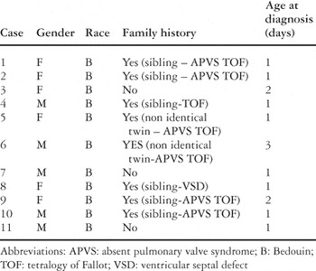
Table 1.
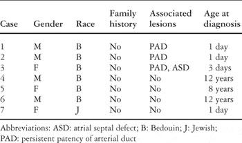
Table 2.
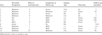
Table 3.
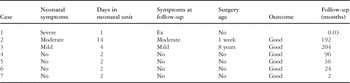
Table 4.

A right ventriculogram, profiled in the antero-posterior view, showing a wide right ventricular outflow tract and huge pulmonary arteries in a four-year-old patient with absent pulmonary valve syndrome and a ventricular septal defect. RV: right ventricle; PA: pulmonary arteries.

A parasternal short axis echocardiogram in a five-year-old patient with absent pulmonary valve syndrome and ventricular septal defect, showing the wide right ventricular outflow tract and enormous dilation of the pulmonary trunk and its proximal branches, which is especially appreciated when the size of these vessels is compared with that of the aorta. AO: aorta; MPA: pulmonary trunk; RPA: right pulmonary artery; LPA: left pulmonary artery.

A parasternal short axis echocardiogram with Doppler color flow mapping showing systolic (a) and diastolic (b) flow at the ventriculo-pulmonary junction in a newborn with “absent pulmonary valve syndrome” and mild pulmonary stenosis but severe regurgitation. MPA: pulmonary trunk.

A right ventriculogram in the lateral position, showing a wide right ventricular outflow and aneurysmal pulmonary arteries, in a 10-year-old patient with absent pulmonary valve syndrome with an intact ventricular septum. RV: right ventricle; PA: pulmonary arteries.
 You have
Access
- 48
- Cited by