Primary cardiac tumours are rare in children with an incidence rate of 0.027–0.08%. Reference Tzani, Doulamis, Mylonas, Avgerinos and Nasioudis1 The most common among these tumours is cardiac rhabdomyoma, followed by cardiac fibroma. Reference Nadas and Ellison2 The overwhelming majority of childhood cardiac tumours are benign. Reference Tzani, Doulamis, Mylonas, Avgerinos and Nasioudis1
Case report
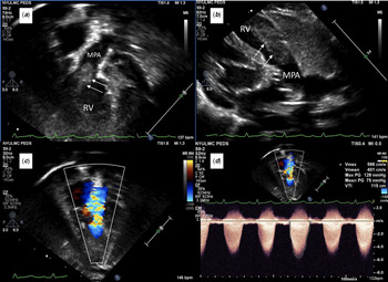
A 2-month-old healthy male infant was referred for cardiology evaluation after his paediatrician detected a systolic murmur during a routine physical examination. Delivery and the post-natal course were uncomplicated and no murmur was noted in the post-natal period. At the consultation, there was no report of cyanosis or dyspnoea and the patient was thriving. He was noted to have a 4/6 harsh ejection systolic murmur at the left upper sternal border and an otherwise normal physical examination. Chest X-ray and electrocardiogram were normal. Transthoracic echocardiogram revealed a pedunculated mass protruding from the distal free wall of the right ventricular outflow tract, crossing the pulmonary valve, and resulting in severe pulmonary stenosis with a peak systolic gradient of 80 mmHg (Fig 1). No pulmonary valve regurgitation or other structural defects were identified. The distal right ventricular outflow tract myocardium appeared thickened. There was normal biventricular function. There was no evidence of extra-cardiac masses on physical examination and ultrasound of other visceral organs. A whole-body computed tomography (CT) scan was not performed.
( a ): An apical four-chamber view showing the tumour extending from the right ventricle into the right ventricular outflow tract. Arrows point to the tumour. ( b ): A parasternal long-axis view showing the tumour extending into the right ventricular free wall. Arrows point to the tumour. ( c ): An apical four-chamber view with colour flow mapping showing aliasing of flow across the right ventricular outflow tract at the level of the pulmonary valve. ( d ): Spectral Doppler showing peak gradient of 125 mmHg and mean gradient of 70 mmHg across the right ventricular outflow tract. RV = right ventricle; MPA = main pulmonary artery.

Given the severity of the right ventricular outflow tract obstruction, surgical excision was elected. The surgery was performed on cardiopulmonary bypass and cardioplegic arrest. The main pulmonary artery was opened. Upon direct surgical inspection, a large spherical mass was seen extending across and obstructing the pulmonary valve. The mass had a broad attachment to the free wall of the distal right ventricular outflow tract. The left and right pulmonary valve cusps were uninvolved; however, the anterior leaflet was encased by the mass. No clear plane between the mass and the right ventricular myocardium was appreciated. The mass was serially shaved to remove as much as possible to relieve the underlying obstruction. The anterior pulmonary cusp was excised. Completion transoesophageal echocardiogram revealed no residual obstruction and moderate pulmonary insufficiency.
The resected specimen was sent for pathological evaluation. Three irregular fragments of white-pink rubbery tissue were received, measuring 2.4 cm in the greatest aggregate dimension.
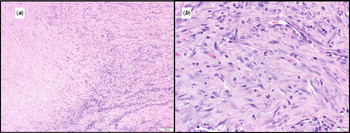
Microscopic evaluation showed nodularity and zonation of the neoplasm along with bland central necrosis. There is a bland proliferation of oval to spindle-shaped myoid tumour cells exhibiting a multi-layered growth pattern. Areas of the tumour exhibited variable cellularity and biphasic population of spindle cells with the plump immature-appearing cells associated with the branching vasculature and the more mature-appearing spindle cells in a whirling pattern in a myxohyaline stroma. Immunohistochemistry stains showed that the lesional cells are diffusely positive for smooth muscle actin and focally positive for Desmin. The tumour cells are negative for anaplastic lymphoma kinase or pan-cytokeratin. The molecular fusion panel study failed to find any gene fusion, including V-rel avian reticuloendotheliosis viral oncogene homolog A gene fusion. These findings were deemed most in keeping with that of a myofibroma (Fig 2).
( a ): This low-power photomicrograph exhibits the biphasic population of spindle cells, including the plump immature-appearing cells associated with branching vasculature and the more mature-appearing spindle cells arranged in a whirling pattern throughout a myxohyaline stroma. ( b ): This high-power photomicrograph further exhibits the plump and immature spindle cells.

The infant was discharged on post-operative day 6 without any complication. He has been followed by outpatient paediatric cardiology and has been doing well with no recurrence of the cardiac mass or right ventricular outflow tract obstruction. We do not plan to perform a whole-body CT scan, unless he shows signs of systemic involvement.
Discussion
A recent systematic review showed that the majority of paediatric cardiac tumours are benign. Cardiac rhabdomyomas were the most common type (constituting 33%) followed by cardiac fibromas (18%), cardiac myxoma (18%), and teratoma (8%). Reference Tzani, Doulamis, Mylonas, Avgerinos and Nasioudis1 Infantile myofibroma is rare but one of the most common benign fibrous tumours of infancy. Reference Mashiah, Hadj-rabia and Dompmartin3 It usually presents in the first 2 years of life and can be solitary (myofibroma), multi-centric (myofibromatosis), or generalised. Reference Ludomirsky, Vargo, Murphy, Gresik, Ott and Mullins4 The solitary form is most common, accounting for 50–80% of all cases and usually involves the head or neck, presenting as a flesh-coloured dermal or subcutaneous nodule. Reference Oudijk, den Bakker and Hop5 Approximately 60% of cases occur in boys. Reference Mashiah, Hadj-rabia and Dompmartin3 There has been a previous report of a cardiac tumour in the right ventricular outflow tract of an infant with a similar clinical presentation. This tumour had a similar gross pathological appearance to that seen in our patient. However, it was ultimately classified as an undifferentiated sarcoma. Reference Ludomirsky, Vargo, Murphy, Gresik, Ott and Mullins4
Solitary and multi-centric infantile myofibromatosis generally have a benign course and do not metastasise. There is spontaneous regression of the tumour over 1–2 years in most cases. In lesions that do not regress or are symptomatic, local resection of the lesions seems to be sufficient. Reference Oudijk, den Bakker and Hop5 The risk of recurrence of solitary infantile myofibromatosis is around 7%. Reference Sargar, Sheybani, Shenoy, Aranake-Chrisinger and Khanna6
Generalised infantile myofibromatosis may involve visceral organs. This has a higher mortality rate, and treatment with a combination of methotrexate and vinblastine is recommended. In patients with generalised multi-centric infantile myofibromatosis and visceral disease, the heart is the third most commonly involved site. Reference Wu, Mccavit, Cederberg, Galindo and Leavey7
The differential diagnosis of a soft-tissue mass in the heart of children includes rhabdomyoma, inflammatory myofibroblastic tumour, congenital infantile fibrosarcoma, fibromatoses, haemangioma, neurofibroma, soft-tissue sarcoma, metastatic neuroblastoma, and histiocytosis. Biopsy with histology is the gold standard for diagnosis of this lesion, with the other possible lesions, in this case, having been ruled out by a combination of histological appearance and immunohistochemistry. Reference Oudijk, den Bakker and Hop5,Reference Sargar, Sheybani, Shenoy, Aranake-Chrisinger and Khanna6 If indicated, molecular testing may also be helpful in the evaluation of soft-tissue tumours. For example, inflammatory myofibroblastic tumour, a major differential, in this case, shows rearrangement of 2p23 (anaplastic lymphoma kinase) in ~50% of cases. Reference Antonescu, Suurmeijer and Zhang8 Inflammatory myofibroblastic tumour is a rare possibility and was included on our differential. Reference Burke, Li, Kling, Kutys, Virmani and Miettinen9 In the end, however, the sparse inflammatory backdrop, the biphasic population of cells, and the overall pattern fit better for a myofibroma. In summary, this represents the first report of an apparently solitary myofibroma in the heart of an infant, expanding the differential diagnosis of cardiac tumours.
Conclusion
Infantile fibromyoma or myofibromatosis is a rare entity. Solitary myofibromas are generally benign and can be treated by surgical resection if large, non-regressing, or symptomatic.
Financial Support
This research received no specific grant from any funding agency, commercial, or not-for-profit sectors.
Conflicts of Interest
None.