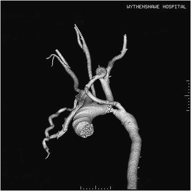
A 58-year-old man was admitted with a troponin-positive acute coronary syndrome. Apart from longstanding ischaemic heart disease with previous percutaneous intervention, his medical history included adult coarctation repair. CT angiography showed dilated bilateral internal thoracic arteries with the left internal thoracic artery being anastomosed beyond the previous coarctation repair (Fig 1). The left internal thoracic artery was significantly dilated at 16 mm just distal to its takeoff and 9 mm lower down. Cardiac MRI showed focal tight narrowing of the proximal descending aorta associated with turbulent flow immediately distal to the left subclavian artery consistent with residual coarctation.
CT aortogram showing residual coarctation of the aorta with dilated bilateral internal thoracic arteries. The left internal thoracic artery is seen anastomosed beyond the area of coarctation.

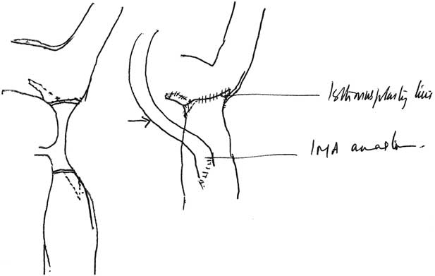
We managed to retrieve the patient’s medical case notes to confirm that he had undergone combined resection and isthmusplasty with implantation of the left internal thoracic artery on the descending thoracic aorta distal to the repair 30 years ago; a hand-drawn operative diagram was also filed in the surgical section of his records (Fig 2). A brief technical report was published at the time from our institution including a total of two patients in whom this technique was carried out.Reference Nair, Moussalli and Beton 1 Beyond the importance of this report from an institutional heritage perspective, the patency of the internal thoracic graft three decades down the line reconfirms the excellent natural characteristics of this vessel as a vascular conduit.
Scanned copy of the original operative diagram following primary surgical repair 30 years ago. As depicted, the patient had been treated by a combined resection and isthmusplasty with implantation of the left internal thoracic artery to the distal aorta.

Acknowledgements
None.
Financial Support
This research received no specific grant from any funding agency or commercial or not-for-profit sectors.
Conflicts of Interest
None.