FOCUS POINTS
• Borderline personality disorder (BPD) is considered to have a pervasive pattern of chronic instability in interpersonal relationships, self-image and affect, with episodes of affective and impulsive dyscontrol
• Reduced amygdalar volume has been demonstrated in patients with BPD, relative to healthy controls, but the same number of studies found no differences between BPD patients and healthy controls
• Reduction in amygdalar volume has also been observed in patients with PTSD, a disorder that is comorbid in 60–70% of BPD patients
• Reduced amygdalar volume may characterize patients with BPD but not patients with a comorbid presentation of BPD and PTSD
Introduction
Borderline personality disorder (BPD) is described as a pervasive pattern of chronic instability in interpersonal relationships, self-image and affect, with episodes of affective and impulsive dyscontrol.1 Because this personality disorder is associated with significant functional impairment, researchers have attempted to identify its biological underpinnings with the hope of understanding the mechanism by which BPD symptoms are expressed. One candidate researchers have considered is impairment in the amygdala, as this brain structure plays a role in emotion regulation.Reference Johnstone, van Reekum, Urry, Kalin and Davidson2 Reduced amygdalar volume has been demonstrated by four studies in patients with BPD, relative to healthy controls,Reference Driessen, Herrmann, Stahl, Zwann, Meier and Hill3–Reference Weniger, Lange, Sachsse and Irle6 but the same number of studies found no differences between BPD patients and healthy controls.Reference Brambilla, Soloff, Sala, Nicoletti, Keshavan and Soares7–Reference Zetzsche, Frodl, Preuss, Schmitt, Seifert and Leisinger10 We conducted a previous meta-analysis in which we pooled results from individual magnetic resonance imaging (MRI) studies and corroborated a reduction in both left and right amygdalar volume in BPD patients.Reference Nunes, Wenzel, Borges, Porto, Caminha and de-Oliveira11
Reduction in amygdalar volume has also been observed in patients with PTSD, a disorder that is comorbid in 60–70% of BPD patients.Reference Zanarini, Frankenburg, Dubo, Sickel, Trikha and Levin12, Reference Zimmerman and Mattia13 Although placed on different axes of the DSM classification system, there are a number of commonalities shared by BPD and PTSD such as etiological (eg, exposure to traumatic events) and biological factors (eg, aberrant patterns of activation in prefrontal and limbic regions), as well as symptomatology (eg, states of dissociation, hyperarousal).Reference Schmahl, Berne, Krause, Kleindienst, Valerius and Vermetten9, Reference Pagura, Stein, Bolton, Cox, Grant and Sareen14 Thus, it is possible that comorbid PTSD could explain the heterogeneity in the results obtained by studies that investigate amygdalar volume in BPD patients relative to healthy controls. Specifically, it could be the case that reduced amygdalar volume is most pronounced in patients with comorbid BPD and PTSD, relative to patients with BPD alone.
The present study had two aims. First, we updated our previous meta-analysisReference Nunes, Wenzel, Borges, Porto, Caminha and de-Oliveira11 by adding one study we had missedReference New, Hazlett, Buchsbaum, Goodman, Mitelman and Newmark8 and two others published after analyses were completed.Reference Weniger, Lange, Sachsse and Irle6, Reference Schmahl, Berne, Krause, Kleindienst, Valerius and Vermetten9 In order to investigate the degree to which comorbid PTSD could account for the heterogeneity observed among individual studies and in our previous meta-analysis of published studies of amygdalar volume in BPD, we compared amygdalar volume in the subset of BPD patients with and without PTSD.
Methods
A systematic review of the literature to identify empirical studies that examined amygdalar volume in BPD patients and healthy controls was conducted. Inclusion criteria for primary studies were the following: (1) be carried out in a population predominantly diagnosed with BPD; (2) compare adult BPD patients with healthy controls; (3) provide the means and standard deviations or variance estimates that could be converted back to such values (eg, standard errors) for both the BPD and healthy control groups; and (4) for the BPD+PTSD comorbidity analysis, discriminate BPD patients with and without PTSD.
Electronic database search of studies included the Medline, PsycINFO®, Biological Abstracts, and Web of Science databases. All publications available up to December 2010 were considered. Medline search was performed using the terms “Amygdal*” and “Borderline Personality Disorder.” When necessary, the authors of studies were contacted for further clarification. The references of all studies considered relevant were carefully examined, including meta-analyses and reviews, in order to locate any additional articles not identified in the above-mentioned search.
Three independent reviewers (C.P.A., C.S.C., and I.R.O.) evaluated the abstracts, decided which of those papers fulfilled the criteria for inclusion, and extracted the data.
Disagreement at any stage was resolved by consensus.
All volumes were converted to cm3 before entering them into our data set. All calculations were performed by means of the Comprehensive Meta-Analysis Software, version 2.0.Reference Borenstein, Hedges, Higgins and Rothstein15 As amygdalar volume is a continuous outcome measure, Hedges’ g statistic was used as a formulation for the standardized mean difference, and a random effects model was used in order to yield more conservative estimates than a fixed effect model. Homogeneity of studies was assessed using the Q test, and data were considered statistically heterogeneous when p < .05.
A meta-regression was conducted in order to assess the relation between one or more covariates (ie, moderators) and a dependent variable.Reference Borenstein, Hedges, Higgins and Rothstein15 Because meta-regression, especially with multiple covariates, is not recommended when the number of studies is small, we decided to use only one covariate, taking a percentage of PTSD patients as moderator.
Findings
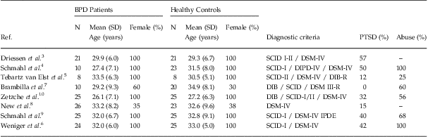
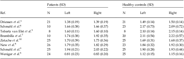
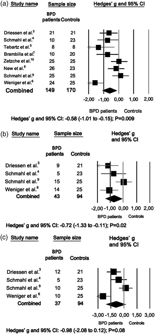
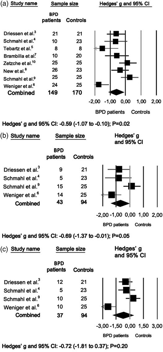
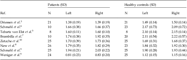
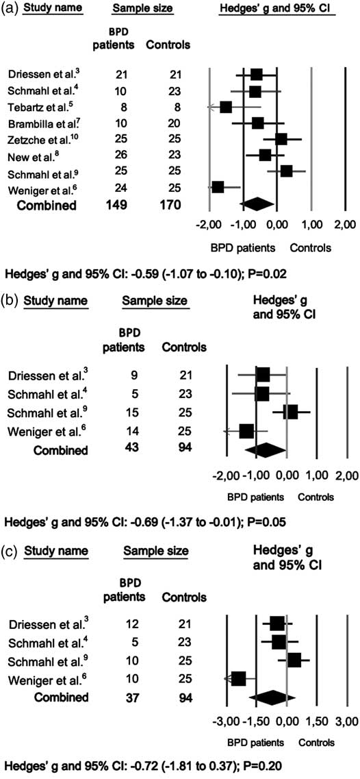
Eight articlesReference Driessen, Herrmann, Stahl, Zwann, Meier and Hill3–Reference Schmahl, Berne, Krause, Kleindienst, Valerius and Vermetten9, Reference Zetzsche, Preuss, Frodl, Schmitt, Seifert and Münchhausen16 involving 149 BPD patients and 170 healthy controls, fulfilled the inclusion criteria. Table 1 describes the demographic and clinical variables of patients with BPD relative to healthy controls, and Tables 2 and 3 the amygdalar volumes according to individual studies. Results pertaining to left amygdalar volume are shown in Figure 1 and to right amygdalar volume in Figure 2. The Hedges’ g pooled effect sizes found statistical significance for the left amygdala (g = −.58, 95% CI = −1.01 to −.15, p = .009; Panel a), and for the right amygdala (g = −.59, 95% CI = −1.07 to −.10, p < .02; Panel a), compared to healthy controls.
Demographic and clinical variables of patients with borderline personality disorder and healthy controls
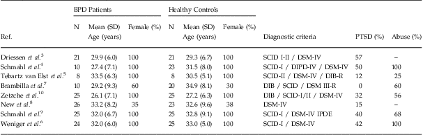
SCID-I = Structured Clinical Interview for DSM-IV Axis I Disorders; SCID-II = Structured Clinical Interview for DSM-IV Personality Disorders; DIPD-IV = Diagnostic Interview for DSM-IV Personality Disorders; DIB = Diagnostic Interview for Borderline Patients; DIB-R = Revised Diagnostic Interview for the Borderline Patients; MDD = Major depressive disorder; PTSD = Post-traumatic stress disorder; IPDE = International Personality Disorder Examination.
Amygdalar volumes (cm3) according to individual studies
Left amygdala: volume differences between BPD patients with and without PTSD compared to healthy controls. Panel (a) includes all BPD patients, Panel (b) BPD patients without PTSD, and Panel (c) BPD patients with PTSD.
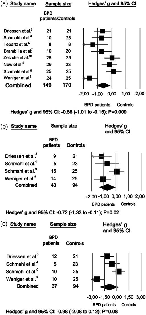
Right amygdala: volume differences between BPD patients with and without PTSD compared to healthy controls. Panel (a) includes all BPD patients, Panel (b) BPD patients without PTSD, and Panel (c) BPD patients with PTSD.
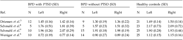
Amygdalar volumes (cm3) in BPD patients with and without PTSD and healthy controls
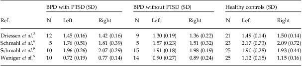
Because the test of heterogeneity was significant for both the left and the right amygdala (Q ≥ 24.00, df = 7; p ≤ .001), we undertook meta-regression calculations using a percentage of PTSD patients as moderator. This analysis did not predict any significant variation in amygdalar volumes in the BPD samples. However, data from the four studiesReference Driessen, Herrmann, Stahl, Zwann, Meier and Hill3, Reference Schmahl, Vermetten, Elzinga and Bremner4, Reference Weniger, Lange, Sachsse and Irle6, Reference Schmahl, Berne, Krause, Kleindienst, Valerius and Vermetten9 that discriminated BPD patients with and without PTSD indicated that amygdalar volumes were reduced in BPD patients without PTSD relative to controls (left: g = −.72, 95% CI = −1.33 to −1.11, p < .02, Panel b; right: g = −.69, 95% CI = −1.37 to −.01, p = .05, Panel b). In contrast, amygdalar volume was not significantly smaller in BPD patients with PTSD relative to controls (left: g = −.98, 95% CI = −2.08 to .12, p = .08, Panel c; right: g = −.72, 95% CI = −1.81 to .37, p = .20, Panel c).
Discussion
Analysis involving all studies in this review indicates a smaller amygdalar volume in both left and right sides in adult patients with BPD in comparison with healthy controls, confirming our previous results,Reference Nunes, Wenzel, Borges, Porto, Caminha and de-Oliveira11 and raising the possibility that reduced amygdalar volume is a biological substrate that underlies symptoms in this personality disorder. Indeed, analyses conducted in subsamples of BPD patients with and without PTSD suggest that reduced amygdalar volume characterizes patients with BPD only, but not patients with comorbid BPD and PTSD. These analyses also raise the possibility that amygdalar volume reductions observed in patients with BPD might be attenuated by the presence of comorbid PTSD. Thus, it is possible that the heterogeneity of results in previous studies investigating amygdalar volume in BPD patients can be accounted for by the presence of comorbid PTSD. However, results from this study fit broadly into the larger literature showing that PTSD is not associated with decreased amygdalar volumeReference Woon and Hedges17 despite the fact that it would be logical for abnormalities in the amygdala to play a role in this pathology because of its central involvement in fear conditioning and emotional responding.
It is unclear why the presence of comorbid PTSD would “protect” the amygdala. However, in interpreting this pattern of results, it is important to consider the possibility that there is not a direct correspondence between the presence or degree of structural abnormalities and the presence of degree of functional abnormalities associated with areas of the brain such as the amygdala. For example, much research with PTSD patients points to excessive amygdalar activity in these patients, such that impaired medial prefrontal cortex functioning impairs amygdala inhibition, which is in turn associated with amygdala hyperactivity, increased subjective distress, and deficient emotion regulation.Reference Koenigs and Grafman18, Reference Shin19 In other words, the body of literature on PTSD suggests that this pathology is not associated with structural abnormalities in the amygdala but that it is associated with significant functional abnormalities in the form of amygdala hyperactivity.
Empirical research has also demonstrated that BPD patients demonstrate stronger amygdalar activity than healthy controls and that amygdalar activity correlates with self-reported deficits in emotion regulation and BPD symptom severity.Reference Koenigsberg, Siever, Lee, Pizzarello, New and Goodman20, Reference Niedtfeld, Schulze, Kirsch, Herpertz, Bohus and Schmal21
When functional abnormalities in the amygdala are considered in patients with comorbid PTSD and BPD, a pattern of results arises that is different than that obtained in the present meta-analysis. For example, patients with comorbid BPD and PTSD show more pronounced amygdala deactivation than patients with BPD alone when they are faced with pain provocation.Reference Kraus, Esposito, Seifritz, Seifritz, Di Salle, Ruf and Valerius22 Thus, there is an emerging body of literature that suggests that comorbid BPD and PTSD is associated with particularly abnormal amygdala activity.
To make sense out of the discrepant results that emerge from studies that examine amygdalar volume and studies that examine amygdalar function in patients with BPD and PTSD, it would behoove researchers (1) to determine the degree to which structural and functional abnormalities are associated with one another, (2) to identify causal factors that can account for the etiology of these abnormalities using longitudinal designs, (3) to link these abnormalities with specific clinical consequences such as deficits in emotion regulation, and (4) to determine whether these abnormalities are etiological in nature or secondary to other processes such as the inability to tolerate stress. Although all of these investigations would be of considerable expense, they would move beyond the mere description of the brain abnormalities in these patients and toward a more precise determination of the pathophysiology of these disorders. Moreover, it may eventually be determined that examining the amygdala as a single unit is too broad to be linked to specific symptomatic presentations,Reference Sotres-Bayon, Bush and LeDoux23 as there is some evidence that the basal amygdala plays a role in defensive aggression, that the central nucleus of the amygdala plays a role in dissociation, and that the lateral nucleus of the amygdala determines which specific structure is activated and, therefore, which specific symptom is expressed.Reference Berdahl24 Both defensive aggression and dissociation are characteristic of many patients with BPD and PTSD.
Results from this study must be considered in light of its strengths and limitations. A major strength of this study is its use of meta-analysis, which pools results across studies to obtain more stable estimates of effect sizes than can be obtained in any one investigation and identifies the presence of heterogeneity that points to the operation of moderating variables. However, a limitation of this meta-analysis is the studies included in analyses each enrolled very small sample sizes of approximately 20 patients with BPD and 20 healthy controls. The sample of BPD patients was further divided into those with and without comorbid PTSD. Even when a powerful tool such as meta-analysis is used, small sample sizes can preclude the detection of significant results (e.g., increase Type II error), and they do not allow for finer grained analysis of other factors that might be associated with heterogeneity in BPD and contribute to the observed pattern of results such as trauma exposure,Reference Schmahl, Vermetten, Elzinga and Bremner4 depressive symptoms,Reference Zetzsche, Frodl, Preuss, Schmitt, Seifert and Leisinger10 dissociative symptoms, substance use, disease duration and severity, and handedness. Analyses of amygdalar volume as a function of all of these variables would be a significant addition to the empirical literature. Unfortunately, insufficient information in the articles included in this meta-analysis precludes further analyses of such confounders.
Conclusion
In all, results from this study raise the possibility that reduced amygdalar volume characterizes patients with BPD but not patients who are characterized by a comorbid presentation of BPD and PTSD. However, the heterogeneity of the BPD sample limits the conclusions that can be drawn. We encourage future researchers to measure both amygdalar volume and activity in the same sample of patients in order to identify the particular type of amygdala abnormality that is most relevant in characterizing and, ultimately, explaining the symptom presentation of BPD patients.
Acknowledgments
We thank Dr. Driessen and Dr. Schmahl for providing data regarding BPD and PTSD comorbidity, not present in their original articles.
Dr. de Oliveira and Dr. Quarantini are, respectively, professor and associate professor in the Programa de Pós-graduação em Medicina e Saúde, Faculdade de Medicina da Bahia, Universidade Federal da Bahia, Salvador, BA, Brazil, Miss Almeida is a master degree student, Mrs. Powell is a PhD student in the Programa de Pós-graduação em Medicina e Saúde, Faculdade de Medicina da Bahia, Universidade Federal da Bahia, Salvador, BA, Brazil, and Dr. Araújo-Neto is associate professor in the Department of Medicine, at the Federal University of Bahia in Salvador, Brazil. Dr. Wenzel is in the Department of Psychiatry, University of Pennsylvania, Philadelphia, PA, USA.
Faculty Disclosures: Dr. de Oliveira has received grant support/honoraria from AstraZeneca, Bristol-Myers-Squibb, Eli Lilly, Janssen-Cilag, Pfizer, Servier and Roche.
Miss Almeida, Dr. Wenzel, Mrs. Seixas, Mrs. Powell, Dr. Araújo-Neto, and Dr. Quarantini report no affiliation with or financial interest in any organization that may pose a conflict of interest.