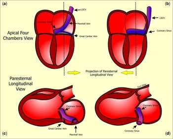
The oblique vein of the left atrium, also known as Marshall’s vein, is the only part of the embryonic left superior caval vein that persists in the normal heart. In most normal individuals, the distal portion of the vein involutes during foetal life,Reference Marshall1 persisting as a ligamentous structure, known as Marshall’s ligament, which raises a pericardium fold along the postero-inferior left atrial wall.Reference Marshall1 The proximal portion of the venous channel persists, running obliquely along the postero-inferior wall of the left atrium between the left pulmonary veins and the orifice of the appendage and draining into the coronary sinus. Indeed, the orifice of the vein is often considered to mark the beginning of the coronary sinus, and is usually guarded by a valve with one or two leaflets, the valve of Vieussens. In about one-twentieth of those with congenitally malformed hearts, however, the entirety of the venous channel persists as a left-sided superior caval vein.
Earlier studies have recognised the importance of the persisting left-sided superior caval vein, but more recently the focus is on Marshall’s vein itself, especially concerning the genesis of atrial fibrillation, with some noting that, whenever they identified the vein using venography, arrhythmogenic focuses were more frequent.Reference Kurotobi, Ito and Inoue2 To the best of our knowledge, little is known concerning the echocardiographic identification of the vein. We have recently interrogated four patients in whom a vascular structure was noted to protrude into the left atrial cavity. We discuss here the evidence supporting the notion that this structure is the oblique vein of the left atrium, and discuss its relationship to the persistent left superior caval vein, noting that, contrary to our expectations, the latter structure does not always occupy the entirety of the left atrioventricular groove.
Description of cases
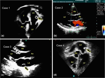
Our first patient was aged 22 years, female, and had previously been diagnosed as having a common arterial trunk. During the ongoing transthoracic echocardiographic evaluation as part of her follow-up, the four-chamber view revealed a vascular structure protruding into the interior of the left atrium, with no relation to the atrioventricular junction (Fig 1a). A left superior caval vein was not apparent during examination, although that we did not positively exclude its presence.
(a) Case 1 – Transthoracic echocardiogram, an apical four chambers view. The arrow shows the circular structure inside the left atrium and above the atrioventricular junction. (b) Case 2 – Transthoracic echocardiogram, a parasternal long-axis view with colour flow mapping. Flow in blue can be seen inside the protruding vascular structure. (c) Case 3 – Transthoracic echocardiogram, a parasternal long-axis view. A similar structure is seen inside the left atrium. (d) Case 4 – Transthoracic echocardiogram, an apical five-chamber view. The arrow shows the same structure inside the left atrium. Ao, aorta; LA, left atrium; LV, left ventricle; RV, right ventricle; T, common arterial trunk.

The second patient, also female, was aged 9 years, and was known to have stenosis of the distal pulmonary arteries associated with a perimembranous ventricular septal defect, an atrial septal defect within the oval fossa, and persistent patency of the arterial duct. During transthoracic echocardiography, we noted a vascular structure that bulged into the left atrial lumen and drained into the coronary sinus. Interrogation in the longitudinal parasternal view revealed that the structure was circular and was attached to the postero-inferior wall of the left atrium. Colour mapping showed that the flow in the vascular structure was opposite to that within the left atrial cavity (Fig 1b). As in the first case, we made no positive search for persistent patency of the left superior caval vein.
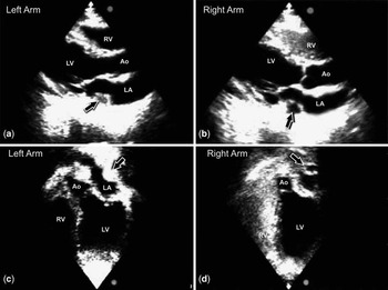
Our third patient, also a female aged 43 years, had no structural abnormalities of the heart. She had been referred for stress dobutamine echocardiography. We noted a vascular structure within the left atrium similar to those encountered in the previous patients (Fig 1c), but on this occasion the findings suggested associated persistence of the left superior caval vein. To confirm this possibility, we injected saline through the right and left antecubital veins. When injected in the right arm, the contrast appeared in the right atrium and ventricle (Fig 2b, d). When injected in left arm, the contrast initially opacified the venous structure, and subsequently the right atrium, thus confirming the diagnosis of persistent patency of the left superior caval vein (Fig 2a, c), even in the absence of a brachiocephalic vein.
(a) Transthoracic echocardiogram, parasternal long-axis view. After injection of saline solution in the left arm antecubital vein, the arrow shows the structure being filled by the contrast. (c) Transthoracic echocardiogram, an apical five-chamber view. The structure is completely filled by the bubbles (arrow), but the right chambers are still free of contrast. (b) and (d) It is possible to see the contrast inside the right chambers when the saline solution is injected in the right arm, but the structure (arrows) in the left atrium is free of contrast. Ao, aorta; LA, left atrium; LV, left ventricle; RV, right ventricle.

Our fourth patient, aged 79 years, was a man who had previously suffered a myocardial infarction, and was now referred for a routine follow-up transthoracic echocardiogram. We again noted a vascular structure within the left atrium (Fig 1d), which could be followed to drain into the right atrium through the coronary sinus. In this case, interrogation from the suprasternal notch was highly suggestive of persistent patency of the left superior caval vein.
The course of the persistent left superior caval vein
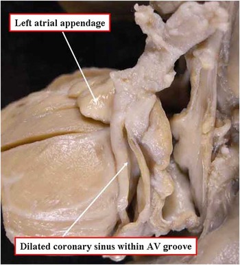
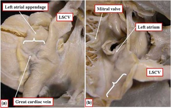
In a preliminary analysis of 10 cardiac specimens with persistent left superior caval vein, obtained randomly from the anatomical collection of our institution, we noted two discrete relationships of the vein as it joined the coronary sinus in the inferior part of the left atrioventricular groove. In the most frequent pattern, found in seven hearts, the persistent left superior caval vein joined the coronary sinus at the obtuse margin of the ventricular mass, the dilated coronary sinus then occupying the entire inferior part of the left atrioventricular groove (Fig 3). In the remaining three hearts, in contrast, the coronary sinus deviated from its anticipated location within the atrioventricular groove, crossing obliquely to meet the persistent left superior caval vein between the left pulmonary veins and the left atrial appendage, with the great cardiac vein occupying the lateral part of the inferior atrioventricular groove (Fig 4a). When a section was taken across the inferior margin of the left atrioventricular groove in the hearts with this second pattern, no dilated venous channel was observed within the atrioventricular groove (Figs 4b and 5).
The most frequent pattern of drainage of the left superior caval vein, found in seven of the series of ten hearts studied from our archive. Note that the dilated venous structure occupies the entirety of the inferior part of the left atrioventricular groove.

The less frequent pattern of drainage of the left superior caval vein, found in three of our ten hearts with persistent left superior caval vein. Note that the persistent caval vein deviates away from the atrioventricular junction (a, white bracket. Opening the heart inferiorly through the atrioventricular groove (b) shows the absence of any dilated venous channel.

Schematic drawing showing the two patterns for location of the persistent left superior caval vein as seen in the apical four-chamber view (a and b), and the parasternal longitudinal view (c and d). We believe that when the vein takes an oblique course, it can be visualised at the posterior wall of the left atrium, as shown in panels a and c.

Discussion
Depending on the definition used, the coronary sinus is considered to commence either at the site of the valve of Vieussens, or at the point of union of the great cardiac vein with the oblique vein of the left atrium. The morphology of the venous valve itself, even if present, is markedly variable.Reference Zawadzki, Pietrasik, Petrasik, Marchel and Ciszek3 Because of this variability, we believe that it is more consistent to use the union of the oblique and great cardiac veins as the anatomic origin of the coronary sinus.
During cardiac development, it is the left cardinal vein that occupies the site of the oblique vein. This anatomic arrangement persists in postnatal life when a left-sided superior caval vein drains into the coronary sinus. Such persistence of the left superior caval vein is said to vary from 0.3% to 0.5% in the absence of other associated congenital cardiac defects, but to occur in up to one-twentieth of patients with congenitally malformed hearts.Reference Campbell and Deuchar4, Reference Fraser, Dvorkin, Rossall and Eidem5
Dilation of the coronary sinus is an indirect sign of persistence of the left superior caval vein. Moreover, individuals with such anatomical findings are more susceptible to arrhythmias and sudden cardiac death. Anderson et alReference Anderson and Latham6 suggested that the persistence of the left-sided venous channel may influence other anatomic features such as abnormalities of the sinus node and fibrosis of the atrioventricular bundle.
Patients requiring pacemakers or implantable cardioverter defibrillators have been noted to have a slightly higher prevalence of the persistent left superior caval vein.Reference Biffi, Boriani, Frabetti, Bronzetti and Branzi7
We have described the protrusion of the left superior caval vein as it courses along the postero-inferior wall of the left atrium to drain into the coronary sinus. Indeed, some authors suggest that its presence can obstruct flow to the left ventricular inlet.Reference Gilard, Mansourati and Etienne8 Of our four patients, unequivocal evidence of a persistent left superior caval vein was found in only one. We cannot rule out the possibility of this finding in the other patients, as none had dilation of the venous channel in the inferior atrioventricular groove as seen in the parasternal longitudinal echocardiographic section. This could be the result of all the described patients having the least common pattern of drainage of the persistent left superior caval vein, as revealed by the preliminary examination of a cohort of hearts from our archive of anatomical specimens. According to our anatomical findings, in two-fifths of hearts having a left superior caval vein draining into the coronary sinus, the left superior caval vein itself occupies the site of Marshall’s vein in the left atrial wall, with the coronary sinus deviating from its anticipated location within the inferior part of the left atrioventricular groove, and hence producing the situation in which a dilated venous channel is lacking within the lateral part of the groove.
Our present purpose, therefore, is no more than to emphasise our finding of a curious venous structure protruding into the left atrial cavity. In all four patients, we found the circular image laterally half way between the atrioventricular groove and the atrial roof. By rotating the transducer, we were able to follow its course to the right atrial orifice of the coronary sinus. Contrast of saline, performed only in our third patient, confirmed the presence of a left-sided superior caval vein and absence of a communicating brachiocephalic vein.
We suggest that further clinical studies are essential to investigate whether, in all cases when a left superior caval vein is present, a dilated venous structure will be found in the left atrioventricular groove. Had we systematically interrogated this region in all our patients, we might have discovered a dilated coronary sinus, but our initial study of hearts with persistent left superior caval veins suggests that the venous channel may not have been within the atrioventricular groove.
We have been unable to find any previous investigation related to echocardiographic identification of Marshall’s vein. This vein is usually of small calibre, and, as far as we can establish, has not previously been noted echocardiographically. In recent years, intracardiac echocardiography has been used to guide eletrophysiological procedures, such as left ventricular pacing or resynchronisation via the coronary sinus.Reference Shalaby9 This approach has proved that it is possible to recognise the valve of Vieussens.Reference Scholten, Szili-Torok, Thornton, Roelandt and Jordaens10 To the best of our knowledge, no one has reported the visualisation of the left superior caval vein.
It is now our opinion that all four of our patients must have had persistence of the left superior caval vein, although it has not definitively shown in three of them. Another point, warranting discussion, is the possibility that the bulging venous structure can obstruct the inflow to the left ventricle. Impaired left ventricular filling and partial occlusion of the mitral valve have previously been related to dilation of the coronary sinus.Reference Cochrane, Marath and Mee11 In our patients, however, we found no evidence, as judged by Doppler interrogation, for obstruction of flow to the left ventricle.