Introduction
Genome editing directly in mammalian embryos (Meyer et al., Reference Meyer, Martin, Wurst and Kühn2010; Flisikowska et al., Reference Flisikowska, Kind and Schnieke2013) is a powerful new technique that promises to radically streamline the genetic modification of large animal species, such as pigs, and so provide exciting new resources for biomedical research. However such an approach requires a reliable supply of developmentally competent porcine embryos. The first successful birth of a piglet derived by in vitro fertilization (IVF) of porcine oocytes flushed from the reproductive tract was reported almost 3 decades ago (Cheng et al., Reference Cheng, Polge and Moor1986), and there have since been numerous reports of live births in pigs by IVF (Grupen, Reference Grupen2014), but obtaining in vivo matured oocytes is costly and time consuming (Nakai et al., Reference Nakai, Kashiwazaski, Takizawa, Hayashi, Nakatsukasa, Fuchimoto, Noguchi, Kaneko, Shino and Kikuchi2003). Moreover, unlike cattle, non-surgical ovum pick up is not practical in pigs due to their anatomy. Developing in vitro production (IVP) of pig embryos including in vitro maturation (IVM) has however proved problematic, and the systems available are still unacceptably inefficient, especially compared with other livestock species such as cattle (Grupen, Reference Grupen2014). In vitro matured porcine oocytes have problems at several stages in the process. They have reduced ability to be fertilized in vitro, to block polyspermy, to undergo normal pronuclear formation upon sperm penetration (Abeydeera & Day, Reference Abeydeera and Day1997; Abeydeera, Reference Abeydeera2002) and culture conditions are suboptimal for supporting embryo development beyond the initial cleavage divisions (Grupen, Reference Grupen2014).
Pig ovaries are usually obtained from slaughterhouses, which is convenient, but variations between animals and their handling can significantly affect the quality of the tissue arriving in the laboratory. For example, porcine oocytes are highly susceptible to temperature stress, and transient temperature shocks can occur in vivo during slaughter (Tong et al., Reference Tong, Heng, Chen, Yip and Ng2004), or ovaries be exposed to low temperature after removal (Yuge, Reference Yuge2003). These stresses result in poor maturation and subsequent embryo development and are often outside the control of the experimenter. A simple, reliable means of identifying cumulus–oocyte complexes (COC) with high maturational competence before IVM culture would reduce an important source of variability and aid the improvement and standardisation of subsequent IVM conditions.
As with many mammalian species, porcine COCs are usually graded by visual assessment of morphological features such as the thickness and compactness of the cumulus investment, ooplasm homogeneity (Akshey et al., Reference Akshey, Malakar, De, Jena, Sahu and Dutta2011; Garg et al, Reference Garg, Dutta, Malakar, Jena, Kumar, Sahu and Prakash2012) and the size of follicles (Hyttel et al., Reference Hyttel, Fair, Callesen and Greve1997; Hendriksen et al., Reference Hendriksen, Vos, Steenweg, Bevers and Dielemans2000) or oocytes (Coticchio et al., Reference Coticchio, Sereni, Serrao, Mazzone, Iadarola and Borini2004). Based on light microscopy, mammalian COCs surrounded by several layers of cumulus cells and with evenly granulated ooplasm have higher developmental competence in vitro than oocytes with irregularly granulated ooplasm and fewer cumulus layers (Wang et al., Reference Wang, Sun, Hosoe, Shioya and Day1997; Sirard et al., Reference Sirard, Richard, Blondin and Robert2006; Miyano & Manabe, Reference Miyano and Manabe2007). Many other different selection criteria have also been reported (Galeati et al., Reference Galeati, Modina, Lauria and Mattioli1991; Gandhi et al., Reference Gandhi, Lane, Gardner and Krisher2001; Qian et al., Reference Qian, Shi, Ding, Sha and Fan2003).
The cumulus layer is undoubtedly vital for oocyte development, for example it plays an important role in the distribution of cortical granules, and thus the ability to undergo sperm penetration (Cheng et al., Reference Cheng, Chen, Lee, Pai, Huang, Huang and Lee2013). However visual observation provides only limited information about its functional competence. Alternative methods of analysing cumulus cells are available, including evaluation of telomere length (Lee et al., Reference Lee, Joo, Na, Yoon, Choi and Kim2001), cumulus cell apoptosis (Corn et al., Reference Corn, Hauser–Kronberger, Moser, Tews and Ebner2005; Assou et al., Reference Assou, Haouzi, De Vos and Hamamah2010) and gene expression profiling using microarray analysis (Ruppert-Lingham et al., Reference Ruppert-Lingham, Paynter, Godfrey, Fuller and Shaw2006), but are too time consuming and expensive for routine laboratory use. Propidium iodide staining has also been used as a non-invasive method of determining cumulus cell integrity (Uchikura et al., Reference Uchikura, Nagano and Hishinuma2011), but this requires fluorescence microscopy, which may not be available for some researchers and may be potentially mutagenic.
Here we report the use of lissamine green B (LB) dye to visually assess the quality of COCs used for in vitro culture. LB is used in ophthalmology to evaluate ocular membrane integrity (Hamrah et al., Reference Hamrah, Alipour, Jiang, Sohn and Foulks2011), but to our knowledge has never been used previously in reproductive technology. LB is a non-toxic synthetic organic acid dye that can be used as a supravital stain and, unlike propidium iodide, is directly visible, making staining rapid and simple. LB offers an efficient, objective, non-invasive and directly visible means of selecting good quality COCs and oocytes.
Materials and methods
Chemicals, cell culture media and supplements
Chemicals, reagents and culture media were purchased from Sigma Chemical Co., St. Louis, MO, USA unless otherwise indicated. Media were prepared fresh and sterilised by 0.2 μm membrane filtration.
Collection of oocytes
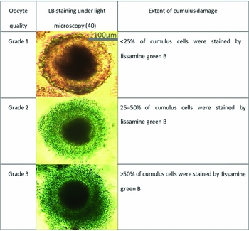
Slaughterhouse ovaries were brought to the laboratory within 1 h of collection in a temperature-controlled box maintained at 39°C. Ovaries were thoroughly washed with pre-warmed (39°C) Dulbecco's phosphate-buffered saline (DPBS) solution containing 0.1% polyvinyl alcohol and penicillin, streptomycin solution. Follicles with a diameter of 3–8 mm were aspirated using an 18-gauge needle and a 5 ml syringe. Aspiration medium consisted of HEPES-buffered TCM199 (M 2520) with 26 mM NaHCO3 (S 5761), 1 mM l-glutamine (G8540), 0. 5 mg/ml polyvinyl alcohol (P 8136), 50 μg/ml gentamicin (G1264) and 20 U/ml heparin (H3149). Oocytes (COCs) were retrieved in the same medium without heparin using a stereomicroscope. Oocytes with even, dark cytoplasm and more than one compact layer of cumulus cells were selected. In total, 767 oocytes (COCs) were obtained from ~70 ovaries. These were divided randomly into two groups, one to study nuclear maturation and one to study parthenogenetic activation. Each one was further divided randomly into two sub-groups, one was graded by the morphology of their surrounding cumulus investments: grade A, at least three layers; grade B, less than three layers; or grade C, only corona radiata. The other sub-group was LB stained and scored according to a system previously used for propidium iodide staining (Ruppert-Lingham et al., Reference Ruppert-Lingham, Paynter, Godfrey, Fuller and Shaw2006; Uchikura et al., Reference Uchikura, Nagano and Hishinuma2011), with grade 1 having <25% cumulus cells stained; grade 2, 25–50% cumulus cells stained; and grade 3, >50% cumulus cells stained, as shown in Fig. 1.
Classification of COCs by lissamine green B staining.

Optimization of lissamine green B staining
A stock solution of 1% LB (199583) in DPBS (pH = 7.0) was prepared and further diluted into TCM199 medium. All staining was performed at 39°C. Suitable staining conditions to detect cumulus cell damage were determined using COCs artificially damaged by treatment with 0.1% hyaluronidase (H3506) for 30 min at room temperature. Treated COCs were placed in groups of 25 in 500 μl droplets at four different LB concentrations (1, 0.5, 0.25 or 0.1%) under mineral oil for 2.5, 5 or 10 min. Each treatment was repeated three times. Following staining, COCs were washed in phosphate-buffered saline (PBS) to remove residual stain, observed by zoom stereomicroscope, and the number of stained COCs was recorded.
Determination of nuclear maturation after IVM culture
After quick assessment of cumulus membrane integrity, oocytes were washed twice in in vitro maturation (IVM) medium and placed in groups of 40–60 in a final volume of 500 μl IVM medium in 4-well dishes (Nunc GmbH, Co. KG, Germany). Oocytes were cultured at 39°C under 5% CO2 in air and maximum humidity for 46 h. The maturation medium consisted of TCM 199 (M 2154) with 1 mM l-glutamine, 1 mM sodium pyruvate (P3662), 0.1 mM 2-mercaptoethanol (M7522), 50 ng/ml epidermal growth factor (E4127), 50 μg/ml gentamicin and 0.1% (w/v) BSA (A3311). During the first 24 h of IVM, the medium was supplemented with 10 IU/ ml PMSG and 5 IU/ ml hCG (MSD Tiergesundheit, Germany) and 5 μl/ml ITS (recombinant human insulin, human transferrin, and sodium selenite, I3146) (Sjunnesson et al., Reference Sjunnesson, Morrell and González2013).
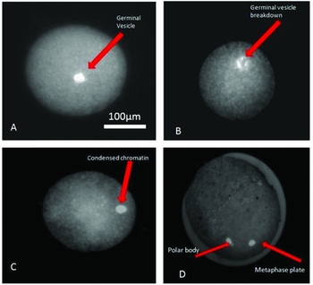
After IVM culture, cumulus investments were removed from the oocytes by gentle pipetting. Oocytes were then fixed for 30 min in 0.5% glutaraldehyde (500 μl PBS + 10 μl 25% glutaraldehyde G-5882), washed in 500 μl PBS for 5 min then transferred to 500 μl Hoechst (B2261) stain solution (0.1%). The nuclear status was evaluated using an epifluorescence microscopy (ultraviolet (UV) light filter; Carl Zeiss) using standard measures like germinal vesicle breakdown, condensation of chromatin and appearance of the first polar body as confirmation of nuclear maturation (Fig. 2).
Progression of lissamine green B stained oocytes through different stages of maturation. (A) Immature oocyte. (B) Germinal vesicle breakdown. (C) Appearance of first polar body. (D) Mature oocyte with visible polar body and metaphase plate.

Parthenogenetic activation of porcine oocytes
Oocytes were activated chemically to assess their developmental competence. After 46 h IVM culture, COCs were manually denuded by mouth pipetting, then activated by incubating in IVM medium containing 5 μM Ca-ionophore A23187 (C7522) for 5 min at 39°C under 5% CO2 in air. Oocytes were washed three times in IVM culture medium and incubated in 500 μl droplets containing 2 mM 6-dimethylaminopurine (6-DMAP, D2629) and covered with mineral oil in an incubator at 39°C under 5% CO2 in air for 4 h. After activation, oocytes were cultured in 500 μl porcine zygote medium (PZM 5 IFP) in 4-well dishes (Nunc GmbH, Co. KG, Germany) overlaid with 500 μl mineral oil (M8410). The dish was incubated undisturbed at 39°C in 5% CO2 in air for 7 days. Embryo development was observed microscopically and cleavage assessed at 48 h post-activation.
RNA isolation and reverse-transcription
Apoptotic gene expression was evaluated in groups of 50–60 COCs. Total RNA was isolated using an RNeasy Mini Kit (Qiagen, Europe) according to the manufacturer's instructions. Sample purity was assessed using the A260/A280 nm ratio with expected values between 1.8 and 2.0, RNA samples were treated with TURBO DNA-free™ Kit (Life Technologies) to remove genomic DNA. Reverse-transcription reactions were performed with 0.5–1 μg total RNA using the SuperScript® III First-Strand Synthesis System for RT-PCR (Life Technologies) and random hexamer as a primer, according to the manufacturer's instructions. For each sample, negative controls lacking reverse transcriptase were prepared using the identical procedure.
Quantitative real-time PCR
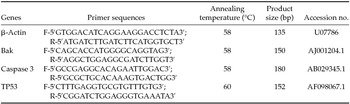
Gene expression was assessed by quantitative RT-PCR. Primers for BAK, CASP3 and TP53, and a housekeeping gene ACTB (beta-actin) were designed using ‘Primer3’ software. The predicted fragment sizes are listed in Table 1. Real-time PCR amplification was conducted using an ABI 7300 real-time PCR System (Applied Biosystems). A KAPA SYBR FAST q PCR Kit (Kapa Biosystems) was used to provide real-time quantification of the amplified product. Three replicates of each reaction were measured and the mRNA level of each sample was normalised to that of beta-actin. Relative mRNA levels were determined by the ΔΔCt method (Livak & Schmittgen, Reference Livak and Schmittgen2001).
Primer sequences and annealing temperature

Measurement of DNA fragmentation
The terminal deoxynucleotidyl transferase-mediated dUTP nick end labelling (TUNEL) procedure was used to fluorescein–dUTP label 3′-OH ends of DNA fragments generated by apoptosis, allowing detection of apoptotic cells by fluorescence microscopy. Fixed and permeabilized embryos were subjected to the TUNEL assay procedure using an in situ apoptosis detection kit (TaKaRa Bio Inc., Japan) according to the manufacturer's instructions. COCs were fixed for 30 min at room temperature in PBS (pH 7.4), permeabilized for 5 min on ice in permeabilization buffer, and then washed once in PBS. COCs were incubated with terminal transferase and labelled nucleotide solution in a humidified, sealed chamber in the dark at 37°C for 1 h. Slides were overlaid with a coverslip and the edges sealed with quick drying nail polish, then evaluated using an epifluorescence microscopy (green excitation filter, Carl Zeiss). Oocytes showing discrete pinpoint fluorescent green signals were judged to be positive for DNA fragmentation. The apoptosis score was expressed as a percentage of the total.
Statistical analysis
Data were analysed using GraphPad Prism (GraphPad Software, Inc. USA) after arcsin transformation of percentage values. Differences between mean percentages were analysed by two-way analysis of variance (ANOVA). A value of P < 0.05 was considered to be statistically significant.
Results
Lissamine B staining
A staining regimen of 0.50% LB for 5 min was found to be optimum for staining in terms of specificity and easily detectable coloration. Under light microscopy, damaged cumulus cells were stained greenish blue, while healthy cumulus cells remained unstained. The ooplasm of denuded oocytes that incurred damage to the zona pellucida stained greenish blue.
Nuclear maturation
Of the 372 COCs in the nuclear maturation group, 191 were graded by morphology, with 63 assessed as grade A (32.99%), 67 grade B (35.07%) and 61 grade C (31.93%); 181 were graded by LB staining as 59 grade 1 (32.59%), 65 grade 2 (35.91%, and 57 grade 3 (31.49%). Nuclear maturation of each oocyte was scored by the attainment of metaphase II, as judged by the presence of condensed chromosomes in an equatorial position and extrusion of the first polar body (Fig. 2). Nuclear maturation data (Table 2) showed no significant difference between morphological grade A (79.9 ± 3.82%), grade B (86.48 ± 3.07%), or grade C (82.00 ± 3.00%) oocytes. The nuclear maturation rates of LB graded oocytes were: grade 1 (84.70 ± 3.36%), grade 2 (83.61 ± 3.44%) and grade 3 (62.26 ± 3.69%). The difference between grade 3 and grades 1 and 2 was significant (P < 0.05).
Effects of cumulus morphological categories on maturational and developmental ability of porcine oocytes

a,b Values with different superscripts differ significantly (P < 0.05). Values are means ± standard deviation (SD).
Developmental competence of oocytes
The parthenogenetic activation group comprised 395 oocytes. 204 were graded by morphology as 67 grade A (32.84%), 71 grade B (34.80%) and 66 grade C (32.35%); 191 were graded by LB staining as 64 grade 1 (33.50%), 65 grade 2 (34.03%), and 62 grade 3 (32.46%). Development was scored as the number of embryos consisting of two to eight evenly sized blastomeres 48 h after parthenogenetic activation. There was no significant difference between morphological grade A (84.55 ± 4.45%), grade B (85.06 ± 4.49%), or grade C (81.60 ± 4.64%) oocytes. The rates of parthenogenetic activation in LB graded oocytes were: grade 1 (87.71 ± 2.46%), grade 2 (82.72 ± 3.39%) and grade 3 (62.88 ± 3.85%). Again this shows a statistically significant difference between grade 3 and grades 1 and 2 (P < 0.05) (Table 2).
TUNEL assay for oocytes and cumulus cells
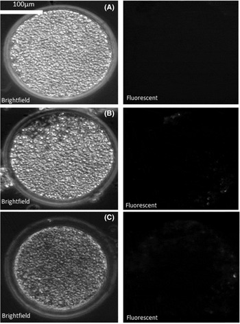
In total, 176 COCs were graded by either morphology or LB staining. For each grade a TUNEL score was determined as the percentage of observed COCs showing signs of DNA fragmentation, as indicated by a fluorescent signal (Table 3 and Fig. 3). Throughout, fluorescent signals were observed only in the ooplasm and not in the cumulus cells. There was no significant difference between morphological grade A (20.97 ± 3.52%) and grade B (22.69 ± 4.05%) oocytes, but grade C oocytes showed significantly higher (38.42 ± 4.03%) incidence of DNA fragmentation (P < 0.05). The TUNEL score in LB graded oocytes was similar: grade 1 (21.66 ± 4.08%), grade 2 (21.82 ± 5.85%), with grade 3 significantly higher (37.08 ± 2.76%) (P < 0.05). There was no significant difference in TUNEL score between morphological grade C and LB grade 3 COCs.
Rate of DNA fragmentations as detected by TUNEL assay in different category of porcine oocytes

a,b Values with different superscripts differ significantly (P < 0.05). Values of DNA fragmentation are means ± standard deviation (SD).
DNA fragmentation detection by TUNEL assay. (A) Porcine oocyte with no detectable DNA fragmentation. (B) Porcine oocyte with moderate DNA fragmentation. (C) Porcine oocyte with extensive DNA fragmentation.

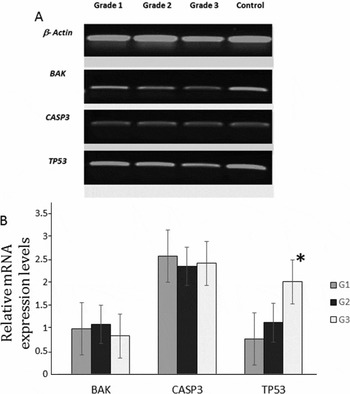
Expression of stress and apoptosis-related genes in lissamine B graded porcine COCs.
Differential expression of the stress-associated gene TP53 and two genes related to apoptosis, BAK (BCL2-antagonist/killer) and CASP3 (caspase 3), was analysed by quantitative real-time RT-PCR in three pools of 50–60 COCs representing LB-stained grades 1, 2 and 3 (Fig. 4). Three replicates were conducted for each experiment. Figure 4 shows that BAK and CASP3 expression was similar in all grades of LB-stained COCs, but TP53 expression was significantly greater (P < 0.05), in grade 3 COCs than grades 1 and 2.
(A) Reverse-transcription PCR of apoptosis-associated genes in lissamine B stained oocytes. Relative gene expression levels of lissamine B stained porcine oocytes. (B) The mRNA levels of BAK, Caspase 3 and TP53 were analysed in porcine oocytes. Results are displayed as fold changes in different grades of lissamine B stained oocytes relative to each other. Significant difference is marked with an asterisk (*P < 0.05).

Discussion
We evaluated the usefulness of lissamine green B (LB) staining for non-invasive prediction of maturational competence of porcine oocytes. LB preferentially stains membrane-damaged or devitalised cells (Kim & Foulks, Reference Kim and Foulks1999). Studies using LB to stain rabbit and human corneal epithelial cells in vitro show that it does not stain healthy, proliferating cells and has a minimal effect on cell viability (Kim, Reference Kim2000). Evidence to date indicates no carcinogenic or cytotoxic properties (Brantom et al., Reference Brantom, Creasy and Gaunt1987; Clode, Reference Clode1987; Moorhouse et al., Reference Moorhouse, Creasy and Gaunt1987).
Our data indicate that LB staining of COCs immediately after collection is superior to morphological grading as a predictor of oocyte viability, nuclear maturation after IVM culture, and successful parthenogenetic activation and development. Parthenogenesis was used as a convenient and valid functional assay of developmental competence (Rougier & Werb, Reference Rougier and Werb2001; McElroy et al., Reference McElroy, Byrne, Chavez, Behr, Hsueh, Westphal and Pera Reijo2010).
While higher rates of nuclear maturation, fertilization and development after IVP have been reported in bovine oocytes graded on the basis of the thickness of the cumulus cell layers, our data show that this cannot automatically be extended to porcine oocytes. Our findings clearly reaffirm the importance of a cumulus layer for oocyte maturation and development (Davachi et al., Reference Davachi Dadashpour, Kohram and Zainoaldini2012; Prates et al., Reference Prates, Nunes and Pereira2014), but suggest that measures of cumulus cell integrity are more useful than morphological parameters alone.
Our investigation of the role of apoptosis in COC quality revealed some interesting findings. As expected, DNA fragmentation measured by TUNEL assay showed that LB grade 3 COCs were more damaged than higher grade COCs, consistent with their developmental properties and also in line with morphological grade C COCs. DNA fragmentation in immature oocytes (in particular, GV and MI stages) could be a consequence of stress during maturation of the ovarian follicle or hypoxia resulting from compromised microcirculation and correlate with aneuploidy or other chromosomal abnormalities (Van Blerkom, Reference Van Blerkom1996). It is notable that we found no evidence of DNA fragmentation in the cumulus layer in immature porcine COCs. This is consistent with a similar finding in cattle in which no TUNEL signal was obtained in cumulus cells of immature oocytes (Yuana et al., Reference Yuana, Van Sooma, Leroya, Dewulfa, Van Zeverenb, de Kruifa and Peelman2005).
Gene expression analysis of three genes chosen for their well known roles in stress and apoptosis however revealed an interesting and unexpected distinction. The expression of TP53, a stress response gene induced by DNA damage, was significantly greater in LB-stained grade 3 COCs, consistent with TUNEL assay findings and the poorer developmental parameters of this grade. Studies in humans and mice have revealed a clear relationship between in vitro culture related stress and TP53 expression in embryos (Chandrakanthan et al., Reference Chandrakanthan, Li, Chami and O’Neill2006). In mice, genetic inactivation of Mdm2, a negative regulator of p53, results in early embryonic lethality (Jones et al., Reference Jones, Roe, Donehower and Bradley1995) that can be reversed by deletion of Trp53 (Luna et al., Reference Luna, Wagner and Lozano1995), demonstrating that the p53 response pathway plays a direct role in embryonic loss.
It was expected that BAK and CASP3 expression would also be greater in the lowest quality COCs, but this was not the case. BAK and CASP3 are members of two important regulatory families involved in apoptosis. BAK is a pro-apoptotic member of the Bcl-2 family whose cytoplasmic elevation is sufficient to cause induction of oocyte apoptosis (Morita & Tilly, Reference Morita and Tilly1999). The lack of any clear correlation between the RNA level of these genes and COC grade, despite evident DNA damage, could be due to a variety of reasons. One is that there is abundant maternal CASP3 mRNA in the oocytes (Weil et al., Reference Weil, Jacobson, Coles, Davies, Gardner, Raff and Raff1996), and this may mask any differential expression in the cumulus cells of different grades. Another is that regulation of caspase 3 activity is known to occur at the protein level, by modification of an inactive proenzyme. In which case direct immunocytological detection of the active form of caspase-3 may be a more reliable measure. It is also known that caspase cleavage and DNA fragmentation do not take place simultaneously (Yuana et al., Reference Yuana, Van Sooma, Leroya, Dewulfa, Van Zeverenb, de Kruifa and Peelman2005). However, our findings are also consistent with similar observations in bovine embryos treated with the apoptosis inducer staurosporine, which showed no difference in the expression of several apoptosis-related genes including CASP3 and other Bcl family genes (Vandaele et al., Reference Vandaele, Goossens, Peelman and Van Soom2008).
Cumulus cells play a critical role in oocyte maturation and fertilization by releasing and mediating signals to oocytes (Vandaele et al., Reference Vandaele, Goossens, Peelman and Van Soom2008), but the relationship between cumulus cell apoptosis and the development potential of the oocyte is unclear. There are in fact contradictory reports in various species as to whether definitive apoptosis actually takes place in COCs (Tanghe et al., Reference Tanghe, Soom, Nauwynck, Coryn and de Kruif2002). For example, some studies have shown that human and bovine cumulus cells undergo apoptosis (Lee et al., Reference Lee, Joo, Na, Yoon, Choi and Kim2001, Mikkelsen et al., Reference Mikkelsen, Host and Lindenberg2001; Yuana et al., Reference Yuana, Van Sooma, Leroya, Dewulfa, Van Zeverenb, de Kruifa and Peelman2005), While others report the absence of apoptosis in cumulus cells of rats (Zeuner et al., Reference Zeuner, Muller, Reguszynski and Jewgenow2003), pigs (Szoltys et al., Reference Szoltys, Tabarowski and Pawlik2000), and cattle (Manabe et al., Reference Manabe, Imai, Ohno, Takahagi, Sugimoto and Miyamoto1996).
The simplicity and non-invasive nature of LB staining makes it straightforward to adopt as a quality control test for embryological labs. LB staining can give valuable information about the quality of oocytes in terms of maturational ability, developmental potential and extent of DNA damage. Further studies are clearly required to assess any long-term effects of LB on embryo survival and fetal health.
Acknowledgements
We would like to thank Steffen Löbnitz for help collecting samples. This work was financially supported by grant 16EX1024B; ‘Platform Advanced Preclinical Animal Models’ from the German Federal Ministry of Education and Research (Bundesministerium für Bildung und Forschung) as part of the m4 cluster initiative Personalized Medicine and Targeted Therapies. RD was supported by a scholarship from the German Academic Exchange Service (Deutscher Akademischer Austausch Dienst). The authors are members of COST Action BM1308 ‘Sharing Advances on Large Animal Models’ (SALAAM).