Introduction
Myringoplasty is defined as the repair of the tympanic membrane alone without inspection of the ossicular chain or removal of disease from the middle ear.Reference Merchant, Nadol and McKenna1 The surgical closure of tympanic membrane perforations was first described by Zoellner and Wullstein in 1952. Since then, there have been considerable advances in surgical approaches, techniques and choice of graft materials.
At present, myringoplasty and tympanoplasty most commonly utilise temporalis fascia and cartilage grafts. A temporalis fascia graft is primarily used in the popular underlay technique via an endaural or postaural approach. Cartilage is used as a chondroperichondrial island graft in high-risk tympanic membrane perforations and middle-ear atelectasis.Reference Sperkelsen, Hildmann and Sudhoff2 This too is employed in an underlay technique, mostly via a postaural approach. For small perforations, a transcanal adipose plug myringoplasty yields satisfactory results in most cases.Reference Ozgursoy and Youralmaz3
This study aimed to evaluate the feasibility and outcomes of myringoplasty in select cases using a recently described technique (chondroperichondrial clip myringoplasty) performed via a transcanal approach.Reference Fernandes4
Patients and methods
Inclusion criteria
All patients presenting at the Combined Military Hospitals, Rawalpindi and Quetta, Pakistan from January 2008 to January 2011 with dry, small to medium-sized central tympanic membrane perforations and a wide ear canal were eligible for inclusion in the study. The study setting was an urban academic tertiary care facility, to which most patients are referred by primary care or general otolaryngology practitioners.
Exclusion criteria
Patients were excluded from the study if, at initial assessment in the clinic, they were under 11 years of age, had tympanic membrane perforations greater than 50 per cent of the pars tensa or were adjacent to anterior annulus, narrow or tortuous ear canals, only hearing ear, air–bone gap than 35 dB or an absent air-bone gap, or if there were extensive adhesions connecting the tympanic membrane and/or malleus with the promontory.
Clinical data
The following information was recorded for each patient: age, gender, relevant comorbid otolaryngological conditions and status of the opposite ear. Pure tone audiometry was performed to record bilateral masked air and bone conduction thresholds at 0.5, 1, 2 and 4 kHz, and the average air–bone gap was calculated. The results of the post-operative audiogram carried out at last follow up were included in the analysis.
Patients with dry ears were observed for six months to allow the possibility of spontaneous closure. For wet ears, a diligent effort was made to render each ear dry. Active allergic symptoms and any upper respiratory tract infection were optimally controlled prior to surgery.
All patients were counselled in detail about the procedure, and written informed consent was obtained. They were informed about the possibility of surgical failure which may require an alternative procedure at a later date.
Surgical technique
All operations were performed under general anaesthesia by the same surgeon (the first author, AS). Intravenous ceftazidime was given at induction, followed by local infiltration with lignocaine and adrenaline at the tragal graft donor site. Meticulous asepsis techniques were employed.
The margins of the tympanic membrane perforation were de-epithelialised circumferentially and the mucosa underneath was roughened to facilitate healing. Any granulation tissue at the edge of the perforation was also removed. The size of the perforation in relation to the tympanic membrane (pars tensa) was reviewed to ensure that it did not exceed 50 per cent of the surface area. A precise impression of the size of the perforation was then taken using a Silastic® template.
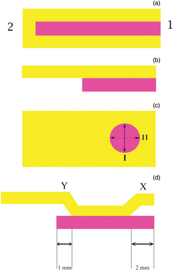
A standard chondroperichondrial island graft was harvested from the ipsilateral tragus (Figure 1a) including the tragal dome. An effort was made to keep the cartilage component of the graft just larger than the size of the freshened perforation with the help of the Silastic impression mould. The perichondrium was completely elevated off one side of the cartilage (Figure 1b). The cartilage component of the graft was carefully cut in relation to the size of the perforation according to the requirements of the technique (Figure 1c). A ‘clip’ was created between the perichondrium and cartilage at its anterior and posterior borders, which is the hallmark of this technique (Figure 1d). Care was taken to keep the graft continually wet with saline during sculpting and preparation to prevent the separation of cartilage from the perichondrium. Small pieces of Gelfoam (Upjohn Laboratories, Michigan, USA) were placed into the middle ear through the perforation.
Stylised representation of the sequential steps of the chondroperichondrial clip myringoplasty technique. (a) Composite cartilage perichondrial island graft after harvesting. Tragal cartilage is shown in pink and perichondrium in yellow. 2 = tragal dome end; 1 = cut end of graft. (b) Perichondrium completely elevated off one surface of the cartilage. (c) Vertical height of cartilage component of the graft (I) is same as that of the freshened tympanic membrane perforation, but antero-posterior length (II) is about 3 mm more than that of the perforation. (d) Perichondrium elevated to a width of about 2 mm from the anterior margin of the graft (X) and to a width of about 1 mm from the posterior margin (Y), creating a ‘clip’ both anteriorly and posteriorly.

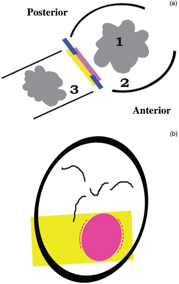
The graft, once ready, was carefully held from the perichondrium overlying the cartilage component with the help of fine cupped forceps, and introduced parallel to the plane of the tympanic membrane and at right angles to the long axis of the ear canal. The anterior edge of the graft was introduced first so that the clip (the slit between the cartilaginous and perichondrial components of the graft) engaged the anterior edge of the perforation. Gentle rocking of the graft anteroposteriorly also engaged the posterior margin of the perforation at the posterior clip (Figure 2a).
Final position of the graft at closure of the tympanic membrane perforation. (a) The cartilage is medial, the perichondrium is lateral and the ‘clip’ is on the plane of the tympanic membrane remnant (broken blue line). Graft is supported by Gelfoam (1) in the middle ear (2) and ear canal (3). (b) Another view of the graft in position. Cartilage (pink oval) occupies the perforation with the perichondrium attached to its lateral surface, while its anterior and posterior edges (dashed pink lines) lie medial to the tympanic membrane. The perichondrium component of the graft (yellow) is lateral to the tympanic membrane.

The overlying perichondrium was then spread out over the tympanic membrane. Gelfoam pledgets placed over the graft and filling the bony ear canal, together with those in the middle ear, helped to prevent graft displacement by supporting from both sides (Figure 2a and b). At the end of the procedure, the operating time (defined as the time from graft harvesting to the insertion of Gelfoam in the ear canal) and details of any additional procedure performed on the operated or contralateral ear were noted.
All patients were discharged within 24 hours with antibiotic steroid ear drops and antibiotic ointment for the graft donor site. They were advised against forceful nose blowing and sniffing, lifting heavy weights, and air travel for three weeks. Post-operative follow up included examinations of any graft donor site morbidity or other complications. The final assessment of surgical outcome was made after a minimum period of 12 months.
Results
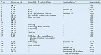
The final study group comprised 23 patients (excluding 5 who were lost to follow up early on) ranging in age from 11 to 50 years (mean 24.4 years), including one 11-year-old child. The male to female ratio was 14:9. In 56.5 per cent of cases (13 out of 23), an associated or predisposing comorbid otolaryngological condition was identified (Table I); in 69 per cent of cases (9 out of 13), these conditions included chronic rhinosinusitis, upper respiratory allergy and smoking. Evidence of some previous middle-ear surgery in the opposite ear was observed in 23 per cent of cases. An equal proportion had previously sustained blunt ear trauma in the ipsilateral ear, resulting in a persistent perforation. One patient (7.7 per cent) was unique in presentation, as he had a residual perforation from a previous underlay tympanoplasty.
Summary of patient and surgery data

*Performed at the same time as the chondroperichondrial clip myringoplasty. Pt = patient; no = number; y = years; procs = procedures; surg dur = duration of surgery; M = male; F = female; CRS = chronic rhinosinusitis; VT = ventilation tube; prev = previous; URT = upper respiratory tract
During surgery, an additional ear procedure was performed in the ipsilateral and/or contralateral ear in 26 per cent of cases (6 out of 23). The most common procedure was myringotomy with ventilation tube placement. One patient (7.7 per cent), however, required adipose plug myringoplasty in the opposite ear. An analysis of the operative duration revealed that the procedure lasted less than 30 minutes in all those in whom no additional ear procedure was performed (70 per cent of cases, 16 out of 23). More time was needed for surgical procedures that entailed ipsilateral, contralateral or bilateral myringotomy with ventilation tube placement. The procedure took the longest (50–60 minutes) in the one patient (7.7 per cent) in whom a contralateral adipose plug myringoplasty was also performed.
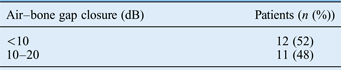
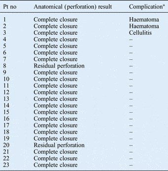
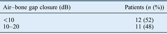
A final assessment of surgical outcome was carried out in the follow-up period which ranged from 12 to 26 months (mean 13 months). Analysis of post-surgery hearing thresholds revealed closure of the air–bone gap to within an acceptable range in all patients (Table II). Complete graft ‘take up’ resulting in total closure of the perforation was seen in 91.3 per cent of patients (21 out of 23). A smaller residual perforation was seen within a few months in 8.7 per cent of patients (2 out of 23). After an interval of six months, adipose plug myringoplasty was carried out in these (two) patients, resulting in complete closure of the perforation.
Post-surgery audiological results
Only a few minor complications were seen in the post operative period (Table III). There were no late failures, these included haematoma and/or cellulitis at the tragal graft donor site in 13 per cent of patients (3 out of 23) in the early days following surgery. There were no late failures, middle-ear infections or other complications at the site of the graft placement up to the time of last follow up.
Surgical outcomes

*At graft donor site. Pt no = patient number
Discussion
Central perforations of the tympanic membrane are a common middle-ear condition. A host of surgical techniques and approaches have been described to manage this condition. These are well established and regularly yield reliable results in expert hands. Such methods include the overlay technique, the more popular underlay technique and the fairly recently described multi-layered sandwich graft tympanoplasty.Reference Farrior and Wiet5 These can be carried out via transcanal, endaural and postaural approaches. All of them have their unique advantages and disadvantages.Reference Wehrs6 Temporalis fascia is the most commonly used graft material. The use of cartilage and fat is limited to specific situations only.Reference Merchant, Nadol and McKenna1, Reference Ozgursoy and Youralmaz3
The present study is unique as it included a highly select group of patients with central tympanic membrane perforations, and adhered to stringent inclusion and exclusion criteria. Although type I tympanoplasty has a good success rate in children,Reference Bajaj, Bais and Mukherjee7 they were generally excluded from this study because any transcanal technique is not feasible with a smaller ear canal. An 11-year-old girl with unusually wide ear canals was the only paediatric patient.
Central perforations of the pars tensa are often secondary to infections, allergic and non-specific inflammatory foci in the upper respiratory tract leading to impaired mucociliary function, and/or eustachian tube dysfunction. Blunt ear trauma is also recognised as a possible aetiological factor.Reference Manolidis, Glasscock and Gulya8 In the current study, 39 per cent of patients (9 out of 23) showed evidence of chronic rhinosinusitis, upper respiratory tract allergies or smoking. Eustachian tube dysfunction is generally inferred from the state of middle-ear mucosa, frequent middle-ear infections and the status of the opposite ear. There is, however, an enormous variation in the degree and nature of involvement of either ear.Reference Farrior and Wiet5 Intra-operatively, 17.4 per cent of patients in the current study (4 out of 23) required ventilation tube placement or adipose plug myringoplasty in the contralateral ear, which suggests the possibility of eustachian tube dysfunction.
Anatomically, a successful myringoplasty is defined as 100 per cent graft take up with complete closure of the tympanic membrane perforation.Reference Fernandes4 High take rates (ranging from 86 to 97 per cent) with standard underlay and overlay techniques using temporalis fascia have been reported in several series.Reference Mousel, Nguyen and Haberman9 Fat is an alternative graft material for small perforations (more than 4 mm). It is used in a transcanal approach, and high success rates (82.4 per cent) have been reported.Reference Ozgursoy and Youralmaz3 Cartilage is used in cartilage–perichondrium composite grafts for middle-ear reconstructive procedures. Its unique rigid and less compliant properties tend to resist resorption and retraction, even in persistent eustachian tube dysfunction.Reference Dornhoffer10 Its use is therefore indicated in revision cases and in patients with: total and subtotal perforations; large atrophic areas; tympanosclerotic plaques; and advanced middle-ear retraction pockets, atelectasis and adhesions.Reference Sperkelsen, Hildmann and Sudhoff2 For these conditions, the cartilage perichondrial graft is used in an underlay fashion, generally via the postaural approach.
This study presents a unique application of the chondroperichondrial graft, which involved modifications in graft design and placement technique, as well as employing a transcanal approach without tympanomeatal flap elevation. As the technique did not involve any removal of disease from the middle ear or ossicular reconstruction, it is referred to as ‘chondroperichondrial clip myringoplasty’ in this study. This is in contrast to another study (Fernandes, 2003), wherein the same technique was called ‘chondro perichondrial clip tympanoplasty’. The success rate reported in the present study was 91 per cent. The study by Fernandes, which was restricted to 15 patients only, reported a 100 per cent success rate.Reference Fernandes4
A striking feature of the present case series was that one of the patients had an earlier failed underlay tympanoplasty performed with a temporalis fascia graft and a smaller residual perforation. The chondroperichondrial clip myringoplasty technique resulted in complete closure of the perforation in this case, an observation which has also been described by Fernandes.Reference Fernandes4 Cartilage is the well-established graft material of choice in revision cases because of its resilience and reduced nutritional requirements which are met by diffusion alone.Reference Merchant, Nadol and McKenna1, Reference Dornhoffer10
The two cases of partial failures in this study had no active upper respiratory tract infections or allergies, and no evidence of any eustachian tube dysfunction. In addition, there was no evidence of any middle-ear infection in the post-operative period. Technical errors during surgery were presumed to be the cause of the failures. In both of these cases, the tympanic membranes healed completely with a subsequent adipose plug myringoplasty.
The chondroperichondrial clip myringoplasty technique aims to combine the advantages of both underlay and overlay techniques, while avoiding their inherent limitations. Compared to the overlay technique, blunting is avoided by not removing the outer squamous epithelial layer of the tympanic membrane, while lateralisation is avoided by means of the graft being anchored by the ‘clip technique’. Compared to the underlay technique, the morbidity and increased operative time involved in tympanomeatal flap elevation are minimised.Reference Fernandes4, Reference Wehrs6 However, the chondroperichondrial clip myringoplasty technique was actually technically more difficult and challenging than both the underlay and overlay techniques. In addition to careful case selection, there are several crucial steps in this technique, all of which are critical to surgical outcome. These include graft design and size, creation of the clip on the graft, and the accurate engaging of this clip to the margins of the perforation. It is therefore suggested that only surgeons experienced in underlay and overlay techniques and the transcanal approach should undertake this procedure.
• Chondroperichondrial clip myringoplasty is an alternative technique for closure of central tympanic membrane perforations
• Stringent criteria for case selection should be adopted for optimal results
• Meticulous chondroperichondrial graft harvesting, design and size modifications, and placement are critical to success
• Complete healing of tympanic membrane perforations was seen in 91 per cent of cases, and all showed satisfactory hearing improvement
• This technique avoids the morbidity of tympanomeatal flap elevation and middle ear entry, with acceptable operating times and few complications
Functionally, the outcome of any middle-ear reconstructive procedure is based on the closure of the air–bone gap at four frequencies before and after surgery.11 This was achieved in more than half of the patients in this study to within 10 dB, which can be considered an optimal result. Moreover, none of the patients had a residual air–bone gap of 20–30 dB. Fernandes reported closure of the air–bone gap to within 10 dB in 100 per cent of cases following chondroperichondrial clip tympanoplasty.Reference Fernandes4
A complication rate of 1–3 per cent has been reported for myringoplasty and tympanoplasty. Complications include sensorineural hearing loss, re-perforations, retractions, canal stenosis, epithelial cysts, middle-ear infections and graft donor site morbidity.Reference Mousel, Nguyen and Haberman9 The incidence of complications was higher in the current study (there were residual perforations in 8.3 per cent of cases, and haematoma or cellulitis at the donor site in 13 per cent of cases). This is probably because chondroperichondrial clip myringoplasty is an evolving technique. In addition, most of the patients in this series (95.6 per cent, 22 out of 23) belonged to the lower socioeconomic class, and chronic malnutrition may have resulted in suboptimal tissue healing and reduced immunity.
Conclusion
The promising results achieved with chondroperichondrial clip myringoplasty in this study are probably due to the strict selection criteria as well as the attention paid to the unique technicalities of several of the steps. This technique avoids a tympanotomy, with virtually no manipulation of middle-ear structures. A reduced operative time and shorter period of hospitalisation render the technique cost effective. Chondroperichondrial clip myringoplasty cannot be considered superior to other myringoplasty and tympanoplasty techniques, but can be considered a cost-effective alternative for small to medium-sized perforations in cases with large ear canals and no ossicular pathology. Further studies involving a larger cohort of patients are needed to further clarify the efficacy of this technique.