INTRODUCTION
Fasciolosis due to Fasciola gigantica is an important disease of ruminants in the tropical countries of Asia and Africa resulting in great economic losses to the livestock industry for more than US $3·2 billion per annum (Sobhon et al. Reference Sobhon, Anantavara, Dangprasert, Viyanant, Krailas, Upatham, Wanichanon and Kusamran1998; Spithill et al. Reference Spithill, Smooker, Copeman and Dalton1999; Torgerson and Claxton, Reference Torgerson, Claxton and Dalton1999; Mas-Coma et al. Reference Mas-Coma, Bargues and Valero2005, Reference Mas-Coma, Valero and Bargues2009). Furthermore, human infections with F. gigantica are also reported in many countries (Ashrafi et al. Reference Ashrafi, Valero, Panova, Periago, Massoud and Mas-Coma2006a , Reference Ashrafi, Valero, Massoud, Sobhani, Solaymani-Mohammadi, Conde, Khoubbane, Bargues and Mas-Coma b ; Le et al. Reference Le, De, Agatsuma, Blair, Vercruysse, Dorny, Nguyen and McManus2007). As a result infection cannot be examined by microscopic detection in the feces due to the absence of the parasite's eggs during the non-reproductive phase of the flukes (Ghosh et al. Reference Ghosh, Rawat, Velusamy, Joseph, Gupta and Singh2005; Kumar et al. Reference Kumar, Ghosh and Gupta2008). Alternatively, diagnosis of fasciolosis in animals has been accomplished by two accesses of immunoassays, i.e. detection of antibody in the serum samples of infected animals (Zimmerman et al. Reference Zimmerman, Nelson and Clark1985; Swarup et al. Reference Swarup, Pachauri, Sharma and Bandhopadhyay1987; Fagbemi and Obarisiagbon, Reference Fagbemi and Obarisiagbon1990; Guobadia and Fagbemi, Reference Guobadia and Fagbemi1995; Sriveny et al. Reference Sriveny, Raina, Yadav, Chandra, Jayraw, Singh, Velusamy and Singh2006) and detection of circulating antigen (Langley and Hillyer, Reference Langley and Hillyer1989; Fagbemi et al. Reference Fagbemi, Obarisiagbon and Mbuh1995; Viyanant et al. Reference Viyanant, Krailas, Sobhon, Upatham, Kusamran, Chompoochan, Thammasart and Prasittirat1997; Velusamy et al. Reference Velusamy, Singh, Sharma and Chanda2004; Anuracpreeda et al. Reference Anuracpreeda, Wanichanon, Chawengkirtikul, Chaithirayanon and Sobhon2009a , Reference Anuracpreeda, Wanichanon and Sobhon b , Reference Anuracpreeda, Chawengkirtikul, Tinikul, Poljaroen, Chotwiwatthanakun and Sobhon2013a , Reference Anuracpreeda, Chawengkirttikul and Sobhon2016b ). The detection of antigen rather than antibodies is considered to be a more reliable method for identifying animals with pre-patent infection, which could not be detected by the usual parasitological test. Moreover, the antigen detection can evaluate the status of infection, which could be used to monitor the efficacy of treatment (Mbuh and Fagbemi, Reference Mbuh and Fagbemi1996; Hillyer, Reference Hillyer and Dalton1999).
In trematode species, fatty acid binding proteins (FABPs) play a vital role in the uptake and transport of lipid molecules, i.e. long chain fatty acids and cholesterol from the host (Meyer et al. Reference Meyer, Myer and Bueding1970). These proteins were first isolated from Fasciola hepatica by affinity chromatography using rabbit antisera against somatic antigen of Schistosoma mansoni (Hillyer et al. Reference Hillyer, del Llano de Diaz and Reyes1977). In addition, a cDNA library has been made for these proteins from F. hepatica (Rodriguez-Perez et al. Reference Rodriguez-Perez, Rodriguez-Medina, Garcia-Blanco and Hillyer1992; Chicz, Reference Chicz1994), and an almost identical group of proteins has been characterized and expressed in Escherichia coli (Smooker et al. Reference Smooker, Hickford, Vaiano and Spithill1997). In comparison with F. hepatica, Grams et al. (Reference Grams, Vichasri-Grams, Sobhon, Upatham, Viyanant, Sirisinha, Chaiyaroj and Tapchaisri2000) had cloned FABP cDNA from F. gigantica by using real-time polymerase chain reaction (RT-PCR). In this study, we produced a monoclonal antibody (MoAb) against recombinant F. gigantica FABP, and use it in a sandwich enzyme-linked immunosorbent assay (sandwich ELISA) for detection of circulating FABP antigen of F. gigantica in the sera of experimentally and naturally infected animals. The use of this MoAb could provide the immunodiagnosis of fasciolosis with high sensitivity, specificity and accuracy.
MATERIALS AND METHODS
Ethics statement
All animal experiments were approved by the Animal Care and Use Committee (SCMUACUC), Faculty of Science, Mahidol University, Thailand and were specifically used for this study. National Laboratory Animal Center, Nakorn Pathom, Mahidol University, Thailand has earned AAALAC international accreditation. On necropsy time, all animals (mice, hamsters and rabbits) were anaesthetized. The thoraco-abdominal cavity of mice and hamsters was opened, whereas the cardiac withdrawal was performed to collect all blood from the rabbits. The cattle sera were obtained from Department of Livestock, Ministry of Agriculture and Co-operatives, Bangkok, Thailand. Also, we have received consent to collect the parasite specimens from animals at the abattoir.
Collection of parasite specimens
Metacercariae of F. gigantica
A method described by Anuracpreeda et al. (Reference Anuracpreeda, Songkoomkrong, Sethadavit, Chotwiwatthanakun, Tinikul and Sobhon2011, Reference Anuracpreeda, Chawengkirttikul and Sobhon2016b ) was used to obtain F. gigantica metacercariae. Briefly, the metacercariae were obtained from experimentally infected snails, Lymnaea ollula. The snails were infected with miracidiae, hatched from the mature eggs, and conceded to develop into sporocysts and cercariae. After 42–56 days, the cercariae were shed from these snails and attached to the cellophane papers and transformed into metacercariae. The metacercariae were brushed off and collected from cellophane papers and washed several times with Hank's balance salt (HBS) solution containing 100 U mL−1 penicillin and 100 mg mL−1 streptomycin and used immediately.
Newly excysted juveniles (NEJ) of F. gigantica
Fasciola gigantica NEJ was obtained by the method according to Anuracpreeda et al. (Reference Anuracpreeda, Songkoomkrong, Sethadavit, Chotwiwatthanakun, Tinikul and Sobhon2011, Reference Anuracpreeda, Chawengkirttikul and Sobhon2016b ). To activate the excystment, the metacercariae were incubated in a solution containing 1% (w/v) pepsin (pepsin A from porcine gastric mucosa, P-7000, Sigma–Aldrich Co.) and 0·4% (v/v) HCl at 37 °C for 45 min. After washing with distilled water, they were incubated in a solution of 0·02 M sodium dithionite (Fluka Biochemika), 0·2% (w/v) taurocholic acid (T-4009, Sigma-Aldrich Co.), 1% (w/v) NaHCO3, 0·8% (w/v) NaCl and 0·005% (v/v) HCl at 37 °C for 45 min and washed with distilled water. The activated metacercariae were excysted in fresh Roswell Park Memorial Institute (RPMI)-1640 medium (Sigma Chemical Co., St. Louis, MO, USA) containing 10% fetal calf serum and 10 µg mL−1 gentamycin at 37 °C overnight. On the following day, the NEJ were collected and washed several times with HBS solution and used immediately.
Juvenile parasites of F. gigantica
A method described by Anuracpreeda et al. (Reference Anuracpreeda, Songkoomkrong, Sethadavit, Chotwiwatthanakun, Tinikul and Sobhon2011, Reference Anuracpreeda, Srirakam, Pandonlan, Changklungmoa, Chotwiwatthanakun, Tinikul, Poljaroen, Meemon and Sobhon2014) was used to obtain juvenile parasites from male Golden Syrian hamsters were orally infected with metacercariae. At 1, 3 and 5 weeks after infections, the infected animals were sacrificed and the liver was teased to collect the parasites. They were washed several times with HBS solution and used immediately.
Adult stages of F. gigantica and other parasites
For the cross-reactivity study, adult trematodes including Fasciola hepatica, Eurytrema pancreaticum, Gigantocotyle explanatum, Cotylophoron cotylophorum, Paramphistomum cervi, Paramphistomum gracile, Fischoederius cobboldi, Gastrothylax crumenifer, Carmyerius spatiosus, Carmyerius gregarious and Schistosoma spindale, adult cestodes including Moniezia benedeni and Avitellina centripunctata, as well as adult nematodes including Haemonchus placei, Trichuris sp. and Setaria labiato-papillosa were collected from the infected cattle or water buffaloes killed at local abattoirs. Adult Opisthorchis viverrini were obtained from hamsters infected orally with metacercariae. Adult Schistosoma sp. including Schistosoma mansoni, Schistosoma japonicum and Schistosoma mekongi were obtained by perfusing mice after being infected with schistosome cercariae. All parasite specimens were washed several times in HBS solution before being processed for further experiments (Anuracpreeda et al., Reference Anuracpreeda, Panyarachun, Ngamniyom, Tinikul, Chotwiwatthanakun, Poljaroen and Sobhon2012, Reference Anuracpreeda, Phutong, Ngamniyom, Panyarachun and Sobhon2015, Reference Anuracpreeda, Chawengkirttikul and Sobhon2016b , Reference Anuracpreeda, Chawengkirttikul and Sobhon c , Reference Anuracpreeda, Chankaew, Puttarak, Koedrith, Chawengkirttikul, Panyarachun, Ngamniyom, Chanchai and Sobhon d ; Panyarachun et al., Reference Panyarachun, Sobhon, Yotsawan, Chotwiwatthanakun, Anupunpisit and Anuracpreeda2010, Reference Panyarachun, Ngamniyom, Sobhon and Anuracpreeda2013).
Preparations of parasite antigens
Whole body (WB) antigens of the parasites
WB antigens of the parasites were carried out according to the method of Anuracpreeda et al. (Reference Anuracpreeda, Wanichanon and Sobhon2008, Reference Anuracpreeda, Chawengkirttikul and Sobhon2016a , Reference Anuracpreeda, Chawengkirttikul and Sobhon b ). Briefly, the WB antigens were obtained by extracting whole parasites (metacercariae, NEJ, 1, 3, 5-week-old juveniles, adults of F. gigantica and other species) in a lysis buffer containing 10 mm Tris-HCl, pH 7·2, 150 mm NaCl, 0·5% (v/v) Triton X-100, 1 mm EDTA and 1 mm PMSF (P-7626, Sigma-Aldrich Co.). The extracted antigens were homogenized, sonicated and rotated for at 4 °C 1 h. Thereafter, the suspensions were centrifuged at 5000 × g , at 4 °C for 20 min to get rid of the parasites’ eggs, and the supernatants were collected, lyophilized and kept at −70 °C until use in subsequent experiments.
Tegumental antigens (TA) of adult F. gigantica
Fasciola gigantica TA was prepared as per the method of Anuracpreeda et al. (Reference Anuracpreeda, Wanichanon, Chaithirayanon, Preyavichyapugdee and Sobhon2006, Reference Anuracpreeda, Chawengkirttikul and Sobhon2016b ). Briefly, adult parasites were incubated in an extracting solution containing 1% Triton X-100 in 0·05 M Tris buffer, pH 8·0, 0·01 M EDTA, 0·15 M NaCl at room temperature for 20 min. Thereafter, the solution was collected and centrifuged at 5000 × g at 4 °C for 20 min. The extracted TA was collected and equilibrated in 0·01 M phosphate buffered saline (PBS), pH 7·2, at 4 °C for 24 h, using Spectra/Por dialysis membrane (Spectrum Medical Industries, Los Angeles, California, USA). Then it was lyophilized, and stored at −70 °C until further use.
Excretory-secretory (ES) antigens of adult F. gigantica
The ES antigens were obtained according to the method of Anuracpreeda et al. (Reference Anuracpreeda, Poljaroen, Chotwiwatthanakun, Tinikul and Sobhon2013b , Reference Anuracpreeda, Chawengkirttikul and Sobhon2016b ). Briefly, live adult worms were individually incubated at 37 °C for 3 h in the sterile RPMI-1640 medium (Gibco) [pH 7·4 with HEPES 20 mm, supplemented with penicillin (50 IU mL−1), streptomycin (50 µg mL−1) and gentamycin (50 µg mL−1)]. Then, the medium was centrifuged at 5000 × g at 4 °C for 20 min to get rid of the eggs. Thereafter, the supernatants containing ES antigens were collected and equilibrated in 0·01 M PBS, pH 7·2, at 4 °C for 24 h, using Spectra/Por dialysis membrane before it was lyophilized and stored at −70 °C until use.
Protein determinations
The protein contents in the parasites’ extracts were determined using Lowry's method (Lowry et al. Reference Lowry, Rosebrough, Farr and Randal1951). These extracts were stored at −70 °C until use.
Preparation of recombinant F. gigantica fatty acid binding protein (rFgFABP)
The method described by Grams et al. (Reference Grams, Vichasri-Grams, Sobhon, Upatham, Viyanant, Sirisinha, Chaiyaroj and Tapchaisri2000) and Sirisriro et al. (Reference Sirisriro, Grams, Vichasri-Grams, Ardseungneon, Pankao, Meepool, Chaithirayanon, Viyanant, Tan-Ariya, Upatham and Sobhon2002) was used to obtain rFgFABP. Briefly, the complementary DNA (cDNA) of adult FgFABP (AdFgFABP) gene was cloned and ampliflied from the adult F. gigantica cDNA library. A fragment of FgFABP was isolated by polymerase chain reaction (PCR) analysis and cloned into pGEM®-T easy vector (Promega, Madison, USA) and prepared for sequencing by Macrogen Inc. (South Korea). The full-length FgFABP cDNA was subcloned into the pET-30b vector (Novagen) and transformed into Escherichia coli BL21 (DE). The rFgFABP expression was induced with isopropyl-β-D-thiogalactoside (IPTG) to 1 mm final concentration at 37 °C. The rFgFABP was purified by using nickel-nitrilotriacetic acid (Ni-NTA) affinity chromatography (QIAGEN). The eluted protein fractions were analyzed by Sodium dodecyl sulphate-polyacrylamide gel electrophoresis (SDS-PAGE) and FABP-containing fractions were pooled and dialysed against 0·01 M PBS buffer, pH 7·4, at 4 °C overnight. The rFgFABP was stored at −70 °C until used in further experiments.
Production of monoclonal antibodies (MoAbs) against rFgFABP
The method described by Anuracpreeda et al. (Reference Anuracpreeda, Srirakam, Pandonlan, Changklungmoa, Chotwiwatthanakun, Tinikul, Poljaroen, Meemon and Sobhon2014, Reference Anuracpreeda, Chawengkirttikul and Sobhon2016b ) was used to obtain MoAbs against rFgFABP. Briefly, MoAbs against rFgFABP were produced by fusion of mouse myeloma cells (P3 × 63-Ag8·653) with the spleen cells from inbred BALB/c mice immunized with rFgFABP using polyethylene glycol (Sigma-Aldrich Inc., Saint Louis, MO, USA). MoAbs, expressed from the hybridoma cells, were screened for recognition of F. gigantica antigens by indirect enzyme-linked immunosorbent assay (indirect ELISA). The highly reactive hybridoma cells were cloned by limiting dilution methods using a feeder layer of spleen cells. The antibody isotypes were screened by a standard ELISA using the SBA Clonotyping™ System/HRP (SouthernBiotech, USA). One hybridoma clone (3A3) producing a high titre of antibody against rFgFABP was selected.
Assessing the reactivity and specificity of MoAb 3A3
Indirect ELISA method described by Anuracpreeda et al. (Reference Anuracpreeda, Srirakam, Pandonlan, Changklungmoa, Chotwiwatthanakun, Tinikul, Poljaroen, Meemon and Sobhon2014, Reference Anuracpreeda, Chawengkirttikul and Sobhon2016b ) was used for assessing the reactivity and specificity of MoAb 3A3. For reactivity study, 100 µL of 100 µg mL−1 of WB, TA and ES of F. gigantica diluted in coating buffer (15 mm Na2CO3, 35 mm NaHCO3, pH 9·6) was added into each well of a flat bottom F96 microtiter plate (Nunc A/S, Roskilde, Denmark) and incubated at 37 °C overnight. After washing three times with 0·05% Tween 20 in normal saline solution (NSST), the nonspecific binding was blocked by adding 100 µL well−1 of a blocking solution containing 0·25% bovine serum albumin (BSA), 0·05% Tween 20 (Sigma) in 0·01 M PBS, pH 7·2 at 37 °C for 1 h. Thereafter, the plate was similarly washed, and 100 µL of undiluted MoAb 3A3 was added and incubated at 37 °C for 2 h. After washing again, the plate was incubated with 100 µL well−1 of horseradish peroxidase conjugated goat anti-mouse immunoglobulin (Sigma-Aldrich Inc.) diluted in blocking solution at 1:6000 at 37 °C for 1 h. Subsequently, the plate was washed with the same buffer, and 100 µL well−1 of 3,3’,5,5’-tetramethyl benzidine (TMB) substrate (KPL, Gaithersburg, USA) was added and incubated for 10 min at room temperature. Then the enzymatic reaction was stopped by the addition of 100 µL 1N HCl. Finally, the optical density (OD) value at 450 nm was read in a microplate reader (Multiskan Ascent, Labsystems, Helsinki, Finland).
In the cross-reactivity study, a flat bottom F96 microtiter plate was coated with 10 µg mL−1 of WB extracts from F. gigantica as well as WB from other trematode, cestode and nematode parasites in 100 µL well−1 of coating buffer, and incubated at 37 °C overnight. The plate was then washed three times with 0·05% NSST, blocked with 100 µL well−1 of a blocking solution at 37 °C for 1 h. After washings of the plate, 100 µL of undiluted MoAb 3A3 and diluted MoAb 3A3 at 1:10, 1:100, 1:500, 1:1000 and 1:10 000 were added to the wells and incubated at 37 °C for 2 h. After washing again, 100 µL well−1 of horseradish peroxidase conjugated goat anti-mouse immunoglobulin (Sigma-Aldrich Inc.) diluted 1:6000 in the blocking solution was added and incubated at 37 °C for 1 h. Thereafter, 100 µL of TMB substrate solution was added to each well, after 10 min the enzymatic reaction was stopped by the addition of 100 µL 1N HCl. Finally, the OD value was measured at 450 nm using a microplate reader (Multiskan Ascent, Labsystems, Helsinki, Finland).
For immunoblotting assay, proteins (10 µg) in WB from F. gigantica as well as WB from other trematode, cestode and nematode parasites were separated by 12·5% SDS-PAGE (Laemmli, Reference Laemmli1970), and transferred to nitrocellulose (NC) membranes (Bio-Rad, Philadelphia, PA, USA) for immunoblotting (Towbin et al. Reference Towbin, Staehelin and Gordon1979). Each NC membrane was blocked with a blocking solution (5% skimmed milk in Tris buffered saline (TBS) pH 7·4 containing 0·05% Tween 20) at room temperature for 2 h, and incubated in undiluted MoAb 3A3 and myeloma culture fluid (as the negative control) at room temperature for 2 h. After washing three times with TBS, bound MoAb 3A3 on the NC membranes was detected by incubation with the horseradish peroxidase-conjugated goat anti-mouse immunoglobulin (Sigma-Aldrich Inc.), diluted to 1:4000 with 1% skimmed milk in TBS, containing 0·05% Tween 20, pH 7·4, at room temperature for 1 h. Thereafter, the NC membranes were washed and developed with specific TMB substrate solution at room temperature for 3–5 min. Finally, the reaction was stopped with distilled water.
Production of polyclonal antibody (PoAb) against rFgFABP
PoAb against rFgFABP was prepared by immunizing New Zealand White rabbits with rFgFABP as per the method of Anuracpreeda et al. (Reference Anuracpreeda, Chawengkirtikul, Tinikul, Poljaroen, Chotwiwatthanakun and Sobhon2013a , Reference Anuracpreeda, Chawengkirttikul and Sobhon2016b ). Briefly, 500 µg rFgFABP in 500 µL PBS solution was mixed with an equal volume of complete Freund's adjuvant (Sigma-Aldrich Inc.), and injected subcutaneously into the rabbits. Two boosters followed at 3-week intervals with 250 µg rFgFABP in PBS emulsified in incomplete Freund's adjuvant (Sigma-Aldrich Inc.) via the same route. Blood samples were collected 1-week after final boost and tested for the antibody titers in the antisera.
Purification of MoAb 3A3 and PoAb against rFgFABP
The IgG fraction of both MoAb and PoAb was purified by 50% saturated ammonium sulphate, dialyzed against an excess of PBS and applied to an affinity chromatography in a Mab trap protein G Sepharose column (Amersham Pharmacia Biotech AB, Uppsala, Sweden). Thereafter, the purified IgG of PoAb was conjugated with biotin using N-hydroxysuccinimidobiotin (Sigma Co) as described earlier by Anuracpreeda et al. (Reference Anuracpreeda, Chawengkirttikul and Sobhon2016b ).
Animals
Experimental mice
One hundred and twenty 5-week-old outbred ICR mice, from National Laboratory Animal Center, Mahidol University, Nakorn Pathom, Thailand, were used in this study. All animals were subdivided into two groups: control and infected groups, 60 control and 60 infected mice were made up of 5 subgroups (12 mice per group). Mice were sacrificed at 1, 4, 7, 21 and 35 days post infection. Each mouse of infected groups was orally infected with 30 metacercariae of F. gigantica, while control groups received only 0·5 mL of 0·85% NaCl solution. At necropsy day, all infected mice were anaesthetized and their peritoneal cavities were opened. The carcasses and organs were carefully examined for any pathological alterations and the presence of worms. The livers were teased and submerged in HBS solution and the parasites were recovered. Blood samples of all animals were collected aseptically into tubes without anticoagulant, and allowed to clot at room temperature for 1 h before being centrifuged at 3500 × g for collection of the sera. For cross-reactivity study, 20 sera were collected from mice infected with S. mansoni and 20 sera from hamsters infected with Opisthorchis viverrini. In addition, 60 sera from non-infected hamsters were collected and used as the negative control.
Natural cattle
Serum samples from cattle with monoinfections of F. gigantica, other trematode, cestode, nematode parasites and non-infected cattle were obtained from fields in many zones of Thailand. Sixty fasciolosis sera were obtained from cattle with confirmed F. gigantica infection using a standard parasitological method. Fecal samples of the animals were examined for Fasciola eggs by sedimentation method (Soulsby, Reference Soulsby1965) and for other parasite eggs by flotation method (Hammond and Sewell, Reference Hammond and Sewell1972). Sera from P. cervi infection (paramphistomosis, n = 50), M. benedeni infection (monieziasis, n = 10), strongylid infections (n = 10), Trichuris sp. infection (trichuriasis, n = 10) and Strongyloides sp. infection (strongyloidiasis, n = 10) were tested for the cross reactivity study. Negative control sera were also collected from non-infected cattle (n = 60) whose stool samples at the time of blood collection contained no parasite eggs.
The lower detection limit and the specificity of sandwich ELISA
The recombinant FABP (rFABP) and WB antigens of metacercariae, NEJ, 1, 3, 5-week-old juveniles, adults and TA as well as ES antigens of adult F. gigantica were serially diluted in a solution containing 1% BSA-0·05% PBST and tested to evaluate the lower detection limit of sandwich ELISA. The OD values were examined and correlated with the amounts of antigen. The end point of detection limit was judged to be the lowest amount of antigen still showing the positive OD values. To determine the specificity of ELISA, WB antigens from other trematode, cestode and nematode parasites were prepared at various concentrations and used to detect possible presence of FABP antigen.
Detection of circulating FABP antigen by sandwich ELISA
The sandwich ELISA was performed as per the method described previously by Anuracpreeda et al. (Reference Anuracpreeda, Chawengkirttikul and Sobhon2016b ). A 50 µL of rabbit anti mouse IgG (Dako A/S, Glostrup, Denmark) in coating buffer (10 µg mL−1) was coated in each well of a flat bottom F96 micro-ELISA plate (Nunc A/S, Roskilde, Denmark), and incubated at 4 °C overnight. To remove excess antibody, the plate was washed with a washing buffer containing 0·05% Tween 20 in normal saline solution (NSST) at room temperature for 1 min. Thereafter, 50 µL of purified MoAb 3A3 diluted in 1% BSA in PBS pH 7·2 (10 µg mL−1) was applied to each well of the precoated plate, and incubated at 37 °C for 3 h. After three washings with the same washing buffer, 150 µL of 5% skim milk in PBS was added and incubated at 37 °C for 1 h to block the unbound sites. The wells were washed three times with washing buffer, and 50 µL of reference antigens or samples in 1% BSA-0·05% Tween 20 in PBS (PBST) was added to triplicate wells, and then incubated at 4 °C overnight. After washing again, 50 µL of biotinylated rabbit IgG antibody against rFgFABP (2 µg mL−1 of 1% BSA-0·05% Tween 20 in PBST) was added to the wells and allowed to react at 37 °C for 90 min. Afterward, the wells were washed and followed by the addition of 50 µL of streptavidin-conjugated peroxidase (Zymed Laboratory Inc.) diluted 1:6000 in 1% BSA-0·05% PBST. The plate was incubated at 37 °C for 1 h and washed as described earlier. A 50 µL of TMB substrate solution was added to each well, and after 10 min the reaction was stopped with 50 µL of 1 N HCl. Finally, the OD was measured at 450 nm using an ELISA reader (Multiskan Ascent, Labsystems, Helsinki, Finland).
Detection of the antibody against FABP by indirect ELISA
Indirect ELISA used for detection of the antibody against FABP in the sera of infected animals was followed according to the method described by Anuracpreeda et al. (Reference Anuracpreeda, Chawengkirttikul and Sobhon2016b ). ELISA plate was coated with 1 µg mL−1 of rFgFABP (100 µL well−1) in coating buffer, and incubated at 4 °C overnight. IgG antibodies were detected with horseradish peroxidase–conjugated goat anti-mouse immunoglobulin.
Assessment of results and statistical analysis
The ELISA cut-off value between negative and positive samples was calculated as the average of the OD of control non-infected sera plus three times the standard deviations (s.d.). Each serum sample was tested in triplicate and expressed as an individual mean OD. All data from the detection of FABP in the serum samples of experimentally infected mice and naturally infected cattle were calculated and analyzed with independent-sample t-test using SPSS for Windows program version 19·0 (SPSS Inc., Chicago, Illinois). The P-value greater than 0·05 was considered not significant and less than 0·05 and 0·01was considered to be highly and very highly significant, respectively. The method of Galen (Reference Galen1980) was used to calculate the diagnostic sensitivity, specificity, positive and negative predictive values, false positive and negative rate, as well as accuracy. These values were calculated using the formulas as follows: sensitivity = [no. of true positives/(no. of true positives + no. of false negatives)] × 100, specificity = [no. of true negatives/(no. of true negatives + no. of false positives)] × 100, positive predictive value = [no. of true positives/(no. of true positives + no. of false positives)] × 100, negative predictive value = [no. of true negatives/(no. of true negatives + no. of false negatives)] × 100, false positive rate = [no. of false positives/(no. of false positives + no. of true negatives)] × 100, false negative rate = [no. of false negatives/(no. of false negatives + no. of true positives)] × 100, and accuracy = [all with true positives and negatives/all test done] × 100. The primary data of the sandwich ELISA are as follows: true negative = number of control samples (other parasitosis and non-infected controls) that show negative result, true positive = number of proven F. gigantica infection samples that show positive result, false positive = number of control samples that show positive result and false negative = number of proven F. gigantica infection samples that show negative result.
RESULTS
Expression and purification of rFgFABP
The cDNA sequence encoding FgFABP was subcloned in the expression vector, pET-30b vector (Novagen) and transformed into E. coli BL21 (DE). The rFgFABP was purified by using Ni-NTA affinity chromatography (QIAGEN) and analyzed by SDS-PAGE. The calculated molecular weight (MW) of rFgFABP appeared at approximately 20 kDa (Supplementary Fig. S1).
MoAbs against rFgFABP
Six stable hybridoma clones of MoAbs against rFgFABP, designated 1B6, 2H8, 3A3, 4G9, 5C3, 6D1, were produced. They were selected and expanded in culture flasks to obtain large volume of MoAb, which were then collected for further experiments. The immunoglobulin isotypes of all MoAbs were found to be IgG1 and κ light chain. Clone 3A3 had the highest titre (up to 3·65 in ELISA OD reading at 450 nm with the cut off point at 0·15); hence, it was used in this study.
Reactivity and specificity of MoAb 3A3
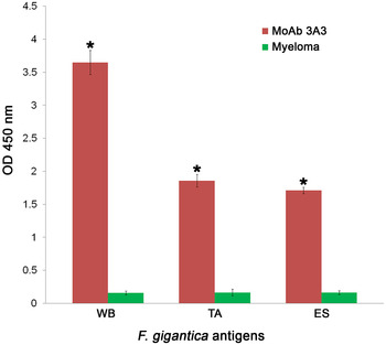
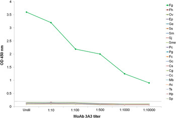
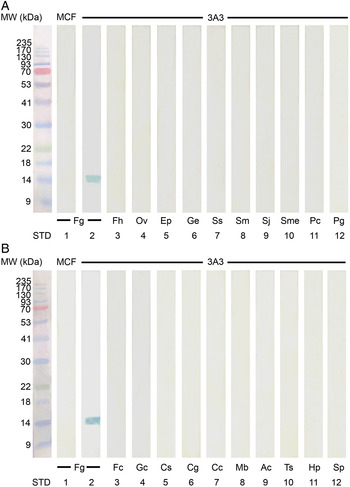
The native FgFABP in WB, TA and ES fractions were reacted with MoAb 3A3 and the relative levels of FgFABP in each fraction were assessed by indirect ELISA. The levels of reactivity of FgFABP in WB, TA and ES with MoAb 3A3 were significantly higher when compared with control myeloma culture fluid (Fig. 1). In both indirect ELISA (Fig. 2) and immunoblotting assay (Fig. 3A and B) were used for studying the specificity of MoAb 3A3. MoAb 3A3 exhibited strong reaction with FABP antigen in WB of F. gigantica at MW of 14·5 kDa, while showing no cross-reaction with WB antigens from 15 trematodes (F. hepatica, O. viverrini, E. pancreaticum, G. explanatum, S. spindale, S. mansoni, S. japonicum, S. mekongi, P. cervi, P. gracile, F. cobboldi, G. crumenifer, C. spatiosus, C. gregarious and C. cotylophorum), from two cestodes (M. benedeni and A. centripunctata), as well as from three nematodes (Trichuris sp., H. placei and S. labiato-papillosa).
The levels of the native FABP in WB, TA and ES fractions of adult F. gigantica was evaluated by its reactivity with MoAb 3A3 using indirect ELISA. Significant increase of OD values representing FABP levels were shown in WB, TA and ES antigens (red column) when compared with the control myeloma culture fluid (green column). The result was considered significant if P-value is less than 0·05 (P < 0·05) represented in asterisk (*). ELISA, enzyme-linked immunosorbent assay; FABP, fatty acid binding protein; OD, optical density; MoAb, monoclonal antibody; WB, whole body; TA, tegumental antigens; ES, excretory-secretory.

Mean ELISA OD values of cross-reactivities studies between MoAb 3A3 and the panel of antigen from various trematode, cestode and nematode parasites. Fg, F. gigantica; Fh, F. hepatica; Ov, O. viverrini; Ep, E. pancreaticum; Ge, G. explanatum; Ss, S. spindale; Sm, S. mansoni; Sj, S. japonicum; Sme, S. mekongi; Pc, P. cervi; Pg, P. gracile; Fc, F. cobboldi; Gc, G. crumenifer; Cs, C. spatiosus; Cg, C. gregarious; Cc, C. cotylophorum; Mb, M. benedeni; Ac, A. centripunctata; Ts, Trichuris sp.; Hp, H. placei; Sp, S. labiato-papillosa. These parasites’ antigens were allowed to react with undiluted MoAb culture fluid (Undil) and MoAb at dilution 1:10, 1:100, 1:500 1:1000 and 1:10 000. Horizontal dotted line is the cut off level, i.e. the mean OD of control sera plus 3 s.d. The OD values greater than this cut-off value are considered to be positive detection. ELISA, enzyme-linked immunosorbent assay; OD, optical density; MoAb, monoclonal antibody.

The specificity of MoAb 3A3 is tested with WB antigens from F. gigantica, other trematode, cestode and nematode parasites by using immunoblotting technique. (A) Immunoblot analysis showing the cross-reactivities of MoAb clone 3A3 with WB antigens from Fg, F. gigantica (lane 2); Fh, F. hepatica (lane 3); Ov, O. viverrini (lane 4); Ep, E. pancreaticum (lane 5); Ge, G. explanatum (lane 6); Ss, S. spindale (lane 7); Sm, S. mansoni (lane 8); Sj, S. japonicum (lane 9); Sme, S. mekongi (lane 10); Pc, P. cervi (lane 11); Pg, P. gracile (lane 12). (B) Immunoblot analysis of the cross-reactivities of MoAb 3A3 with WB antigens from F. gigantica (lane 2), Fc, F. cobboldi (lane 3); Gc, G. crumenifer (lane 4); Cs, C. spatiosus (lane 5); Cg, C. gregarious (lane 6); Cc, C. cotylophorum (lane 7); Mb, M. benedeni (lane 8); Ac, A. centripunctata (lane 9); Ts, Trichuris sp. (lane 10); Hp, H. placei (lane 11); Sp, S. labiato-papillosa (lane 12). Lane 1 of (A) and (B) is WB antigen from F. gigantica (Fg) blotted with the MCF, which is used as the negative control. STD is the lane containing standard protein molecular weight markers. MCF,myeloma culture fluid; MoAb, monoclonal antibody; WB, whole body.

The lower detection limit and the specificity of sandwich ELISA
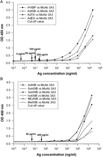
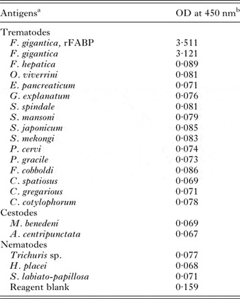
The ELISA cut-off value was 0·15. The lower detection limit of this assay was evaluated from the different concentrations of rFABP, WB, TA and ES antigens of adult F. gigantica. Based on the lowest concentrations of antigen that still exhibited the sensitivity of test, this test could detect rFABP and FABP in WB, TA and ES fractions of F. gigantica at the lowest concentrations of 5, 50, 100 and 200 pg mL−1, respectively (Fig. 4A). Likewise, the lowest dilution of WB in Met and NEJ antigens was at 400 pg mL−1, and for 1-, 3-, 5-week-old juveniles and adult antigens was at 50 pg mL−1 (Fig. 4B). Moreover, this sandwich ELISA described herein was highly specific for FABP antigen, as no cross-reactivity was demonstrated when the assay was employed to detect this antigen at various concentrations of other parasite antigens (Table 1).
The lowest concentrations of rFABP and FABP in WB of various stages of F. gigantica, TA and ES antigens of F. gigantica as examined by the sandwich ELISA assay. (A) Lines with black circle, white circle, black triangle and white triangle indicate the concentration levels of rFABP and FABP in WB, TA and ES antigens of adult F. gigantica. The arrows indicate the lowest concentrations of FABP that could still be detected. (B) Lines with black circle, white triangle, black square, white diamon, black triangle and white hexagon exhibit the concentration levels of FABP in WB fractions of adult, 5-, 3-, 1-week-old juveniles, NEJ and metacercariae (Met) of F. gigantica, respectively. The arrows indicate the lowest concentrations that FABP could still be detected in each sample. ELISA, enzyme-linked immunosorbent assay; rFABP, recombinant fatty acid binding protein; MoAb, monoclonal antibody; WB, whole body; TA, tegumental antigens; ES, excretory-secretory; NEJ, Newly excysted juveniles.

Specificity testing of a sandwich ELISA to various crude preparations from trematode, cestode and nematode parasite antigens

a The protein content of each parasite antigen preparation was adjusted to 20 µg mL−1, and a 50-μL volume was used for analysis.
b Mean OD was determined in triplicates performed on three separate occasions.
Application of sandwich ELISA for detection of circulating FABP antigen in sera from infected animals
Experimentally infected animals
At day 35 post infection, the morbidity rate of infected mice was 100% and no mortality has occurred throughout this study. The parasite recoveries varied from 5 to 19 worms per mouse with an average of 10·92 ± 4·17. For liver pathology, the liver were swollen and covered with large patches of fibrinopurulent exudates that later caused adhesion of all of the liver lobes with adjacent organs in the abdominal cavity. Likewise, numerous white tracks and foci were seen and appeared as irregular lines that scattered throughout the centre of the hepatic lobes.
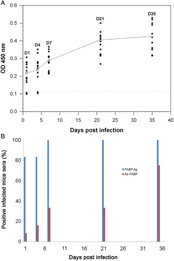
The mean ODs for antigen detection were significantly different from those of control sera at day 1, 4 (P < 0·05), 7, 21 and 35 (P < 0·01) post infection. The levels of detectable antigen in infected sera peaked at day 21 to day 35 post infection (Fig. 5A). The total numbers of F. gigantica metacercariae- infected mice sera and the numbers as well as the percentages of positive sera at day 1, 4, 7, 21 and 35 post infection were 83·33% (10 out of 12), 83·33% (10 out of 12), 100% (12 out of 12), 100% (12 out of 12) and 100% (12 out of 12), respectively (Fig. 5B).
Detection of circulating FABP antigens and antibody against FABP antigens of F. gigantica is evaluated by ELISA assay. (A) Detection of circulating FABP antigens of F. gigantica in the serum samples of the infected mice as compared with non-infected mice by sandwich ELISA. Black stars represent OD values of individual mouse serum; white squares indicate mean OD values of all mice in the experimental group. The horizontal dotted line denotes the cut-off level. Any values above this line are considered positive detection. (B) Comparison between detection of circulating FABP antigens using sandwich ELISA and antibody against FABP antigens using indirect ELISA in the sera of mice infected with F. gigantica. ELISA, enzyme-linked immunosorbent assay; FABP, fatty acid binding protein; OD, optical density.

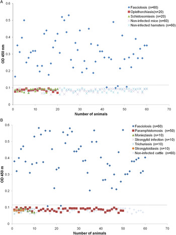
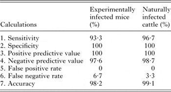
Additionally, sera from, 60 fasciolosis mice, 20 schistosomiasis mice, 20 sera opisthorchiasis hamsters, as well as 60 and 60 non-infected healthy mice and hamsters, were examined. The results revealed that 56 of 60 (93·3%) fasciolosis sera were positive, whereas all 160 (100%) sera from those with other infections and non-infected healthy animals were negative (Fig. 6A). Therefore, this assay showed a sensitivity and specificity of 93·3 and 100%, respectively, with a positive predictive value of 100%, a negative predictive value of 97·6%, false positive rate of 0%, false negative rate of 6·7% and an accuracy of 98·2% (Table 2).
Sandwich ELISA reactivity pattern denotes (A) the relative levels of circulating FABP antigens (OD values) in sera from mice infected with F. gigantica (fasciolosis), S. mansoni (schistosomiasis) and hamster infected with O. viverrini (opisthorchiasis). Serum samples from non-infected mice and hamsters were used as controls. The horizontal dotted line represents the cut-off level for a positive detection. (B) The relative levels of circulating FABP antigens (OD values) in sera from cattle naturally infected with F. gigantica (fasciolosis), P. cervi (paramphistomosis), M. benedeni (Monieziasis), Strongylids (strongylid infection), Trichuris sp. (trichuriasis), and Strongyloides sp.(strongyloidiasis). Sera from non-infected cattle were used as controls. The horizontal dotted line exhibits the cut-off level for a positive detection. ELISA, enzyme-linked immunosorbent assay; FABP, fatty acid binding protein; OD, optical density.

Calculation of diagnostic values of the sandwich ELISA for FABP antigen detection in sera of mice experimentally and cattle naturally infected with F. gigantica

Naturally infected animals
Two hundred and ten sera from cattle were collected and tested by sandwich ELISA. Sixty sera from cattle infected with fasciolosis, 50 from cattle infected with paramphistomosis, 10 from cattle infected with monieziasis, 10 from cattle infected with strongylid infections, 10 from cattle infected with trichuriasis, 10 from cattle infected with strongyloidiasis and 60 from non-infected cattle were examined. The results exhibited that 58 of 60 (96·7%) fasciolosis sera were positive, while all 90 sera from those infected with other parasites, and all 60 sera from non-infected controls were negative (Fig. 6B). Hence, the method showed a sensitivity and specificity of 96·7 and 100%, respectively, with a positive predictive value of 100%, a negative predictive value of 98·7%, a false positive rate of 0%, a false negative rate of 3·3% and an accuracy of 99·1% (Table 2).
Indirect ELISA for the detection of antibody against F. gigantica FABP
The numbers of serum samples from infected mice being tested and percentages of positive detection during the period of 1–35 days post infection were 8·3% (1 out of 12) at day 1, 16% (2 out of 12) at day 4, 33·3% (4 out of 12) at day 7, 33·3% (4 out of 12) at day 21 and 75% (9 out of 12) at day 35 (Fig. 5B). The mean OD for mouse antibody diagnosed fasciolosis at 35 days post infection is significantly different from that of the control sera OD (P < 0·05).
DISCUSSION
The problem of precisely diagnosing fasciolosis using the conventional parasitological tests, including microscopic detection of the fluke's eggs in the feces and detection of serum antibodies, has led to develop a more accurate diagnostic method. In this study, we have detected circulating F. gigantica FABP antigen in the sera from both F. gigantica experimentally infected mice and naturally infected cattle using a MoAb-based sandwich ELISA. The MoAb 3A3 specific to FgFABP exhibited no cross-reactivities with antigens of other adult parasites, including F. hepatica, O. viverrini, E. pancreaticum, G. explanatum, S. spindale, S. mansoni, S. japonicum, S. mekongi, P. cervi, P. gracile, F. cobboldi, G. crumenifer, C. spatiosus, C. gregarious, C. cotylophorum, M. benedeni, A. centripunctata, Trichuris sp., H. placei and S. labiato-papillosa. Hence, we could use this MoAb 3A3 to detect the circulating FABP antigen in both early and late stages of infection. In our earlier study, we have also produced MoAb specific to 28·5 kDa tegumental antigen (28·5 kDa TA), recombinant cathepsin B3 (rCatB3) and recombinant cathepsin L1 (rCatL1) of adult F. gigantica as well as developed reliable MoAb-based sandwich ELISA for diagnosis of fasciolosis by F. gigantica in experimentally infected mice and naturally infected cattle sera (Anuracpreeda et al. Reference Anuracpreeda, Wanichanon, Chawengkirtikul, Chaithirayanon and Sobhon2009a , Reference Anuracpreeda, Chawengkirtikul, Tinikul, Poljaroen, Chotwiwatthanakun and Sobhon2013a , Reference Anuracpreeda, Chawengkirttikul and Sobhon2016b ). Close to our findings, Viyanant et al. (Reference Viyanant, Krailas, Sobhon, Upatham, Kusamran, Chompoochan, Thammasart and Prasittirat1997) produced MoAb against TA of F. gigantica whose target antigen was recognized at MW of 66 kDa. However, they reported that cross reactivity with other parasite antigens from Paramphistomum sp., S. spindale, S. mansoni and S. mekongi was occurred and the MoAb clone was not stable. Fagbemi et al. (Reference Fagbemi, Aderibigbe and Guobadia1997) used MoAbs against whole worm antigens of F. gigantica for the diagnosis of fasciolosis in cattle; however, this MoAb exhibited more cross-reactivities with other antigens from P. microbothrium, Dicrocoelium hospes and Schistosoma bovis. Likewise, Arafa et al. (Reference Arafa, Abaza, El-Shewy, Mohareb and El-Moamly1999) reported that the diagnosis of human fasciolosis was developed using MoAbs against F. gigantica ES antigens at MW of 49·5 kDa. It was found that cross-reactivity with S. mansoni antigen occurred by using these MoAbs. Another study of F. gigantica revealed that MoAb against isoforms of cathepsin Ls of juvenile and adult fluke was used for diagnosis of animal fasciolosis (Wongwairot et al. Reference Wongwairot, Kueakhai, Changklungmoa, Jaikua, Sansri, Meemon, Songkoomkrong, Riengrojpitak and Sobhon2015).
In this study, the results indicated the advantage of using the rabbit anti-mouse IgG to precoat the plate, which helped to enhance the binding of MoAb 3A3, and hence also the binding to antigen. Consequently, the assay was able to detect rFABP and FABP antigens in WB, TA and ES fractions of F. gigantica at the concentrations as low as 5, 50, 100 and 200 pg mL−1, respectively. In addition, the FABP antigen could also be detected in WB of Met and NEJ at 400 pg mL−1, and in WB of 1-, 3-, 5-week-old juveniles and adult antigens 50 pg mL−1. Furthermore, the use of MoAb as the antigen-capturing antibody increased the specific binding of the PoAb, which could help to reduce the background and yielded very low detection limits of the assay. The detection limits of our assay are lower than those of Langley and Hillyer (Reference Langley and Hillyer1989) who detected F. hepatica ES antigens in serum samples of experimentally infected mice at a concentration 0·25 ng mL−1. Also, the detection limits are better than the results reported by Mezo et al. (Reference Mezo, Gonzalez-Warleta and Ubeira2004) who developed the capture ELISA for the detection of F. hepatica ES antigens in fecal supernatants of infected animals, and found that the detection limit was 0·3 (for sheep) and 0·6 ng mL−1 (for cattle). In our previous study, we have developed a sandwich ELISA for the detection of circulating 28·5 kDa TA in the serum samples of mice experimentally infected with F. gigantica. The result revealed that the lower detection limit was 600 pg mL−1 (for TA), 16 ng mL−1 (for WB antigen), and 60 ng mL−1 (for ES antigen), which are considerably higher than in the present study (Anuracpreeda et al. Reference Anuracpreeda, Wanichanon, Chawengkirtikul, Chaithirayanon and Sobhon2009a ). Likewise, a sandwich ELISA was used for detection of circulating CatB3 antigen in the sera of mice and cattle infected with F. gigantica with the lower detection limit at 10 (for rCatB3 antigen), 100 (for Met antigen) and 400 pg mL−1 (for NEJ antigen), which were still higher than in this study (Anuracpreeda et al. Reference Anuracpreeda, Chawengkirtikul, Tinikul, Poljaroen, Chotwiwatthanakun and Sobhon2013a ). Similarly, we have developed and used a sandwich ELISA for detection of rCatL1 and CatL1 in WB and ES fractions of F. gigantica with the lower detection limit at 3, 50 and 100 pg mL−1, respectively. The lowest detection limit for WB in Met, NEJ, 1-week-old juvenile antigens was at 100 pg mL−1, and for 3-, 5-week-old juveniles and adult antigens was at 50 pg mL−1 (Anuracpreeda et al. Reference Anuracpreeda, Chawengkirttikul and Sobhon2016b ). For human fasciolosis, the lowest detection limits reported herein are lower than that reported by Espino and Finlay (Reference Espino and Finlay1994) who used a sandwich ELISA to detect the F. hepatica ES antigens in stool samples of patients, and found that the lower detection limit was 15 ng mL−1, and that of Demerdash et al. (Reference Demerdash, Diab, Aly, Mohamed, Mahmoud, Zoheiry, Mansour, Attia and El-Bassiouny2011) who developed a sandwich ELISA to detect the F. gigantica ES antigens in both serum and stool samples of patients at a concentration level 3 ng mL−1. In the present study, the sensitivity and specificity of this MoAb-based sandwich ELISA were also considered very high as in the sera of experimentally infected mouse at 93·3 and 100%, while in the sera of naturally infected cattle they were 96·7 and 100%, respectively. In addition, the accuracy of this assay in the sera of naturally infected cattle was 99·1%, which is comparable with that of experimentally infected mouse at 98·2%. Our findings indicated that this sandwich ELISA could be successfully applied to naturally infected cattle with a large body size.
In the present study, we have shown that the circulating FABP antigen in the sera of mice experimentally infected with F. gigantica was detectable as early as the first day after infection, with the peak levels occurred during the day 21 and day 35. This data herein is correlated with the reports of our previous studies (Anuracpreeda et al. Reference Anuracpreeda, Chawengkirttikul and Sobhon2016b ). It is possible that the levels of the circulating antigen in infected serum samples were corresponded with the pattern of the parasites’ migration in the host. During the first few weeks post infection, the antigen was slowly and continually released into the hosts’ circulation. Once the worms reached and became mature stage in the biliary system of the liver, they might release a large amount of antigens into the hosts’ circulation. On the other hand, Langley and Hillyer (Reference Langley and Hillyer1989) who used rabbit hyperimmune serum to detect circulating F. hepatica ES antigen in the sera from experimentally infected mouse at the first week after infection. As well, Viyanant et al. (Reference Viyanant, Krailas, Sobhon, Upatham, Kusamran, Chompoochan, Thammasart and Prasittirat1997) who developed a sandwich ELISA for detection of circulating 66 kDa TA in the sera of cattle experimentally infected with F. gigantica at the first week post infection. Another detections were also reported by Fagbemi et al. (Reference Fagbemi, Aderibigbe and Guobadia1995) who developed and utilized a sandwich ELISA for detection of circulating 88 kDa F. gigantica antigen in the sera of experimentally infected cattle at the second and third weeks post infection, and Velusamy et al. (Reference Velusamy, Singh, Sharma and Chanda2004) reported that the circulating 54 kDa F. gigantica antigen was detected in the sera of experimentally infected cattle at the second week post infection.
Concluding remarks
The results obtained in this study clearly indicated that a reliable MoAb-based sandwich ELISA showed high efficiencies and precisions. This assay method could be used as an important diagnostic tool not only for both early and late detections of fasciolosis but also for the seroepidemiological screening of animals, which in turn could contribute to the monitoring and control of the disease from different areas.
SUPPLEMENTARY MATERIAL
The supplementary material for this article can be found at http://dx.doi.org/10.1017/S0031182016001104.
ACKNOWLEDGEMENTS
We are grateful to Assoc. Prof. Dr. David Piedrafita, Federation University, Australia, for providing F. hepatica antigen.
FINANCIAL SUPPORT
This research was financially supported by Research Grants from The Thailand Research Fund and Mahidol University to Panat Anuracpreeda.