Introduction
Exposure to maternal stress during gestation has been linked to a variety of negative pregnancy and postnatal outcomes broadly ranging from miscarriage to increased risk of developing non-communicable diseases in adulthood, including diabetes, cardiovascular disease and metabolic syndrome.Reference Barker 1 – Reference Edwards, Benediktsson, Lindsay and Seckl 4 The mechanisms through which maternal stress affects developmental trajectories leading to the outcomes described are being intensively investigated. Early fetal exposures to high glucocorticoid levels appear to stimulate cell differentiation at the expense of cell proliferation, resulting in fewer total cell numbers in many fetal tissues (e.g. brain, heart and skeletal muscle), intra-uterine growth restriction and reduced birth weight,Reference Seckl 3 – Reference Fowden and Forhead 5 which may account for some of the developmental outcomes observed.
The human fetus is partially protected from exposure to maternal cortisol by a nicotinamide adenine dinucleotide-dependent placental enzyme called 11 β-hydroxysteroid dehydrogenase type 2 (11β-HSD2), which oxidizes bioactive maternal cortisol into bio-inactive cortisone.Reference Edwards, Benediktsson, Lindsay and Seckl 4 , Reference Causevic and Mohaupt 6 In humans, placental 11β-HSD2 has been shown to be present in human pregnancy from as early as the middle of the first trimester until parturition (Table 1).Reference McTernan, Draper and Nicholson 7 – Reference Lopez Bernal and Craft 11 The onset of placental 11β-HSD2 expression, however, has yet to be clearly established. Filling in this knowledge gap is critically important as the earlier an exposure takes place, the larger and more widespread its effects are expected to be.
Cross-sectional studies of changes in placental 11β-HSD2 mRNA expression and activity across uncomplicated human pregnancy

11β-HSD2, 11 β-hydroxysteroid dehydrogenase type 2; mRNA, messenger RNA; PC, post-conception.
For consistency, all gestational periods are listed as approximate weeks PC. Conversion to weeks following the onset of women’s last menstrual periods=weeks PC+2 weeks.
Given the critical protective role this enzyme is hypothesized to have during gestation, we hypothesized that 11β-HSD2 would be expressed during the earliest stages of the placentation process in humans. To that aim, we performed immunocytochemical analysis of placentas collected at 5–8 weeks after the onset of women’s last menstrual period (LMP), which represent, approximately, 3–6 weeks post-conception (PC). Consistent with the hypothesis described above, we predicted that 11β-HSD2 should be expressed very early during placentation, and it should be observed in cells that will be in direct contact with maternal tissue. The placentation process begins at the time of implantation, around days 6–9 during week 2 PC in humans.Reference Baergen 12 Logistically, it is not possible to obtain proto-placental tissue from week 2 PC from naturally occurring pregnancies. Thus, our analyses of placentas from ~3 weeks PC provide the earliest assessment of placental 11β-HSD2 expression in human pregnancy to date (see Table 1). In vitro fertilization (IVF) models and animal studies will be necessary to explore mechanisms that may exist to protect the embryo from maternal cortisol during the first 2 weeks following conception.
Methods
Ethics
Specimen collection and processing protocols were approved by the local Institutional Review Boards under Helsinki convention guidelines. All of the women signed an informed consent (protocol #021-06-972) approved by the ethical committee of the Bnai Zion Medical Center, Haifa, Israel. Processing of de-identified specimens was approved by Yale University’s Human Investigation Committee (HIC protocol #1003006495). Analysis of secondary data was approved by Simon Fraser University’s Research Ethics Board (protocol # 2016s0151).
Subjects and specimens
In total, 18 de-identified, elective pregnancy terminations from otherwise healthy women, ranging from ~3–6 weeks PC (i.e. 5–8 weeks following the onset of LMP), were obtained from the Pathology Department at the Bnai Zion Medical Center (n=2, 4, 8 and 4, respectively in each week). All specimens were formalin-fixed within 5 min of evacuation, paraffin-embedded, serially sectioned (5 μm), and confirmed to be in the early stages of placentation via identification of invasive trophoblasts by pre-screening of the gestational endometrium through staining with hematoxylin and eosin.
Immunocytochemistry
Serial sections (5 μm) from paraffin-embedded tissues were mounted on glass slides previously coasted with a film of 1% poly-d-lysine (30,000–70,000 molecular weight; Sigma), dried for 30 min at <60°C, and stored at room temperatures until analysis. Immunohistochemical staining was performed using EnVision+System-HRP (DAB; Dako North America, Carpinteria, CA, USA) as previously described.Reference Kliman, Feinberg and Schwartz 13 Briefly, we used a polyclonal rabbit anti-human HSD11B2 IgG (HPA056385, Atlas Antibodies, Stockholm, Sweden) as the primary antibody, which has been shown to positively stain cells that are known to express 11β-HSD2, such as cells within human kidney tubules, placental syncytiotrophoblasts and glandular cells within the colon, but not cells that do not express 11β-HSD2, including cells within human kidney glomeruli, decidual cells in the placenta, and endothelial cells within the colon.Reference Uhlen, Fagerberg and Hallstrom 14 , 15 The sections were incubated overnight at 4°C with anti-human HSD11B2 (0.125 μg/ml). Negative control sections were incubated overnight at 4°C with normal rabbit serum (1 μg/ml; Sigma). Blocking, secondary and avidin–biotin complex steps were performed at 22–24°C for 45–60 min. All sections were counterstained with hematoxylin. The presence, location and extent of immunohistochemical staining for 11β-HSD2 was recorded semi-quantitatively by categorizing staining intensity (i.e. undetectable v. light to moderate v. intense) within specific placental features (e.g. villus syncytiotrophoblasts and uterine endometrial glands) and summing the number of features within each staining category. We used these values to estimate the percentage of placental features within each staining category.
Results
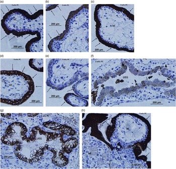
Immunohistochemical staining for human 11β-HSD2 protein expression was present in all specimens collected from 3–6 weeks PC (5–8 LMP). At 3 weeks PC, 11β-HSD2 was expressed by villus syncytiotrophoblasts, but it was absent from cytotrophoblasts and the villous core of chorionic villi (Fig. 1a). The same pattern of 11β-HSD2 protein expression was observed at 4, 5 and 6 weeks PC (Fig. 1b–1d, respectively). Although the vast majority of syncytiotrophoblasts (~98%) exhibited intense staining across early gestation (Fig. 1a–1d), light to moderate staining was observed in ~2% of specimens (e.g. Fig. 1e at 4 weeks PC), but this variation did not appear to be related to gestational age. Although light to moderate 11β-HSD2 staining was observed in the cytoplasm of ciliated columnar epithelial cells surrounding ~50% of uterine endometrial glands from weeks 3–6 PC (Fig. 1f and 1g), the remaining 50% of uterine endometrial glands did not exhibit staining for 11β-HSD2, yet, again, this variation did not appear to be related to gestational age. 11β-HSD2 staining was also not observed in the cytotrophoblasts or core of anchoring villi at 5 or 6 weeks PC, nor in the anchoring trophoblasts in the connecting cell column, early infiltrating trophoblasts or invasive trophoblasts within the maternal decidual basal plate, but was intense within the cytoplasm of surrounding syncytiotrophoblasts (Fig. 1h).
Placental 11 β-hydroxysteroid dehydrogenase type 2 (11β-HSD2) expression by villous and invasive trophoblasts. (a) The majority of 3-week post-conception (PC) chorionic villi exhibited intense syncytiotrophoblast cytoplasmic staining (arrows), whereas the villus core (VC) and cytotrophoblasts (arrow heads) did not. This pattern was similar for the majority of chorionic villi at (b) four (c) five and (d) 6 weeks PC. However, a small proportion of chorionic villi (2%) showed a different pattern, such as the one shown in this chorionic villi from 4 weeks PC (e), where 11β-HSD2 staining was light to moderate within the cytoplasm of syncytiotrophoblasts (arrows). (f) Light to (g) moderate 11β-HSD2 staining was observed in the cytoplasm of ciliated columnar epithelial cells (*) surrounding ~50% of three- to 6-week PC maternal uterine endometrial glands (UEG), as shown in these UEGs from 3 and 5 weeks PC, respectively. (h) Although intense 11β-HSD2 staining was observed in the cytoplasm of syncytiotrophoblasts (arrows) surrounding anchoring villi at 5 weeks PC, it was not observed in the VC, cytotrophoblasts (arrow heads), anchoring trophoblasts of the connecting cell column (CC), early infiltrating trophoblasts ({ }), or invasivpe trophoblasts (*) in the maternal decidual basal plate (BP).

Discussion
Recently we described how maternal cortisol increases immediately after conception.Reference Nepomnaschy, Salvante and Zeng 16 Early exposures to high cortisol levels appear to have important effects on fetal development with critical consequences for the postnatal phenotype in terms of health, cognitive capacities and behavior.Reference Barker 1 – Reference Edwards, Benediktsson, Lindsay and Seckl 4 , Reference Van den Bergh and Marcoen 17 , Reference Weinstock 18 Thus, as 11β-HSD2 is hypothesized to act as a placental barrier to maternal cortisolReference Edwards, Benediktsson, Lindsay and Seckl 4 , Reference Causevic and Mohaupt 6 , we predicted that its expression should begin early and should be most conspicuous in cells that are in intimate contact with the mother’s circulatory system.
Consistent with our predictions, 11β-HSD2 expression is observable in our earliest placental specimens at 3 weeks PC (5 weeks LMP) within placental villus syncytiotrophoblasts. At this time, maternal cortisol levels are gradually increasing with respect to their pre-conception levels.Reference Nepomnaschy, Salvante and Zeng 16 Our results are consistent with a small number of studies that evaluated 11β-HSD2 expression during very early pregnancy. McTernan et al.Reference McTernan, Draper and Nicholson 7 , for example, found relatively low levels of 11β-HSD2 messenger RNA (mRNA) in placental samples collected during weeks 4–6 PC. Lopez Bernal and CraftReference Lopez Bernal and Craft 11 also observed 11β-HSD2 activity in the placenta during the 5th and 6th week of human pregnancy, PC. McTernan et al. Reference McTernan, Draper and Nicholson 7 also found the expression of 11β-HSD2 mRNA to be similarly low during the late first trimester (7–10 weeks PC) compared with later in gestation (11–15 weeks PC). Furthermore, they reported a 12-fold increase in 11β-HSD2 mRNA expression by the early third trimester (25–32 weeks PC) and a 56-fold increase at term (35–40 weeks PC) compared with weeks 4–6 PC.Reference McTernan, Draper and Nicholson 7 Previous studies have reported low levels of 11β-HSD2 activity in the second half of the first trimester (weeks 5–10 PC).Reference Shams, Kilby and Somerset 10 , Reference Lopez Bernal and Craft 11 We were not able to evaluate 11β-HSD2 enzyme activity in the current study. Therefore, while 11β-HSD2 protein was expressed within the placenta as early as week 3 PC, it is still unknown whether this enzyme is metabolically active or whether its activity varies by gestational week during the first few weeks of placental development.
As is the case in humans, in non-human mammalian species 11β-HSD2 expression and activity has yet to be thoroughly investigated during very early gestation. Unlike in humans where 11β-HSD2 appears to increase across the whole gestational period, in the non-human mammalian species studied, placental expression of 11β-HSD2 mRNA peaks approximately two-thirds of the way through gestation, then declines towards term.Reference Fowden and Forhead 5 In mice, for example, placental 11β-HSD2 mRNA expression peaks at embryonic days 13–15 (E13–15), then declines significantly thereafter, remaining low through term (E19).Reference Condon, Ricketts, Whorwood and Stewart 19 Similar expression patterns have been observed in albino Wistar ratsReference Mark, Augustus, Lewis, Hewitt and Waddell 20 , rabbitsReference McArdle, Denton and Maduwegedera 21 and guinea pigsReference Sampath-Kumar, Matthews and Yang 22 , with placental 11β-HSD2 activity following the same pattern, but with a delay of 1–2 days.Reference Condon, Ricketts, Whorwood and Stewart 19 , Reference Sampath-Kumar, Matthews and Yang 22 Understanding this pattern will require a longitudinal exploration of maternal stress axis activity across gestation as well as the role of glucocorticoids in fetal maturation and the existence of complementary mechanisms that may modulate glucocorticoid exposure in each species. Non-human mammalian studies that include longitudinal profiles of placental expression of 11β-HSD2 and its activity from the very moment of conception may help us advance in our understanding not only of the current protective role this enzyme appears to play, but also of the evolution of its role in mammalian species in general and primates in particular.
In terms of the location of placental 11β-HSD2 expression, our observations in human specimens are consistent with previous studies that have primarily localized it within syncytiotrophoblasts slightly later during week 6 PC.Reference Shams, Kilby and Somerset 10 , Reference Krozowski, MaGuire and Stein-Oakley 23 Syncytiotrophoblasts invade the maternal endometrium during placentation and form the outer continuous surface of the placental villi where the majority of maternal–fetal exchange takes place. As humans exhibit hemochorial placentation, once the placenta is fully formed, maternal blood is in direct contact with syncytiotrophoblasts.Reference Baergen 12 As predicted, 11β-HSD2 expression was highest in these cells, which are properly positioned to act as a fetal barrier to maternal cortisol.
Maternal blood flow to the placenta is extremely limited until the end of week 6 PC when it begins to gradually increase, becoming fully established at approximately weeks 9–10 PC when the trophoblast plugs blocking the spiral arteries within the uterus degrade, allowing unrestricted maternal blood flow into the intervillous space.Reference Burton, Jauniaux and Watson 24 , Reference Jauniaux, Hempstock, Greenwold and Burton 25 Coincident with the changes in maternal blood flow, the placental environment transitions from a relatively anoxic to a normoxic environment that is comparable, in terms of oxygen pressure, to the surrounding endometrium.Reference Rodesch, Simon, Donner and Jauniaux 26 Before the establishment of maternal blood flow into the intervillous space, the uterine endometrial glands are an important source of nutrients for embryonic development. As early as day 17 PC (~2.5 weeks PC) and continuing throughout the first trimester, these glands secrete nutrients, including glycogen and proteins like glycodelin A and mucin-1, into the placental intervillous space, which are taken up into syncytiotrophoblasts by phagocytosis (histiotrophic nutrition).Reference Burton, Jauniaux and Charnock-Jones 27 The limited maternal blood flow during this period of development is likely to reduce the risk of overexposure of the embryo to maternal cortisol. However, whether cortisol is present in other maternal fluids that come in contact with the placenta or embryo, during this time, such as the secretions of the uterine endometrial glands, it is not known. We observed light to moderate 11β-HSD2 staining in the ciliated columnar epithelial cells encircling approximately half of uterine endometrial glands between 3 and 6 weeks PC (Fig. 1f and 1g). The presence of 11β-HSD2 within these cells suggest that this enzyme may help to inactivate maternal cortisol entering the glands before the release of their secretions into the intervillous space, thereby potentially protecting the embryo from excess maternal cortisol exposure during the first trimester. Further studies with larger sample sizes are needed to directly examine why only half of the uterine endometrial glands expressed 11β-HSD2 during very early gestation, how much this percentage varies among individuals, and whether this variation has an impact on developmental outcomes. Importantly, glucocorticoid metabolism is also influenced by the expression of 11 β-hydroxysteroid dehydrogenase type 1 (11β-HSD1). This enzyme reduces bio-inactive cortisone into bioactive cortisol.Reference Murphy and Clifton 8 Like 11β-HSD2, the onset, localization and extent of placental 11β-HSD1 expression during very early pregnancy has yet to be clearly established. 11β-HSD1 expression was not measured in the current study, preventing us from having a complete picture of early placental glucocorticoid dynamics.
In sum, 11β-HSD2 is expressed in the human placenta as early as 3 weeks after conception. The localization of 11β-HSD2 in areas where the majority of early maternal–fetal exchange occurs, that is, epithelial cells surrounding maternal uterine endometrial glands and syncytiotrophoblasts at the surface of the placental villi, is consistent with its hypothesized role as a protective barrier to modulate embryonic/fetal exposure to cortisol of maternal origin. It remains unclear whether the human embryo is sensitive to maternal cortisol during very early gestation. Recent studies in rats and mice suggest that murine blastocycts may have functional glucocorticoid receptors and may be sensitive to fluctuations in maternal glucocorticoid levels.Reference Kakourou, Jaroudi and Tulay 28 , Reference Korgun, Dohr and Desoye 29 A recent human study, however, did not observe glucocorticoid receptor gene (NR3C1) expression in blastocysts from IVF patients.Reference Kakourou, Jaroudi and Tulay 28 Additional studies are needed to further investigate whether the pre-implantation human embryo is responsive to maternal cortisol. Additional studies will also be necessary to evaluate the extent of 11β-HSD2 expression during the first 3 weeks immediately following conception. For example, evaluation of 11β-HSD2 mRNA expression in the transcriptomes of fertilized, pre-implantation embryos could help determine whether this protective enzyme is produced immediately after fertilization by the embryos themselves.Reference Reich, Klatsky, Carson and Wessel 30 The concurrent expression of 11β-HSD1 should also be evaluated across early gestation as the location of this enzyme’s expression and the extent of its activity will also influence cortisol dynamics and fetal development. Furthermore, future studies should also investigate the existence of other potential mechanisms that protect the embryo from maternal cortisol before the placenta can begin to produce 11β-HSD2. These studies would likely involve using animal or IVF models.
Acknowledgments
The authors thank all the staff in the Department of Pathology at the Bnai Zion Medical Centre for help with the collection and processing of the tissue specimens used in this study.
Financial Support
This study was funded by a Michael Smith Foundation for Health Research Career Investigator Scholar Award to PAN.
Conflicts of Interest
None.
Ethical Standards
The authors assert that all procedures contributing to this work comply with the Code of Ethical Conduct for Research Involving Humans, the Ethical Principles and Guidelines for the Protection of Human Subjects and the Helsinki Declaration of 1975, as revised in 2008, and has been approved by the institutional committees of the Bnai Zion Medical Center, Yale University and Simon Fraser University.