Introduction
Animal visual pigments, and the proteins on which they are based, have long served as model systems for learning how protein variation affects function and sensory phenotypes. Composed of a protein, opsin, coupled to a chromophore derived from a member of the vitamin A family, visual pigments are the light-sensitive molecules that initiate the visual signaling cascade and thus serve as the foundation on which visual sensitivity depends. Among different animals, a diversity of visual pigments, ranging in number from a single class to more than a dozen spectral types, can be expressed in a single retina. The quality of color vision and the ability of a given species to use the available spectrum of environmental light are determined both by the spectral diversity of visual pigments and by the particular wavelengths of maximal absorbance (λ max) to which they are tuned. For the purposes of this research, we define ultraviolet-sensitive visual pigments as those with λ max ranging from 300 to 400 nm, middle-wavelength-sensitive pigments as those with 400–490 nm, and long-wavelength-sensitive pigments as those with greater than 490 nm.
The evolution of the protein component of visual pigments (e.g., opsin) and its role in generating visual pigment spectral diversity has been investigated most extensively in three taxonomic groups: vertebrates, cephalopods, and insects (Morris et al., Reference Morris, Bowmaker and Hunt1993; Yokoyama, Reference Yokoyama2000; Bowmaker & Hunt, Reference Bowmaker and Hunt2006; Frentiu et al., Reference Frentiu, Bernard, Cuevas, Sison-Mangus, Prudic and Briscoe2007a). These taxa represent the two major groups of photoreceptors and correlated opsin classes: the vertebrate ciliary photoreceptors and opsins and the rhabdomeric photoreceptors and opsins found in cephalopods, arthropods, and other nonvertebrate groups. These groups also represent a range of variation both in numbers of expressed visual pigments and in numbers of spectral absorption, ranging from visual systems based on a single visual pigment (deep-sea fishes) to ones based on five or more (e.g., birds, dipterans). Comparative genomic studies in the mosquito Anopheles gambiae (12 opsin genes) and the fruit fly Drosophila melanogaster (8 opsin genes) as well as evolutionary expression studies in butterflies show that there has been considerable species-specific expansion via duplication of the opsin genes in insects (Briscoe, Reference Briscoe2001; Hill et al., Reference Hill, Fox, Pitts, Kent, Tan, Chrystal, Cravchik, Collins, Robertson and Zwiebel2002; Frentiu et al., Reference Frentiu, Bernard, Sison-Mangus, Brower and Briscoe2007b).
The most complex visual system known in terms of photoreceptor and spectral diversity is found in stomatopod crustaceans (mantis shrimps). Crustacean compound eyes are composed of separate optical units called ommatidia, each containing a group of eight photoreceptor cells. Based on microspectrophotometric and physiological characterization, the retinas of single stomatopod species contain up to 16 different spectral classes of photoreceptors. This large spectral diversity in the retina is due to a number of specialized ommatidial classes found only in stomatopods. In particular, up to six rows of ommatidia form an equatorial region called the midband, where tiered receptors with structural specializations contain several ultraviolet-sensitive receptor types, eight photoreceptor classes optimized for multichannel color vision, and a set of polarization-specific receptors. Additionally, the ommatidia outside the midband contain photoreceptors with a middle-wavelength class of visual pigments devoted to spatial vision (Cronin & Marshall, Reference Cronin and Marshall1989a, Reference Cronin, Marshall and Prete2004; Cronin et al., Reference Cronin, Marshall and Caldwell2000). Based on previous studies of the relationship between photoreceptor spectral sensitivity and visual pigment expression, we hypothesize that each spectral class of stomatopod photoreceptors expresses a different opsin gene and therefore a different visual pigment, leading to the expression of up to 16 different opsins in a single retina.
Stomatopods are particularly attractive for studying the evolution of visual pigment structure and function, as their ecology and visual physiology are well investigated, and the hypothesis of such a large number of visual pigments offers an unparalleled opportunity to track the genetic basis of visual system evolution. Although several recent studies have successfully expressed rhabdomeric opsins in in vitro systems, arthropod opsins are still extremely difficult to express in vitro, with honeybee ultraviolet- and blue-sensitive visual pigments the only examples to date (Koyanagi et al., Reference Koyanagi, Takano, Tsukamoto, Ohtsu, Tokunaga and Terakita2008; Terakita et al., Reference Terakita, Tsukamoto, Koyanagi, Sugahara, Yamashita and Shichida2008). It is the large number of hypothesized visual pigments, however, that makes stomatopods ideal for investigating the molecular mechanisms involved in the spectral tuning rhabdomeric visual pigments. Furthermore, there is variation throughout the order Stomatopoda in terms of habitat (shallow vs. deep marine) and at the level of eye complexity, with some species completely lacking the specialized midband, while others have midbands with two, three, or six ommatidial rows. This diversity at several levels allows for the comparison of opsin genes from multiple spectral classes within a single retina and from a single spectral class across species with varying habitats and eye complexities.
While stomatopod crustaceans contain the most complex visual system known in terms of photoreceptor diversity, this extreme example of vision has not been well characterized molecularly nor has its evolution from earlier arthropod ancestors been investigated. The extensive receptor spectral variation is thought to be founded on a similar level of opsin genes, but to date, only one study has investigated the underlying genetics of visual pigments in any species of mantis shrimp, characterizing 3 of the 16 expected opsin transcripts from the retina of the Caribbean species Neogonodactylus oerstedii (Brown, Reference Brown1996).
Here, we present the first large-scale investigation of opsin expression, diversity, and evolution in the stomatopod crustaceans. We selected five species for study, representing three of the seven recognized superfamilies of Stomatopoda, and amplified transcripts within their retinas using primers developed for typical crustacean middle- and long-wavelength-sensitive (M/LWS) opsins. Based on microspectrophotometric studies, stomatopods contain up to six photoreceptors with M/LWS visual pigments, and correspondingly, we expected that the degenerate primers used would isolate up to six unique opsin transcripts per species. To investigate the evolutionary history of stomatopod opsin diversity, a representative of each unique transcript was used in phylogenetic analyses. Finally, evolutionary trace analysis was used to identify sites that are divergent among stomatopod opsin groups, implying modes of functional diversification.
Materials and methods
Taxon sampling
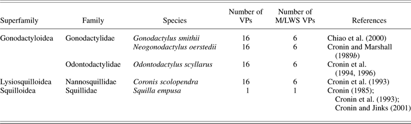
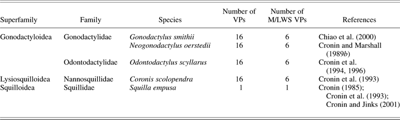
Determination of stomatopod phylogenetic relationships and taxonomic diversity followed the classification of Ahyong and Harling (Reference Ahyong and Harling2000). The five species used for this research included representatives from three of the seven extant superfamilies within the Stomatopoda (Table 1). These three superfamilies—the Gonodactyloidea, Lysiosquilloidea, and Squilloidea—contain ~87% of extant stomatopod species biodiversity. Furthermore, the chosen species span a range of taxonomic distances, so that sequence comparisons can be made across species within a family, families within a superfamily, and across superfamilies.
Summary of the taxonomy, the expected number of visual pigments (VPs) expressed in the retinas of stomatopod species characterized in this study, the expected number of middle- and long-wavelength-sensitive (M/LWS) VPs in each species, and the reference for the microspectrophotometric studies for each species leading to the hypothesized numbers
RNA extraction, polymerase chain reaction, and sequencing
We expected to recover a large amount of variation in opsin sequences because we were dealing with a large number of potentially similar genes in each species. In addition, we have no a priori hypotheses concerning the diversity of alleles for each gene. This poses a difficult situation to characterize using polymerase chain reaction (PCR) methods where the potential for artifacts among similar genes is high. Therefore, we have taken every precaution available to minimize the potential for error. Genetic variation among individuals was eliminated by using a single individual for each species. Because we expect a large amount of opsin diversity with high sequence similarity, the potential for PCR artifacts due to recombination (e.g., chimeras) is high (Bradley & Hillis, Reference Bradley and Hillis1997). The creation of PCR-generated chimeras is correlated with, among other factors, the type of polymerase used for amplification, the number of PCR cycles used, and the diversity of the sequence targets in the sample (Qiu et al., Reference Qiu, Wu, Huang, McDonel, Palumbo, Tiedje and Zhou2001). To minimize the occurrence of chimeric artifacts during PCR cycling, we used Taq polymerase, which has a lower percentage of recombination events than other types of polymerase (Zylstra et al., Reference Zylstra, Rothenfluh, Weiller, Blanden and Steele1998). The occurrence of chimeric artifacts and Taq incorporation errors were also minimized using phylogenetic analyses (see next section below). This methodology was verified using a high-fidelity polymerase as described below in a single species (N. oerstedii) for comparison of clone sequence variation. A final confirmation of our recovered sequence diversity was accomplished by designing gene-specific primers for a N. oerstedii sequence from each major phylogenetic group and amplifying full-length sequences for each selected sequence from complementary DNA (Fig. 1, underlined sequences). These full-length sequences were amplified using Taq polymerase and 30 cycles at 95°C for 10 s, 55°C for 30 s, and 68°C for 90 s.
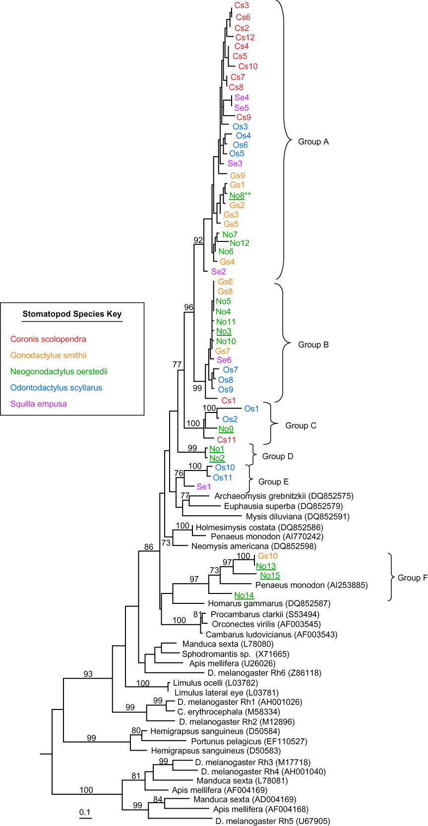
Phylogenetic tree of stomatopod and selected invertebrate opsins (out-groups not shown, and see text for out-groups used). Tree was reconstructed using a maximum likelihood analysis of amino acid sequences. Numbers on branches represent bootstrap proportions from 100 replicates; only bootstrap values above 70% are indicated, and for clarity, values are not shown in clades A and B due to short branch lengths. Opsin sequences from the five species investigated are labeled by a species designation (Cs, Coronis scolopendra; Gs, Gonodactylus smithii; No, Neogonodactylus oerstedii; Os, Odontodactylus scyllarus; Se, Squilla empusa) and a sequence number. The sequence used for three-dimensional homology modeling is indicated by asterisks, and N. oerstedii sequences independently confirmed using gene-specific primers are underlined.

The eyes of each target species were dissected to isolate the retinas, which were either immediately processed or preserved in RNAlater (Qiagen, Valencia, CA) and stored at −20°C until processing. Total RNA was isolated from whole retina using Trizol (Invitrogen, Carlsbad, CA) and used to generate cDNA using 3′ Rapid Amplification of cDNA Ends (RACE) methods. First-strand cDNA synthesis was performed using a poly(T) primer with an added adapter sequence (Oakley & Huber, Reference Oakley and Huber2004) and Superscript III Reverse Transcriptase (Invitrogen) according to the manufacturer’s instructions. Second-strand sequences were generated using HotMaster Taq (Eppendorf, Westbury, NY) with a primer for the adapter sequence and a degenerate primer designed from crustacean middle-wavelength opsin sequences (LWF1a: TGG TAY CAR TWY CCI CCI ATG AA; Porter et al., Reference Porter, Cronin, McClellan and Crandall2007) and the following PCR cycling parameters: 94°C (30 s) for denaturation, 49°C (30 s) for annealing, and 70°C (2 min) for extension for 45–50 cycles. These cycling parameters resulted in just detectable products of the expected size when run on 1% agarose gels and visualized with ethidium bromide. To screen for the possibility of the expression of multiple opsin genes, 3′ RACE products were purified using the MinElute Gel Extraction Kit (Qiagen) and cloned into the pCR2.1-TOPO vector using the TA Cloning Kit (Invitrogen). Colonies were screened for inserts by PCR using vector primers and the following PCR conditions: denaturation at 94°C for 1 min, primer annealing at 55°C for 1 min, and chain extension at 72°C for 1.5 min for 30 cycles. Amplified clone products of the expected size were purified using a QIAquick PCR Purification Kit (Qiagen). In order to sequence the entire insert, purified products were sequenced using the ABI Big Dye Primer Cycle Sequencing Ready Reaction Kit (Applied Biosystems, Foster City, CA) with the LWF1a primer and the 1080_mod2F internal degenerate opsin primer (TI MRI GAI CAR GCI AAR AAR ATG, modified from Oakley & Huber, Reference Oakley and Huber2004) on an ABI PRISM 3100 Automated Capillary Genetic Analyzer (Applied Biosystems, Foster City, CA) automated sequencer. All recovered clone sequences were confirmed to be an opsin by similarity searches using the GenBank BLAST software (http://www.ncbi.nlm.nih.gov/BLAST/). Confirmed opsin sequences were deposited in GenBank (accession numbers GQ221703-GQ221756).
The error rate for this methodology of characterizing transcript sequences was assessed by repeating the procedure with a high-fidelity proofreading polymerase for a single species, N. oerstedii. Briefly, cDNA was generated with the Accuscript high-fidelity reverse transcriptase (Stratagene, LaJolla, CA), and second-strand products were generated with the PrimeSTAR high-fidelity DNA polymerase (Takara Bio Inc., Madison, WI) and the following PCR conditions: initial denaturation at 95°C for 2 min, followed by 30 cycles at 95°C for 15 s, 55°C for 15 s, and 68°C for 90 s. Amplified products were cloned as previously described. Colonies containing inserts were grown in overnight cultures, and the plasmids were purified with the QIAprep Spin Miniprep Kit (Qiagen). Plasmid DNA was sequenced directly using the same primers and sequencing protocols as previously described. One hundred additional clones from this high-fidelity procedure were sequenced as previously described, and the variation and overlap in sequence diversity from a single individual were compared between the original and the high-fidelity methods. We found that these 100 clones from the high-fidelity methods recovered the same set of sequences as the original methods, which validates the original techniques (data not shown).
Phylogenetic methods
To account for the possibility of chimeras in our recovered sequences, we analyzed all the representative sequences from each individual species using the chimera detection program Bellerophon (Huber et al., Reference Huber, Faulkner and Hugenholtz2004). Sequences identified as possible chimeras were manually checked to confirm chimeric origins. Using this procedure, three sequences of the total recovered genes were identified as chimeric and removed from subsequent analyses. To estimate the potentially high Taq incorporation errors in our sequences, the high-fidelity clone sequences were compared to the original sequences obtained from N. oerstedii, a neighbor-joining phylogeny was constructed, and a percent sequence difference was calculated for each cluster of sequences on the tree. Using this approach, we calculated the Taq error to be ≤2%. To account for this potential error, all the isolated stomatopod opsin sequences for each species were analyzed using DOTUR v1.53 (Schloss & Handelsman, Reference Schloss and Handelsman2005), implementing the furthest neighbor-clustering algorithm to delineate sequence clusters at the 98% similarity level (e.g., a sequence distance of 2%). Each of these sequence clusters was considered to represent copies of a unique opsin transcript, and a single sequence from each cluster was chosen for use in subsequent analyses.
The representative sequences of each species were combined with other arthropod opsin sequences from GenBank, translated to amino acids, and aligned using ProbCons v1.08 (Do et al., Reference Do, Mahabhashyam, Brudno and Batzoglou2005), resulting in a final alignment of 330 amino acid positions. To root the tree, a range of out-groups were used, including cephalopod visual opsins (Loligo subulata—Z49108, Octopus dofleini—X07797, and Sepia officinalis—AF000947), vertebrate melanopsins (Homo sapiens—AF147788 and Mus musculus—AF147789), vertebrate visual (Bos taurus—NM_001014890) and nonvisual (Gallus gallus pinopsin—U15762) opsins, and G protein–coupled receptors (GPCRs) from the rhodopsin GPCR family (class A) that are closely related to the opsins (H. sapiens GPR52—NM_005684 and H. sapiens MTNR1A—NM_005958). The best fit model of protein evolution was determined using ProtTest v1.4 (Abascal et al., Reference Abascal, Zardoya and Posada2005), and an amino acid maximum likelihood tree was reconstructed using PhyML (Guindon & Gascuel, Reference Guindon and Gascuel2003; Guindon et al., Reference Guindon, Lethiec, Duroux and Gascuel2005). Branch support values were estimated from 100 PhyML bootstrap replicates as bootstrap proportions (BPs). BP values greater than or equal to 70% were considered strong support for a clade (Hillis & Bull, Reference Hillis and Bull1993).
Without information about the stomatopod genome, it is difficult to distinguish allelic diversity from gene copy diversity. The most similar duplicated opsin gene copies in model organisms exhibit amino acid similarities in the region from helix (H) I to HVIII, ranging from 75% (D. melanogaster Rh3 and Rh4) to 95% (human red and green cone opsins) to 100% (A. gambiae GPRop1, GPRop2, GPRop3, and GPRop4; Hill et al., Reference Hill, Fox, Pitts, Kent, Tan, Chrystal, Cravchik, Collins, Robertson and Zwiebel2002). Additionally, we are not aware of any studies that investigate allelic versus gene copy variation in invertebrate opsin genes, making predictions of the expected allelic variation difficult. In this study, we provide a conservative estimate of the number of opsin gene copies in the genome of each species by dividing the number of unique transcripts (with odd numbers rounded up to the next even number) in each monophyletic clade of stomatopod opsins by two (assuming two alleles per locus) and then summing this number across all monophyletic clades of stomatopod opsins in the phylogeny.
Duplicated gene functional constraints and divergence analyses
The functional divergence of stomatopod opsins was investigated using evolutionary trace analysis (Lichtarge et al., Reference Lichtarge, Bourne and Cohen1996). The evolutionary trace method identifies functionally important amino acid residues by evaluating patterns of sequence conservation across clusters of related proteins and has been useful in investigating opsin functional divergence in vertebrates as well as GPCR functioning in general (Madabushi et al., Reference Madabushi, Gross, Philippi, Meng, Wensel and Lichtarge2004; Carleton et al., Reference Carleton, Spady and Cote2005). Clusters are identified as monophyletic groups on a phylogeny. Within each cluster, every site was identified as either completely conserved among all proteins or variable if even one protein contained a different residue. These traces of each cluster were then compared, and each site was classified as conserved if the residue was identical at a site across all clusters, functionally divergent if the same within but different among clusters, or as not functionally important (e.g., variability in residue identity at a site results in no discernable pattern of conservation by cluster). In order to compare patterns of residue conservation among clusters of stomatopod opsin sequences, we defined six clusters of stomatopod opsins (see Results). Group D was excluded from the evolutionary trace analysis because it only contained two sequences, with an amino acid similarity of 98.2% (1.8% sequence difference). The remaining five groups each contained ≥15% sequence difference.
To investigate structural and functional impacts of divergence analyses, a homology model of a clade A stomatopod opsin (Fig. 1, sequence with asterisk) was constructed. The missing 5′ portion of the stomatopod opsin sequence chosen for modeling was completed using 5′ RACE methods and confirmed using a single set of primers designed to isolate the full-length opsin sequence, so that a full-length stomatopod opsin sequence was used to create the homology model. The stomatopod opsin homology model was constructed using the interactive mode of the 3D-JIGSAW v2.0 server (www.bmm.icnet.uk/servers/3djigsaw/; Bates & Sternberg, Reference Bates and Sternberg1999; Bates et al., Reference Bates, Kelley, MacCallum and Sternberg2001; Contreras-Moreira & Bates, Reference Contreras-Moreira and Bates2002) based on a crystal structure of the squid, Todarodes pacificus, rhodopsin at 2.5 Å (structure 2z73; Murakami & Kouyama, Reference Murakami and Kouyama2008). Since the constructed stomatopod homology model did not contain information on the orientation of the chromophore within the binding pocket, we aligned the new stomatopod structure with the original squid structure (2z73.pdb), and the chromophore location and orientation from the squid model were reproduced in our model for structural reference to the location of the binding pocket. Sites identified by evolutionary trace analysis were mapped onto the stomatopod homology model using Swiss-Pdb Viewer v3.9b1 (http://www.expasy.org/spdbv/; Guex & Peitsch, Reference Guex and Peitsch1997). Throughout the remaining text, amino acid site numbers will be given based on our alignment, followed by the bovine rhodopsin site number (indicated by Rho #) for comparison.
Results
Characterization of stomatopod opsins
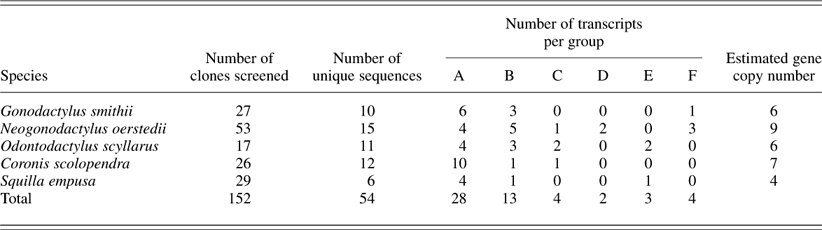
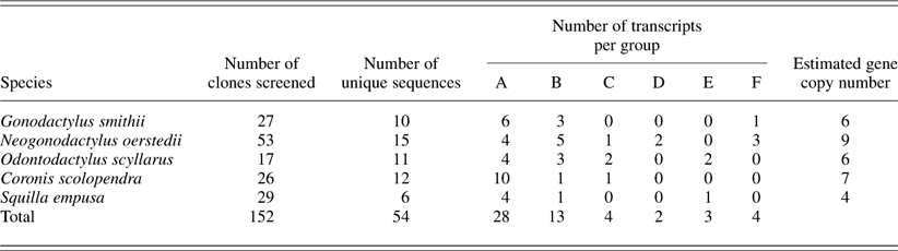
Transcripts of retinally expressed opsins from five stomatopod species were sequenced from the beginning of transmembrane HI through the end of the 3′ UTR. Because the C-terminus of the opsin protein and the 3′ UTR are highly variable and therefore unalignable, only the 305 amino acid residues encompassing HI through HVIII were included in subsequent phylogenetic and functional divergence analyses. The C-terminus and 3′ UTR sequences were useful, however, in confirming the uniqueness of the transcripts. We considered variation among transcripts in these regions as additional support for our methods of assessing clone uniqueness. In total, 152 clones were screened, resulting in 54 unique opsin transcripts among all the species (Table 2). Using primers designed from crustacean M/LWS opsins, the number of unique sequences found in a single species ranged from 6 (Squilla empusa) to 15 (N. oerstedii).
Summary of the number of clones screened per species, the number of unique stomatopod opsin transcripts found in each species investigated, and the distribution of the transcripts in each species among phylogenetically distinct groups (A–F)
All the sequenced transcripts in our five target species contained characteristic opsin features, including the chromophore-binding site (Rho K296), the (E/D)RY motif at the cytoplasmic end of HIII, and a glutamic acid at the site of the hypothesized ancestral counterion (Rho E181; Terakita et al., Reference Terakita, Koyanagi, Tsukamoto, Yamashita, Miyata and Shichida2004). The isolated transcripts also included typical rhabdomeric sequence features, such as a tyrosine at the vertebrate counterion site (Rho E113) and an ~14-amino acid indel in cytoplasmic loop (C) III (Porter et al., Reference Porter, Cronin, McClellan and Crandall2007). In phylogenetic analyses, all the isolated stomatopod sequences clustered with each other and with other crustacean opsins in a group sister to insect opsins that form long-wavelength-sensitive visual pigments (Fig. 1).
Patterns of gene duplication in stomatopod opsins
Within the main clade of crustacean opsins, our 54 unique stomatopod sequences form six well-supported monophyletic clusters (Fig. 1). Group A (BP = 92) contains the largest number of stomatopod opsin sequences (n = 28), followed by group B (BP = 99) with 13 sequences. Group C (BP = 100) is composed of only four sequences from three species (Coronis scolopendra, N. oerstedii, and Odontodactylus scyllarus). Group D (BP = 99) is the smallest group, composed of only two sequences from N. oerstedii, corresponding to two of the transcripts originally isolated by Brown (Reference Brown1996). Sequence groups A–D form a large monophyletic clade of only stomatopod opsins (BP = 64). Group E (n = 3, BP = 76), composed of sequences from two species (O. scyllarus and S. empusa), is clustered with opsins representing two additional orders of crustaceans (Euphausiacea and Mysida). Finally, group F is a well-supported cluster (BP = 97) containing four sequences from two stomatopod species (Gonodactylus smithii and N. oerstedii) as well as an opsin from a decapod shrimp (Penaeus monodon). Group F, together with additional decapod opsins, forms the basal lineage in the main clade of crustacean opsins. Our sequencing efforts did not recover representatives of all six opsin groups in any of the stomatopod species. Both groups A and B, however, contained transcripts from all five species investigated, while groups C–F consisted of sequences from three or fewer species. The largest number of expressed transcripts was observed in groups A and B, where many of the sequences form clusters of closely related sequences from single species.
In this study, up to 15 different opsin transcripts in the crustacean M/LWS group were identified from a single species. Using a conservative estimate of gene copy number, that is, our sequence variation represents two alleles at each locus, the number of opsin gene copies recovered in our set of species ranged from four (S. empusa) to nine (N. oerstedii), with three species containing more opsin copies than predicted (N. oerstedii, C. scolopendra, and S. empusa; Table 2). In particular, the recovery of four gene copies (six transcripts) distributed throughout three phylogenetically distinct sequence clusters from the retina of S. empusa is significant as based on studies of photoreceptor anatomy and spectral sensitivity, and S. empusa eyes were hypothesized to contain a single M/LWS visual pigment (Cronin, Reference Cronin1985).
Functional constraints and divergence in stomatopod opsins
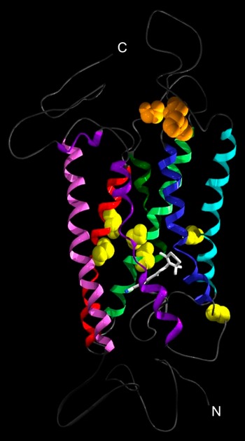
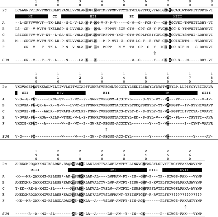
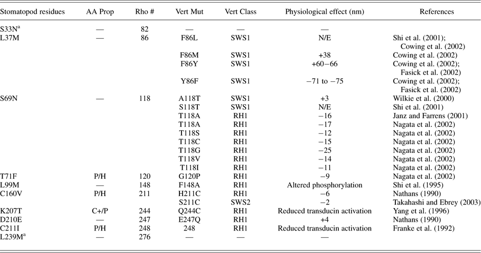
Based on evolutionary trace analysis, 29.4% of the amino acid residues in the region spanning from HI to HVII were conserved in all the currently characterized stomatopod opsin sequences. A total of 10 sites were identified as functionally divergent in stomatopod opsins (Table 3). These form two clusters, one of four sites at the cytoplasmic face of the protein and the other of six sites surrounding the chromophore-binding pocket (Fig. 2). The functional sites in the chromophore-binding pocket are located in HII (two sites), HIII (two sites), HV (one site), and HVI (one site) (Table 3). The stomatopod opsin homology model indicates that all six of these sites are on helical turns facing the binding pocket. The four sites on the cytoplasmic face of the opsin are clustered at loop–transmembrane boundaries in CII and CIII.
Three-dimensional homology model of a stomatopod opsin based on the 2.5 Å squid rhodopsin structure 2z73.pdb (Murakami & Kouyama, Reference Murakami and Kouyama2008). Residues identified as functionally divergent among stomatopod opsins within the chromophore-binding pocket are colored yellow, and those at the cytoplasmic surface are colored orange; for clarity, none of the side chains are depicted. Transmembrane helices are colored as follows: H1, pink; HII, red; HIII, light green; HIV, dark green; HV, light blue; HVI, dark blue; HVII and HVIII, purple. Nontransmembrane domains (e.g., loops, terminal regions) are colored gray. The ends of the protein have been indicated by “C” for C-terminal and “N” for N-terminal. The chromophore from squid rhodopsin 2z73.pdb is shown for reference.

Evolutionary trace analysis of stomatopod opsin groups A, B, C, E, and F (Fig. 1). The first row contains the opsin amino acid sequence from Procambarus clarkii (labeled Pc; accession number: S53494) as a representative crustacean opsin for comparison. The trace for each opsin group represents a consensus sequence for all of the transcripts within that group. If a particular site is classified as functionally divergent among opsin sequence groups, the residues are highlighted in grey; if an identified site also contains a replacement with significant differences in amino acid property, the site is highlighted in black. In the consensus sequence trace for each opsin group, conserved sites are indicated by an amino acid, dashes show the sites variable with respect to residue identity, ?s specify missing data, and a * designates a sequence gap. The summary trace represents the pattern of conservaed and variable residues among the traces of all the opsin groups. In the summary trace (SUM), functionally divergent sites are indicated by an “X”. The major opsin domains (HI–VIII), transmembrane helices; CI–III, cytoplasmic loops; EI–III, extracellular loops are indicated. The amino acid numbers correspond to our alignment of crustacean opsins, not to bovine rhodopsin numbers. However, the vertebrate counterion site (Y64 in this study), the hypothesized ancestral opsin counterion site (E132), and the Schiff base linkage (K258) are indicated by arrows
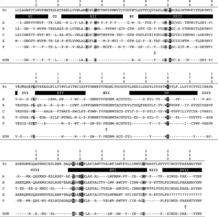
Of the 10 identified sites, 4 differentiate group F from the other stomatopod opsins (residues 37, 160, 207, and 210), with two of the four amino acid replacements corresponding to significant changes in amino acid properties (C160V and K207T). Two of these four sites are within the chromophore-binding pocket, both corresponding to sites previously identified as affecting spectral tuning in vertebrates (Table 4). The remaining two sites are at the cytoplasmic interface and have been shown to affect spectral tuning (D210E, +4 nm) and transducin activation (K207C) in vertebrate visual pigments (Table 4). Two sites (L99M and C211I) differentiate group C from other stomatopod opsins, with both sites found at the cytoplasmic interface of the protein. Both sites have been shown to be functionally important in bovine rhodopsin, affecting either phosphorylation or transducin activation (Table 4). The remaining sites differentiate groups A and B from C, E, and F (S33N) or groups A–C from E and F (S69N, T/F71C, and L/I239M). Although two of these sites have no known function in vertebrates, sites 69 and 71 both have been shown to affect spectral tuning. Additionally, in a comparative evolutionary study of pancrustacean opsin sequences, site 69 (Rho T118) was identified as a site under destabilizing positive selection (Porter et al., Reference Porter, Cronin, McClellan and Crandall2007).
The stomatopod opsin sites exhibiting functional divergence as identified by evolutionary trace analysis. The position and residues of each site, significant property differences (AA Prop), the homologous site number in bovine rhodopsin (Rho #), and previous mutational studies in vertebrates including the site mutated (Vert Mut), the spectral class affected (Vert Class), and the physiological effect (either spectral or related to signal transduction) are listed. Spectral effects are given as shifts in nanometer of the position of the λmax relative to the corresponding wild-type pigment. Amino acid properties are indicated as follows: P, polar; H, hydrophobic; C+, positively charged. N/E, mutation has no spectral effect
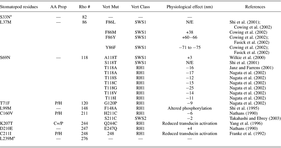
a No mutagenesis studies at homologous sites in vertebrates could be found for these sites.
Discussion
Patterns of gene duplication in stomatopod opsins
Stomatopods contain complex retinas hypothesized to contain up to 16 different visual pigments, with 6 of these having sensitivity to middle or long wavelengths of light. In this study, up to 15 different opsin transcripts in the crustacean M/LWS opsin class were identified from a single species. The isolated transcripts from each species form clusters of highly similar sequences, indicating that there have been many rounds of opsin gene duplication throughout the evolution of stomatopod visual systems, including many recently duplicated functionally similar copies that are still expressed in a single retina. Most of the recent duplication appears to be concentrated in only two of the identified stomatopod opsin clades, groups A and B. This pattern is similar to opsin expression in ostracods, where Oakley and Huber (Reference Oakley and Huber2004) found eye-specific opsin expression of multiple loci in the median versus the compound eyes. Within the isolated stomatopod opsin sequences, the most remarkable examples of multiple opsin expression within a retina are the S. empusa opsins. Relative to the most complex stomatopod eyes, S. empusa exhibits a reduced eye morphology containing only two midband rows. Furthermore, microspectrophotometric studies of S. empusa visual pigments implied that in the retina, all peripheral and midband photoreceptors contain a single visual pigment (Cronin, Reference Cronin1985). Our study, however, recovered six S. empusa opsin transcripts, which are distributed throughout three phylogenetically distinct sequence clusters. Conservatively, these transcripts represent four opsin copies in the genome. Because S. empusa contains at most three morphologically distinct photoreceptor types, these opsins must be coexpressed in some parts of the eye. Finding the expression of multiple opsin gene copies in a retina previously thought to contain a single visual pigment implies that the molecular components of the S. empusa visual system are unexpectedly complex. Similar unexpected opsin sequence diversity has been found in both the crab Hemigrapsus sanguineus (Sakamoto et al., Reference Sakamoto, Hisatomi, Tokunaga and Eguchi1996) and the deep-sea lophogastrid Gnathophausia ingens (Frank et al., Reference Frank, Porter and Cronin2009), where in both species, two expressed opsin copies were found in photoreceptors without physiological evidence for the presence of multiple visual pigment spectral types. In contrast to insect opsins, multicopy opsin expression within a single photoreceptor, possibly of many recently duplicated loci, may represent a common occurrence in crustacean visual systems. The functional consequence for crustacean vision of multiple opsin expression in single photoreceptors is one that requires further research.
The stomatopod opsin clades also do not cluster together. In particular, the phylogenetic position of clade F at the base of the crustacean opsin cluster indicates that many of the gene duplication events leading to the visual pigment diversity in extant species are the product of ancient duplications early in crustacean evolution before the divergence of stomatopods, mysids, decapods, and euphausiids. This is in agreement with the hypothesis of Briscoe and Chittka (Reference Briscoe and Chittka2001) that the major pattern of crustacean visual systems containing one or two visual pigments represents a derived state, with copies of the gene having been lost.
Functional constraints and divergence
Based on evolutionary trace analysis, 29.4% of the amino acid residues are conserved in all the stomatopod opsin sequences, indicating that roughly a quarter of amino acid sites are highly constrained, probably in order to maintain proper protein function. Of the variable residues, 10 sites forming two distinct clusters within the protein exhibited patterns of amino acid difference consistent with functional divergence in stomatopod opsins, that is, residue identity was conserved within a group but varied among groups. The two distinct clusters of divergent sites—one in the chromophore-binding pocket and one at the cytoplasmic face—indicate two different types of potential functional divergence for stomatopod opsins. Identified residues in the chromophore-binding pocket are likely to be involved in tuning spectral sensitivities of the resulting visual pigments. Of the six sites within the chromophore-binding pocket, four correspond to homologous sites in vertebrate opsins that have been shown to affect spectral tuning via site-directed mutagenesis in vitro expression studies, controlling tuning in vertebrate short-wavelength-sensitive cone opsins (sites 37, 69, and 160) and in vertebrate rod opsins (sites 69, 71, and 160) (Table 4). Site 69 (Rho T118) has been identified previously in an evolutionary trace analysis of vertebrate, mollusk, and arthropod visual opsins with bioamine, chemokine, and olfactory receptors by Madabushi et al. (Reference Madabushi, Gross, Philippi, Meng, Wensel and Lichtarge2004) as a residue of global importance to GPCRs affecting ligand binding and by a study of pancrustacean opsins that used comparative methods to identify this residue to be under positive destabilizing selection for the amino acid property of compressibility (Porter et al., Reference Porter, Cronin, McClellan and Crandall2007). This implies that some of the same sites are important in the function and spectral tuning of both ciliary and rhabdomeric opsins, as has been illustrated previously in the evolution of butterfly opsins (Briscoe, Reference Briscoe2001). The two remaining sites (33 and 239) have not been identified in previous studies of vertebrate spectral tuning, implying they are potentially novel sites for tuning crustacean visual pigments. Interestingly, four of the sites within the chromophore-binding pocket occur in clusters of two potentially interacting residues. The sites in these clusters occur within 4 Å of each other, with sites 33 and 37 occurring ~1 helical turn apart and sites 69 and 71 occurring ~1/2 a helical turn apart. These clusters suggest that some sites may tune the spectral sensitivity of crustacean visual pigments in a coordinated manner.
The second potential region of stomatopod opsin functional divergence occurs at the cytoplasmic interface, where the four identified sites are clustered around the loop–transmembrane boundary in both CII and CIII. This portion of rhabdomeric opsins is unique, in that there is an ~14-amino acid indel within CIII not present in ciliary opsins (Porter et al., Reference Porter, Cronin, McClellan and Crandall2007). Elucidation of the crystal structure in squid rhodopsin demonstrates that, unlike bovine rhodopsin, transmembrane HVI extends into the cytoplasmic medium as a rigid structure that interacts with the C-terminal end of the protein (Murakami & Kouyama, Reference Murakami and Kouyama2008). In vertebrates, structural and mutational studies of CII and CIII suggest that these loops play a role in metarhodopsin II interactions with the cognate G protein and rhodopsin kinase (Konig et al., Reference Konig, Arendt, McDowell, Kahlert, Hargrave and Hofmann1989; Franke et al., Reference Franke, Konig, Sakmar, Khorana and Hofmann1990, Reference Franke, Sakmar, Graham and Khorana1992; Palczewski et al., Reference Palczewski, Buczylko, Kaplan, Polans and Crabb1991; Borjigin & Nathans, Reference Borjigin and Nathans1994; Shi et al., Reference Shi, Osawa, Dickerson and Weiss1995; Yang et al., Reference Yang, Farrens, Hubbell and Khorana1996), thermal stability, and denaturation of opsin proteins (Landin et al., Reference Landin, Katragadda and Albert2001) and may form intrarow contacts among rhodopsin dimers (Liang et al., Reference Liang, Fotiadis, Filipek, Saperstein, Palczewski and Engel2003). Furthermore, the residues at the loop–helix interface appear to be the most important for transducin activation (Natochin et al., Reference Natochin, Gasimov, Moussaif and Artemyev2003). Emphasizing the functional importance of the CIII–HVI boundary, studies of HVI demonstrate that light activation causes a movement of this helix (Altenbach et al., Reference Altenbach, Yang, Farrens, Farahbakhsh, Khorana and Hubbell1996) and that key residues in HVI influence conformation of CIII and affect opsin folding, assembly, and/or function (Ridge et al., Reference Ridge, Ngo, Lee and Abdulaev1999). The concentration of sites in these domains of the opsin protein suggests that the second type of stomatopod opsin functional divergence is related to signal transduction via the movement of the cytoplasmic end of HVI and metarhodopsin II function. In particular, group C contains one unique residue at site 211 (HVI), while group F has two unique residues, all clustered at the cytoplasmic interface CIII–HVI, with the group C site (211) and one group F site (207) exhibiting significant changes in amino acid property.
Conclusions
Based on previous morphological and spectral characterization of stomatopod retinas, up to six different expressed M/LWS visual pigments were predicted to exist in most species. In this study of opsin molecular diversity across a broad taxonomic range of species, however, we show that stomatopods have many more expressed transcripts than expected for this opsin class, which we argue is the result of both recent (within the stomatopod lineage) and ancient (within the stem crustacean lineage) gene duplication events. Much of the recent gene duplication activity characterized here is concentrated in two clades (groups A and B) of stomatopod opsins. This pattern suggests that there are large numbers of opsin genes in the genome and that the total number of expressed retinal opsins is much higher than the number of known physiological photoreceptor classes. Furthermore, the characterized M/LWS stomatopod opsins form six main clades, distributed throughout the known crustacean opsins, indicating that some of the characterized molecular diversity is the result of ancient crustacean gene duplications. The currently diagnosed clades exhibit two possible types of functional divergence: sites diverging with respect to spectral tuning and sites diverging with respect to signal transduction interactions. It is highly likely that as the characterization of stomatopod opsin molecular diversity continues, the number of expressed transcripts will continue to increase. Future studies will focus on mapping the expression of these clusters of opsin copies to particular photoreceptor classes, to link sequence diversity with spectral sensitivity, and to determine if particular photoreceptors exhibit the expression of more than one opsin copy. This study of stomatopod opsin molecular diversity provides the first characterization of the gene duplication events and protein specializations required to construct a visual system containing high photoreceptor diversity, including many different spectral sensitivity classes.
Acknowledgments
We would like to thank R. Caldwell for help in acquiring specimens and K. Arikawa, K. Carleton, K. Dittmar de la Cruz, C. Hofmann, and M. Pérez-Losada for insightful comments on how to improve this manuscript. This study was supported by grants from the National Science Foundation (IOS 0721608) and by the Air Force Office of Scientific Research (02NL253).