Introduction
Patients with major depressive disorder (MDD), when compared to healthy controls (HCs), recall fewer specific autobiographical memories (AMs), defined as episodic memories of personally experienced events, and instead recall more categorical AMs, defined as summaries of recurring events (Williams et al. Reference Williams, Barnhofer, Crane, Hermans, Raes, Watkins and Dalgleish2007). This effect persists despite remission of depressive symptoms in patients with recurrent depression (Mackinger et al. Reference Mackinger, Pachinger, Leibetseder and Fartacek2000; Nandrino et al. Reference Nandrino, Pezard, Poste, Reveillere and Beaune2002; Spinhoven et al. Reference Spinhoven, Bockting, Schene, Koeter, Wekking and Williams2006), seems to be unrelated to symptom severity (Wessel et al. Reference Wessel, Meeren, Peeters, Arntz and Merckelbach2001; Gibbs & Rude, Reference Gibbs and Rude2004) and manifests in healthy individuals at high familial risk for developing depression (Young et al. Reference Young, Bellgowan, Bodurka and Drevets2013a ). These results suggest that overgeneral AM retrieval may act as a vulnerability factor influencing the development of MDD (Brittlebank et al. Reference Brittlebank, Scott, Williams and Ferrier1993; Young et al. Reference Young, Bellgowan, Bodurka and Drevets2013a ).
Neuroimaging studies of AM in depressed MDD samples (dMDD) compared to controls report increased activity in the dorsolateral prefrontal cortex (DLPFC) during negative AM recognition of memories recalled prior to scanning and cued again during functional magnetic resonance imaging (fMRI) (Keedwell et al. Reference Keedwell, Andrew, Williams, Brammer and Phillips2005; Whalley et al. Reference Whalley, Rugg and Brewin2012), increased activity in the ventrolateral PFC (VLPFC) during positive AM recognition (Keedwell et al. Reference Keedwell, Andrew, Williams, Brammer and Phillips2005), decreased activity in the hippocampus/parahippocampus and anterior insula during AM recall in which memories are recalled during scanning (Young et al. Reference Young, Erickson, Nugent, Fromm, Mallinger, Furey and Drevets2012), and increased activity in the medial frontal gyrus, pregenual anterior cingulate cortex (ACC) and lateral orbitofrontal cortex (OFC) during specific AM recall (Young et al. Reference Young, Bellgowan, Bodurka and Drevets2013a ). Although functional anatomical correlates of AM recall in subjects remitted from MDD (rMDD) have not been assessed previously, functional neuroimaging studies of other cognitive domains in rMDD samples show altered activity in structures implicated in encoding and recalling affective stimuli. For example, rMDDs show decreased activity compared to controls in the DLPFC while viewing positive faces (Norbury et al. Reference Norbury, Mannie and Cowen2011), in the hippocampus/parahippocampus during episodic memory testing (Milne et al. Reference Milne, MacQueen and Hall2012), and in the ACC and inferior frontal and medial PFC during encoding of positive words (Arnold et al. Reference Arnold, Fitzgerald, Fernandez, Rijpkema, Rinck, Eling, Becker, Speckens and Tendolkar2011). Additionally, during engagement of emotional regulation strategies, rMDD relative to control subjects show increased blood oxygen level-dependent (BOLD) activity in the lateral OFC and ACC (Kanske et al. Reference Kanske, Heissler, Schonfelder and Wessa2012).
Although behavioral studies suggest that similar abnormalities in AM processing are evident in rMDDs and dMDDs, no study has specifically examined the functional correlates of AM recall in rMDDs. Delineating the functional anatomical correlates of AM deficits in unmedicated rMDDs holds the potential to identify abnormalities in MDD that persist irrespective of depressive symptoms. Therefore, the aim of the current study was to assess differences in the neurophysiological correlates of AM recall among dMDD, rMDD and HC participants. This assessment thus provides data that complement those obtained in our previous study (Young et al. Reference Young, Bellgowan, Bodurka and Drevets2013a ) of the neural correlates of AM recall in healthy individuals at high familial risk for developing MDD, which identified potential trait-like biomarkers present prior to the onset of MDD. We predicted that dMDDs and rMDDs would exhibit similar abnormalities while recalling AMs, such that hemodynamic activity would increase in prefrontal cortical regions implicated previously in emotional control and rumination including the OFC and ACC (Bush et al. Reference Bush, Luu and Posner2000; Hynes et al. Reference Hynes, Baird and Grafton2006), but decrease in hippocampal and medial temporal regions typically recruited by HCs during AM (Svoboda et al. Reference Svoboda, McKinnon and Levine2006).
Method
Participants
Right-handed individuals between ages 18 and 55 years were evaluated for eligibility to enter one of three groups: unmedicated adults with MDD in a current major depressive episode according to DSM-IV-TR (APA, 2000), unmedicated patients remitted from MDD as defined by DSM-IV-TR criteria plus a Hamilton Rating Scale for Depression (HAMD) score < 7 (Frank et al. Reference Frank, Prien, Jarrett, Keller, Kupfer, Lavori, Rush and Weissman1991; APA, 2000), and psychiatrically healthy controls who were gender matched to the depressed groups. Volunteers, recruited from the community through advertisements, underwent medical and psychiatric screening evaluations at the Laureate Institute for Brain Research, which included the Structural Clinical Interview for DSM-IV Disorders (SCID-I/P; First et al. Reference First, Spitzer, Gibbon and Williams2002) and an unstructured diagnostic interview with a psychiatrist.
Exclusion criteria for all subjects included current pregnancy, general MRI exclusions, serious suicidal ideation, psychosis, major medical or neurological disorders, exposure to any medication likely to influence cerebral function or blood flow within 3 weeks, and meeting DSM-IV criteria for drug/alcohol abuse within the previous year or for alcohol/drug dependence (excepting nicotine) within the lifetime. Additional exclusion criteria applied to rMDDs were the presence of any depressive symptom severe enough to impair function or use of a psychotropic drug within 3 months (APA, 2000). HC specific exclusions were current or past history of any major psychiatric disorder, or having a first-degree relative with a mood disorder. After complete study explanation, all participants provided written informed consent as approved by the Western Institutional Review Board (WIRB). Participants received financial compensation for their participation. Although we previously published results obtained in controls and dMDDs using the same experimental paradigm applied in this study, all of the data reported here were obtained from entirely independent subject samples whose data have not been included in our previous publications (Young et al. Reference Young, Bellgowan, Bodurka and Drevets2013a , Reference Young, Bellgowan, Bodurka and Drevets b ).
Intelligence testing was performed using the two-subtest version of the Wechsler Abbreviated Scale of Intelligence (WASI; Wechsler, Reference Wechsler1999). Anxiety and depressive symptoms were rated on the scanning day using the State–Trait Anxiety Inventory (STAI; Spielberger et al. Reference Spielberger, Gorsuch and Lushene1970), the 21-item HAMD (Hamilton, Reference Hamilton1960), the Montgomery–Asberg Depression Rating Scale (MADRS; Montgomery & Asberg, Reference Montgomery and Asberg1979), the Snaith–Hamilton Pleasure Scale (SHAPS; Snaith et al. Reference Snaith, Hamilton, Morley, Humayan, Hargreaves and Trigwell1995) and the Profile of Mood States (POMS; McNair et al. Reference McNair, Lorr and Dropplemen1971).
AM task
Details of the fMRI data acquisition parameters and AM task have been reported previously (Young et al. Reference Young, Bellgowan, Bodurka and Drevets2013a , Reference Young, Bellgowan, Bodurka and Drevets b ). In brief, BOLD fMRI was performed on a 3-T General Electric (GE) Discovery MR750 scanner and eight-channel receive-only head coil. Participants were presented with an emotionally valenced or neutral cue word (60 cues; 20 of each valence) and instructed to recall a past experience, then rate their retrieved memory on specificity and valence. AM recall was compared to a semantic example generation condition in which participants were presented with an emotionally valenced or neutral cue word (30 cues; 10 of each valence) and instructed to think of at least seven examples from the presented category, and then rate the ease with which they generated examples and the number of examples generated. Cue presentation was randomized with the restriction that no two cues from the same valence could be presented sequentially. Following each cue presentation and set of ratings, participants engaged in a riser detection task involving non-word letter strings as a control for visual input/attention (see Supplementary Fig. S1 for a detailed task outline).
Following the scan, participants were presented with all AM cue words and asked to describe the memory for experimenter K.D.Y. to corroborate participants' specificity ratings. The experimenter was blind to diagnosis at the time of rating. In addition to standard memory categorizations of specific (memory for an event that occurred at an identified place and lasted up to 1 day), categorical (category of events containing several episodes without reference to one specific event), extended (extended period without reference to a specific event within the time frame) and semantic (statement of fact without an associated event) (Williams & Dritschel, Reference Williams and Dritschel1988; Williams et al. Reference Williams, Barnhofer, Crane, Hermans, Raes, Watkins and Dalgleish2007), a memory was categorized as ‘can't recall’ if the participant was unable to recall the memory retrieved during fMRI. At this time, participants also used a five-point scale to verbally rate each memory on arousal (very low to very high), vividness (not at all to perfectly clear) and age when memory occurred (childhood, adolescence, after age 18 but greater than 1 year prior to scan, 6 months to 1 year prior to scan, less than 6 months prior to scan).
Assessment of behavioral performance during fMRI
Behavioral data were analyzed using SYSTAT 13 (Systat Software Inc., USA). Potential group differences in age, IQ, depression and anxiety ratings, performance on the riser detection and example generation control tasks, and percentage of memories recalled at each specificity level (specific, categorical, extended, semantic, no memory, can't recall post-scan) were assessed using a one-way ANOVA (entering diagnosis as the independent variable and demographic, mood and specificity variables as dependent variables) and follow-up independent t tests.
Our a priori hypothesis was that rMDDs would exhibit the same AM deficits as previously reported in dMDDs, namely fewer specific and more categorical AMs, and fewer positive AMs relative to controls.
Four repeated-measures ANOVAs were performed to examine group differences in properties of recalled AMs. In each case the between-subjects variable was Diagnosis and the repeated measure was memory Type (specific, categorical). The other repeated measure entered was either Valence (positive, negative, neutral), Arousal (low, medium, high), Vividness (low, medium, high) or Age (childhood, adolescence, adulthood more than 1 year ago, 6 months to 1 year prior to scan, and less than 6 months from scan) for the dependent variable Percent of Memories recalled. Follow-up independent t tests were conducted for significant results within the ANOVAs. The threshold criterion for significance was set at p < 0.05, and post-hoc tests were corrected for multiple comparisons (Bonferroni).
Image processing and analysis
Analysis of fMRI data were performed using AFNI (http://afni.nimh.nih.gov/afni) and consisted of slice acquisition timing correction, within-subject image realignment, co-registration between anatomical and functional images, spatial normalization to the stereotaxic array of Talairach & Tournoux (Reference Talairach and Tournoux1988), and spatial smoothing (4-mm full-width at half-maximum Gaussian kernel). Using 3dDeconvolve for each participant, the evoked hemodynamic response to each event type was modeled as a boxcar function convolved with a cannonical hemodynamic response function. Regressors modeling task and motion parameters were used in the model. The main effects of interest were the 12-s cue word presentations that prompted specific memory recall and example generation. In addition to modeling main effects, each design matrix included regressors modeling rating selection, cue presentation where the memory was not recalled post-scan, cue presentation where non-specific memories were recalled, and even/odd riser question presentations. The non-word letter strings used as stimuli for the riser detection task were modeled as the baseline.
At the group level, 3dANOVA was used to identify regional differences in the BOLD signal between HCs, rMDDs and dMDDs for Specific Memories versus Example Generation and Example Generation versus Riser Baseline. Because the only behavioral difference found between groups was for the valence model, additional contrasts comparing Positive Specific Memories to Positive Example Generation and Negative Specific Memories to Negative Example Generation were also performed. An additional test was performed to compare the BOLD response for each contrast for all three groups combined. The significance criterion for detecting differences was set at p corrected < 0.05 determined using 3dClustSim (cluster size > 30 voxels, thresholded at voxel p < 0.005).
Correlation analysis
For each brain region showing a differential response during fMRI, β weights were extracted for each participant and region. Pearson correlations corrected for multiple comparisons (Bonferroni) were performed to detect associations between the brain imaging and behavioral/clinical data (percentage specific, categorical, positive, negative, recent AMs and HAMD scores). A correlation analysis was also performed between HAMD and AM scores.
Results
Behavioral results
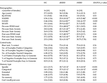
Table 1 lists group demographic and clinical characteristics. The one-way ANOVA showed no significant difference between groups in age or IQ (F 2,45 < 2.20, p > 0.15), but differences were evident for mean MADRS, HAMD, POMS, SHAPS and State and Trait anxiety scores (F 2,45 > 3.74, p < 0.05). dMDDs had higher scores on all ratings compared to both HCs (t 30 > 4.41, p < 0.001) and rMDDs (t 30 > 3.05, p < 0.005) whereas HCs and rMDDs differed on their MADRS and HAMD scores (t 30 > 2.41, p < 0.020) but not on any other rating (t 30 < 1.75, p < 0.090). MADRS and HAMD scores of rMDDs were in the non-depressed range whereas those in dMDDs were in the moderately to severely depressed range. Supplementary Table S1 lists additional clinical characteristics of MDD participants. The rMDDs and dMDDs did not differ in the proportion of subjects manifesting a co-morbid diagnosis, number of major depressive episodes, or number of antidepressant medications taken previously (Fisher's exact test; p > 0.38). rMDDs and dMDDs also did not differ significantly in the time since last antidepressant medication (t 15 = 1.15, p = 0.28).
Participant characteristics by diagnostic group

HC, Healthy control; dMDD, depressed major depressive disorder; rMDD, remitted major depressive disorder; WASI, Wechsler Abbreviated Scale of Intelligence; MADRS, Montgomery–Asberg Depression Rating Scale; HAMD, Hamilton Rating Scale for Depression; SHAPS, Snaith–Hamilton Pleasure Scale; POMS, Profile of Mood States.
Numbers in parentheses indicate one standard deviation of the mean.
a Significant difference from the HC group at p < 0.05.
b Significant difference from the rMDD group at p < 0.05.
Groups did not differ significantly in performance on the Riser Baseline task, the number of examples generated for the differently valenced categories on the Example Generation task, or self-ratings of ease at generating these examples (F 2,45 < 2.71, p > 0.11; Table 1).
Groups differed in the percentage of memories coded as specific (F 2,45 = 15.8, p < 0.001) and categorical (F 2,45 = 18.0, p < 0.001). Controls had more specific and fewer categorical AMs than both rMDDs and dMDDs (t 30 > 4.56, p < 0.001) but the MDD groups did not differ significantly from each other (t 30 < 1.17, p > 0.25). The percentage of memories coded as extended, semantic, no memory, or unable to recall post-scan did not differ significantly between groups (F 2,45 < 1.94, p > 0.17).
Supplementary Table S2 contains the percentage of specific and categorical memories recalled separated by the properties of valence, arousal, vividness and age of occurrence. The analyses in Supplementary Table S2 were exploratory examinations of the self-report ratings of the recalled AMs. Our hypothesis testing focused on the properties of the specific and categorical memories as too few exemplars of other types of AMs were retrieved to allow sufficient power to detect group differences. For the Valence model, there was a main effect of Type (F 1,45 = 39.2, p < 0.001), with more specific than categorical AMs recalled overall (p < 0.001). There was also a main effect of Valence (F 2,44 = 108, p < 0.001), with participants overall recalling more positive than negative or neutral memories (p < 0.001) and more negative than neutral memories (p < 0.001). Importantly, the Type × Valence × Diagnosis interaction was significant (F 2,44 = 6.27, p < 0.004). Follow-up t tests revealed that HCs had more specific positive AMs and fewer categorical negative AMs than both rMDDs and dMDDs (t 30 > 2.97, p < 0.006) but the MDD groups did not differ significantly from each other (t 30 < 1.47, p > 0.15). For the model with Memory Age, there was a main effect of Memory Age (F 4,43 = 35.4, p < 0.001), with participants recalling more memories from recent adulthood than from any other epoch (p < 0.001), in addition to more memories from remote adulthood than from the 6 months to 1 year prior, adolescence or childhood (p < 0.006), and more memories from adolescence than childhood (p = 0.001). Importantly, the Type × Memory Age × Diagnosis interaction was also significant (F 4,43 = 2.78, p = 0.04). Follow-up t tests revealed that dMDDs had more specific AMs from the remote adult time period and fewer specific AMs from the most recent time period than HCs (t 30 > 2.27, p < 0.03) or rMDDs (t 30 > 2.42, p < 0.02) but HCs and rMDDs did not differ from each other (t 30 < 1.62, p > 0.12). The groups did not differ in the percentage of categorical memories recalled from any age range (t 30 < 1.19, p > 0.25). For the Arousal model, there was no main effect for Arousal (F 2,33 = 1.25, p = 0.29) and no interaction with diagnosis (F 2,44 < 0.60, p = 0.52). For the Vividness model, there was a main effect for Vividness (F 2,44 = 32.3, p < 0.001) with more AMs rated as high in vividness than medium or low (p < 0.001), but no difference between the percentage of memories rated as medium and low in vividness (p = 0.15). There was no interaction with Diagnosis for this model (F 2,44 = 2.11, p = 0.10).
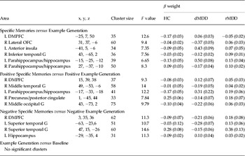
Regions where hemodynamic activity differed significantly between the diagnostic groups for the different contrasts

HC, Healthy control; dMDD, major depressive disorder; rMDD, remitted major depressive disorder; DMPFC, dorsomedial prefrontal cortex; OFC, orbitofrontal cortex; G, gyrus; L, left; R, right;
Coordinates correspond to the stereotaxic array of Talairach & Tournoux (Reference Talairach and Tournoux1988). Cluster size refers to the number of contiguous voxels for which the voxel t value corresponds to p corrected < 0.05. Numbers in parentheses indicate one standard deviation of the mean.
fMRI results
Supplementary Table S3 lists regions where BOLD activity increased in all groups and Table 2 shows group differences in regional BOLD activity. During performance of the Example Generation versus the Riser Baseline task, no significant group difference in regional BOLD activity was identified, suggesting that performance on the control tasks was similar across groups.
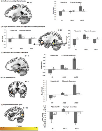
When comparing specific AM recall to example generation, BOLD activity differed significantly across groups (i.e. the Diagnosis × Specific AM Example Generation interaction was significant; Fig. 1), such that BOLD activity was higher in rMDDs than HCs or dMDDs in right lateral OFC, right inferior temporal gyrus and right parahippocampus/hippocampus. In the left DMPFC, anterior insula and parahippocampus/hippocampus, BOLD activity was higher in dMDDs than either rMDDs or HCs, and was higher in rMDDs than HCs.
Group differences in blood oxygen level-dependent (BOLD) activity during specific autobiographical memory (AM) recall. Regions (a) left dorsomedial prefrontal cortex (DMPFC), (b) right orbitofrontal cortex (OFC) and right parahippocampal gyrus, (c) left parahippocampal gyrus, (d) anterior insula and (e) right inferior temporal gyrus are shown where groups had differential BOLD activity during specific AM recall versus example generation (p corrected < 0.05). Using the significant clusters in Table 2 as regions of interest (ROIs), β weights were extracted for the specific AMs versus Riser Baseline conditions, and for the Example Generation versus Riser Baseline conditions. Error bars indicate ±1 standard error of the mean.

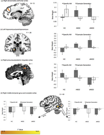
For the positive specific AM recall versus positive example generation contrast (Fig. 2) the mean BOLD activity was higher in rMDDs than HCs or dMDDs in the right middle temporal gyrus and occipital gyrus. In the right precuneus/posterior cingulate, rMDDs and HCs had higher mean BOLD activity than dMDDs. Additionally, BOLD activity was higher in dMDDs than the other groups, and was higher in rMDDs than HCs, in the right DMPFC and left parahippocampus/hippocampus.
Group differences in blood oxygen level-dependent (BOLD) activity during positive autobiographical memory (AM) recall. Regions (a) right dorsomedial prefrontal cortex (DMPFC), (b) left parahippocampal gyrus, (c) right precuneus/posterior cingulate cortex, (d) right middle temporal gyrus and occipital cortex are shown where groups had differential BOLD activity during positive specific AM recall versus positive example generation (p corrected < 0.05). Using the significant clusters in Table 2 as regions of interest (ROIs), β weights were extracted for the specific positive AMs versus Riser Baseline conditions, and the Positive Example Generation versus Riser Baseline conditions. Error bars indicate ±1 standard error of the mean.

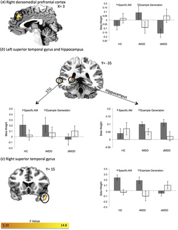
During the negative specific AM recall versus negative example generation contrast (Fig. 3), rMDDs and HCs showed greater BOLD activity than dMDDs in the bilateral superior temporal gyrus, rMDDs had greater activity than HCs and dMDDs in the right DLPFC, and dMDDs manifested greater BOLD activity than rMDDs, who showed greater activity than HCs in the left hippocampus.
Group differences in blood oxygen level-dependent (BOLD) activity during negative autobiographical memory (AM) recall. Regions (a) right dorsomedial prefrontal cortex (DMPFC), (b) left hippocampus and superior temporal gyrus, (c) right superior temporal gyrus are shown where groups had differential BOLD activity during negative specific AM recall versus negative example generation (p corrected < 0.05). Using the significant clusters in Table 2 as regions of interest (ROIs), β weights were extracted for the specific negative AMs versus Riser Baseline conditions and Negative Example Generation versus Riser Baseline conditions. Error bars indicate ±1 standard error of the mean.

Finally, because the average number of neutral memories recalled was less than 10, and the number of categorical AMs recalled by HCs was low, we lacked the statistical power to justify group comparisons involving these parameters.
Correlational analyses
The β weights obtained from the left parahippocampus/hippocampus during specific AM recall were negatively correlated with the percentage of specific AMs recalled (r = − 0.48, p = 0.04), but were positively correlated with the percentage of categorical AMs recalled (r = 0.60, p = 0.001) and with HAMD scores (r = 0.54, p = 0.01). There were no significant correlations between clinical ratings and behavioral performance on the AM task.
Discussion
We replicated previous behavioral findings of fewer specific and more categorical AMs in both dMDDs and rMDDs compared to controls (Williams et al. Reference Williams, Barnhofer, Crane, Hermans, Raes, Watkins and Dalgleish2007), and for the first time demonstrated distinct differences in regional BOLD activity in rMDDs relative to both HCs and dMDDs while recalling specific AMs. Despite scoring in the non-depressed range on depression rating scales, rMDDs showed the same behavioral pattern of AM deficits as dMDDs. We also observed fewer specific positive and more categorical negative AMs in both MDD groups versus controls, in line with previous studies reporting more overgeneral AMs in response to positive cues in dMDDs (van Vreeswijk & de Wilde, Reference van Vreeswijk and de Wilde2004). We also replicated our previous finding of fewer specific AMs from the most recent 6-month epoch in dMDDs versus controls (Young et al. Reference Young, Erickson, Nugent, Fromm, Mallinger, Furey and Drevets2012, Reference Young, Bellgowan, Bodurka and Drevets2013a ). By contrast, rMDDs did not differ significantly from HCs on this parameter. It remains unclear whether the reduction in recent memories recalled by dMDDs reflects an encoding deficit (Ottowitz et al. Reference Ottowitz, Dougherty and Savage2002), or whether they had experienced fewer salient life events due to factors such as inactivity or social isolation (Peeters et al. Reference Peeters, Nicolson, Berkhof, Delespaul and deVries2003).
The fMRI data demonstrated both common and distinct neurophysiological correlates of AM recall across groups. The mean BOLD activity changed in all groups in core components of the AM network (Svoboda et al. Reference Svoboda, McKinnon and Levine2006), including the VLPFC, ACC, posterior cingulate, amygdala, temporoparietal cortex, medial temporal lobe and cerebellum, as participants recalled specific AMs versus example generation (Supplementary Table S3). Differences in BOLD activity between groups were evident in the DMPFC, lateral OFC, anterior insula, precuneus, hippocampus/parahippocampus, and middle, inferior and superior temporal gyrus.
Two regions showed differential activity between groups for every contrast performed: the DMPFC and the left parahippocampus/hippocampus. In the DMPFC, dMDDs had increased BOLD activity versus rMDDs, and both MDD groups showed higher BOLD activity than HCs for specific and positive AMs. This region is putatively involved in executive function, and activity correlates with task difficulty (Kalbfleisch et al. Reference Kalbfleisch, Van Meter and Zeffiro2007). The exaggerated BOLD response in the DMPFC during specific and positive AMs in dMDDs may conceivably reflect the greater difficulty MDDs manifest during retrieval of these AM types. In rMDDs, BOLD activity during AM retrieval in DMPFC was intermediate between HCs and dMDDs, suggesting that rMDDs also experience greater difficulty than controls at recalling specific AMs, albeit to a lesser extent than dMDDs. Of note, when examining negative AMs, rMDDs had increased activity relative to dMDDs, raising the possibility that retrieving negative specific AMs is more difficult for rMDDs than dMDDs.
In the left parahippocampus/hippocampus, the pattern of activity was dMDD>rMDD>HC for every contrast performed. Previous studies reported increased hippocampal metabolism in MDD that decreases towards normative levels with remission (Kennedy et al. Reference Kennedy, Evans, Kruger, Mayberg, Meyer, McCann, Arifuzzman, Houle and Vaccarino2001; Videbech et al. Reference Videbech, Ravnkilde, Pedersen, Egander, Landbo, Rasmussen, Andersen, Stodkilde-Jorgensen, Gjedde and Rosenberg2001), along with decreased hippocampal BOLD activity during word encoding and retrieval in MDD (Werner et al. Reference Werner, Meindl, Materne, Engel, Huber, Riedel, Reiser and Hennig-Fast2009; van Tol et al. Reference van Tol, Demenescu, van der Wee, Kortekaas, Marjan, Boer, Renken, van Buchem, Zitman, Aleman and Veltman2012). Our results suggest that, during AM retrieval, the hippocampal BOLD activity is abnormally elevated in the depressed phase of MDD and only partly restores towards normative levels in remission. These findings are noteworthy in light of evidence of hippocampal volume loss in both dMDDs and rMDDs, suggesting hypotheses that MDDs must work harder to compensate for atrophy in this region (Videbech & Ravnkilde, Reference Videbech and Ravnkilde2004) to successfully retrieve specific AMs of any valence, and/or that excitatory transmission is elevated in the hippocampus in MDD (Duric et al. Reference Duric, Banasr, Stockmeier, Simen, Newton, Overholser, Jurjus, Dieter and Duman2013). The correlation between hippocampal BOLD activity and percentage of specific and categorical AMs, along with the positive correlation between hippocampal activity and severity of depressive symptoms, further supports our hypothesis that participants who have difficulty recalling specific AMs manifest greater hemodynamic activity in the parahippocampus/hippocampus when recalling specific AMs than those who are better able to recall specific AMs. These findings conceivably suggest that the pathophysiology affecting this region in MDD is associated with greater energy utilization to support the neural processing required for specific AM recall.
In addition to our hypothesis that retrieving specific AMs requires more energy utilization in dMDD, the differential pattern of activity in other regions raises the possibility that distinct cognitive strategies are used during AM recall. In the superior temporal gyrus (during negative AMs) and precuneus (during positive AMs), HCs and rMDDs had similar BOLD activity whereas dMDDs had decreased activity compared to the other groups. Notably, both regions have been implicated in self-referential processing, but in reciprocal capacities. In previous reports, BOLD activity in the superior temporal gyrus increased when attention was focused on others versus self (van der Heiden et al. Reference van der Heiden, Scherpiet, Konicar, Birbaumer and Veit2013), whereas precuneus activity increased during self-referential processing and first-person perspective taking (Cavanna & Trimble, Reference Cavanna and Trimble2006). Our results suggest the hypothesis that HCs and rMDDs focus attention on themselves during positive AM recall but shift attention toward others during negative AM recall. Although our study design could not address this hypothesis directly, the absence of this BOLD activity pattern in dMDDs suggests that this pattern might underlie a cognitive strategy during AM recall that plays an adaptive role in maintaining euthymia. The pattern of BOLD activity in the DMPFC, insula and hippocampus paralleled the clinical mood and anxiety ratings; although rMDDs had depression ratings in the non-depressed range, their scores were still significantly higher than those in controls. Our results suggest that, like these clinical ratings, once a patient is in remission, the exaggerated BOLD activity evident during AM retrieval in the disease state decreases incompletely towards normative levels.
The distinct neuroimaging results across groups may be attributable to different cognitive strategies used by each group to achieve a specific memory. As rMDDs manifest the ability to remain in remission without antidepressant medications (suggesting a subphenotype more likely to maintain euthymia), differences between MDD groups may reflect adaptive mechanisms used by rMDDs that promote resilience and prevent illness chronicity. The engagement of regions involved in attentional focus on others to a greater extent in rMDDs, along with engagement of regions involved in self-referential processing and mental effort to a greater extent in dMDDs, suggests the hypothesis that focusing attention away from the self and associated ruminative processing serves as a protective mechanism. Additional support for this hypothesis comes from behavioral studies in which overgeneral AM in rMDDs is more likely when interpersonally relevant content is accessed prior to memory recall (Barnhofer et al. Reference Barnhofer, Crane, Spinhoven and Williams2007), and increased exposure to self-relevant AM cues is associated with decreased AM specificity (Crane et al. Reference Crane, Barnhofer, Mark and Williams2007).
Our results support the assertion that AM overgenerality constitutes a trait-like marker of depression, as overgeneral AM recall is also found in patients no longer suffering significant pathology but at risk for recurrence (found in the current study), and also in healthy individuals at increased familial risk for developing MDD (Young et al. Reference Young, Bellgowan, Bodurka and Drevets2013a ). The lack of correlation between depression severity and AM performance is in line with previous research that has failed to find such a relationship (Wessel et al. Reference Wessel, Meeren, Peeters, Arntz and Merckelbach2001; Gibbs & Rude, Reference Gibbs and Rude2004), and further suggests that AM overgenerality constitutes a trait-like marker of MDD. Nevertheless, it remains conceivable that AM overgenerality only becomes maladaptive and contributes to psychopathology when coupled with increased rumination or self-focus. This hypothesis is supported by recent findings that patients at high familial risk for developing MDD also had increased BOLD activity in regions involved in focusing attention towards others while recalling specific AMs, whereas dMDDs had increased BOLD activity in regions involved in self-referential and ruminative processing (Young et al. Reference Young, Bellgowan, Bodurka and Drevets2013a ). Assessing whether AMs focus more on self or others has not been characterized in studies of AM in MDD. Addressing this question in future studies may prove informative, especially in the context of longitudinal designs examining whether a shift in focus of AMs from others to self occurs during or preceding an onset of a major depressive episode.
Our a priori hypotheses were partially supported. We replicated previous behavioral results (Young et al. Reference Young, Erickson, Nugent, Fromm, Mallinger, Furey and Drevets2012, Reference Young, Bellgowan, Bodurka and Drevets2013a ), and the hypotheses regarding prefrontal cortical activity were supported, as BOLD activity increased in OFC and medial PFC regions in dMDDs and/or rMDDs relative to HCs during AM recall. However, we did not replicate the previously reported differences between dMDDs and HCs in the mesiotemporal cortex obtained by comparing BOLD activity during AM recall against a subtraction control condition. Thus, although we found abnormal activity in the MDD groups in mesiotemporal regions recruited during AM recall when contrasted against the example generation task, the direction of the difference was contrary to our prediction. Differences between the control tasks used in the current study (example generation) versus our previous study (solving subtraction problems) may account for the distinct fMRI findings in dMDDs versus HCs across studies. The subtraction control task used in Young et al. (Reference Young, Erickson, Nugent, Fromm, Mallinger, Furey and Drevets2012) differed from the AM task on several cognitive components, and the benefit of the current design is that, by comparing BOLD activity during recall of specific AMs to activity during example generation, we more specifically examined the hemodynamic correlates of specific episodic memory while controlling for the neural processing related more generally to memory retrieval.
Several limitations of our study merit comment. Each group had more females than males and we were underpowered to investigate sex-by-diagnosis interactions. Gender differences in AM recall have been reported (Young et al. Reference Young, Bellgowan, Bodurka and Drevets2013b ), and thus our results may not generalize to both males and females. Studies with larger samples powered to detect gender and diagnosis interactions are therefore warranted. Furthermore, the ANOVAs used to examine behavioral differences were not corrected for multiple comparisons, increasing the probability that some associations reported here are spurious. Nevertheless, the post-hoc tests applied to assess the significance of regional BOLD differences across groups were corrected for multiple comparisons, and the behavioral results obtained are consistent with previous findings of AM deficits and mood abnormalities in depression. Finally, although our groups did not differ significantly on any demographic characteristic, nominally the HCs and dMDDs differed in age by approximately 7 years, and other factors, such as years of education, were not considered.
AM recall contributes to the generation of adaptive responses to social interactions and stressful circumstances (Williams et al. Reference Williams, Conway and Cohen2008), and allows imagination/prediction of future events (Williams et al. Reference Williams, Ellis, Tyers, Healy, Rose and MacLeod1996). Therefore, the inability to draw on specific positive AMs, along with the tendency to recall overgeneral negative AMs, may interact with daily life situations or mood changes to induce a depressive episode. The results of the current study suggest that existing cognitive therapies for depression might benefit from targeting overgeneralized memories and shifting the focus of AMs from the self to others, particularly during negative AMs, in patients at risk for the development of a major depressive episode based on personal or family history of MDD.
Supplementary material
For supplementary material accompanying this paper visit http://dx.doi.org/10.1017/S0033291714000464.
Acknowledgments
This research was supported by the Laureate Institute for Brain Research through the William K. Warren Foundation. The funders had no influence on the design or conduct of the study, collection, management, analyses, or interpretation of the data, or in the preparation, review or approval of the manuscript.
Declaration of Interest
W.C.D. is currently an employee of Johnson & Johnson, Inc. and has consulted for Myriad/Rules Based Medicine, Inc. and Eisai, Inc. The other authors have no financial conflicts of interest or disclosures to declare. Dr Young takes responsibility for the integrity of the data and accuracy of the data analysis. All authors had full access to all the data in the study.