Introduction
Oocytes play a critical role in the regulation of oogenesis, ovulation rate and fecundity (Eppig et al., Reference Eppig2001; Gilchrist et al., Reference Gilchrist, Ritter and Armstrong2004; McNatty et al., Reference McNatty, Moore, Hudson, Quirke, Lawrence, Reader, Hanrahan, Smith, Groome, Laitinen, Ritvos and Juengel2004; Gilchrist & Thompson, Reference Gilchrist and Thompson2007). Advanced reproductive technologies rely on the basic techniques of oocyte maturation and in vitro fertilization (IVF), so that the identification and development of baseline requirements for these processes are essential. With the advent of transgenic technologies in the pig for biomedical applications such as xenotransplantation (Cooper et al., Reference Cooper, Ye, Rolf, Zudhi, Cooper, Kemp, Reemtsma and White1991, Reference Cooper, Gollackner and Sachs2002; Prather et al., Reference Prather, Hawley, Carter, Lai and Greenstein2003), in vitro maturation (IVM) of porcine oocytes is becoming an important option. Oocytes matured in vitro have compromised developmental competence, which could be due to inappropriate levels of oocyte-secreted factors (OSFs) (Gilchrist et al., Reference Gilchrist, Lane and Thompson2008; Yeo et al., Reference Yeo, Gilchrist, Thompson and Lane2008). Secreted soluble growth factors from the oocyte act on neighbouring follicular cells to regulate a broad range of granulosa cell and cumulus cell (CC) functions such as proliferation, apoptosis, luteinisation, metabolism and expansion (Eppig, Reference Eppig2001; Gilchrist et al., Reference Gilchrist, Ritter and Armstrong2004, Reference Gilchrist, Lane and Thompson2008; Hussein et al., Reference Hussein, Froiland, Amato, Thompson and Gilchrist2005). Presence of denuded oocytes (DOs) in co-culture alters the function of mural granulosa cells and CCs in vitro and, because the two cell types are generally not in physical contact with each other, this situation demonstrates that the effect is mediated by soluble factors secreted into the medium by the DOs (Gilchrist et al., Reference Gilchrist, Lane and Thompson2008). Different species have been employed as experimental models for the study of OSFs, including the mouse (Dong et al., Reference Dong, Albertini, Nishimori, Kumar, Lu and Matzuk1996; Elvin et al., Reference Elvin, Yan, Wang, Nishimori and Matzuk1999; Yan et al., Reference Yan, Wang, DeMayo, DeMayo, Elvin, Carino, Prasad, Skinner, Dunbar, Dube, Celeste and Matzuk2001; Yi et al., Reference Yi, LaPolt, Yoon, Chen, Lu and Lyons2001; Su et al., Reference Su, Wu, O'Brien, Pendola, Denegre, Matzuk and Eppig2004), sheep (Davis et al., Reference Davis, McEwan, Fennessy, Dodds, McNatty and 1992; Galloway et al., Reference Galloway, McNatty, Cambridge, Laitinen, Juengel, Jokiranta, McLaren, Luiro, Dodds, Montgomery, Beattie, Davis and Ritvos2000; Mulsant et al., Reference Mulsant, Lecerf, Fabre, Schibler, Monget, Lanneluc, Pisselet, Riquet, Monniaux, Callebaut, Cribiu, Thimonier, Teyssier, Bodin, Cognie, Chitour and Elsen2001; Souza et al., Reference Souza, MacDougall, Campbell, McNeilly and Baird2001; Wilson et al., Reference Wilson, Wu, Juengel, Ross, Lumsden, Lord, Dodds, Walling, McEwan, O'Connell, McNatty and Montgomery2001; Juengel et al., Reference Juengel, Hudson, Heath, Smith, Reader, Lawrence, O'Connell, Laitinen, Cranfield, Groome, Ritvos and McNatty2002, Reference Juengel, Hudson, Whiting and McNatty2004; Hanrahan et al., Reference Hanrahan, Gregan, Mulsant, Mullen, Davis, Powell and Galloway2004; McNatty et al., Reference McNatty, Hudson, Whiting, Reader, Lun, Western, Heath, Smith, Moore and Juengel2007) and human (Di Pasquale et al., Reference Di Pasquale, Beck-Peccoz and Persani2004; Montgomery et al., Reference Montgomery, Zhao, Marsh, Mayne, Treloar, James, Martin, Boomsma and Duffy2004; Dixit et al., Reference Dixit, Rao, Padmalatha, Kanakavalli, Deenadayal, Gupta, Chakrabarty and Singh2006; Laissue et al., Reference Laissue, Christin-Maitre, Touraine, Kuttenn, Ritvos, Aittomaki, Bourcigaux, Jacquesson, Bouchard, Frydman, Dewailly, Reyss, Jeffery, Bachelot, Massin, Fellous and Veitia2006; Palmer et al., Reference Palmer, Zhao, Hoekstra, Hayward, Webb, Whiteman, Martin, Boomsma, Duffy and Montgomery2006). However, in pigs, little information is known about the regulation by OSFs of cumulus cell expansion and oocyte quality. Previously, it was thought that the oocyte is a passive recipient of cumulus cell functions, but there is increasing evidence that the oocyte is not inactive, but instead could be a principal regulator of its own developmental competence through autocrine and paracrine activities. The concept that there is a critical bidirectional communication axis between the mammalian oocyte and the follicular somatic cells was established by many studies based on the original OSFs bioassays (Salustri et al., Reference Salustri, Ulisse, Yanagishita and Hascall1990a,b; Vanderhyden et al., Reference Vanderhyden, Caron, Buccione and Eppig1990). Current knowledge is still lacking on the interaction between paracrine and gap-junctional signalling within the cumulus–oocyte complex (COC), on interactions between molecules and other lesser known OSFs, as well as with traditional hormonal regulators.
The oocyte's ability to control follicular cell development and function is exerted through paracrine factors such as growth differentiation factor 9 (GDF9) and bone morphogenetic factor 15 (BMP15) (Gilchrist et al., Reference Gilchrist, Lane and Thompson2008). GDF9 and BMP15 are two oocyte-specific growth factors that are recently discovered members of the transforming growth factor-β (TGF-β) superfamily and have been given considerable attention since two landmark studies demonstrated that their absence causes sterility (Dong et al., Reference Dong, Albertini, Nishimori, Kumar, Lu and Matzuk1996; Galloway et al., Reference Galloway, McNatty, Cambridge, Laitinen, Juengel, Jokiranta, McLaren, Luiro, Dodds, Montgomery, Beattie, Davis and Ritvos2000). GDF9 is required for early folliculogenesis, and this factor together with BMP15 are central regulators of GC/CC differentiation, are potential contraceptive targets and may be associated with the pathogenesis of ovarian dysfunction (Gilchrist et al., Reference Gilchrist, Ritter and Armstrong2004; Shimasaki et al., Reference Shimasaki, Moore, Otsuka and Erickson2004; Juengel & McNatty, Reference Juengel and McNatty2005; McNatty et al., Reference McNatty, Hudson, Whiting, Reader, Lun, Western, Heath, Smith, Moore and Juengel2007). GDF9 mediates its effects through activation of the SMAD2/3 signalling pathway in cumulus cells (Gilchrist et al., Reference Gilchrist, Ritter, Myllymaa, Kaivo-Oja, Dragovic, Hickey, Ritvos and Mottershead2006; Diaz et al., Reference Diaz, Wigglesworth and Eppig2007; Dragovic et al., Reference Dragovic, Ritter, Schulz, Amato, Thompson, Armstrong and Gilchrist2007; Su et al., Reference Su, Sugiura, Wigglesworth, O'Brien, Affourtit, Pangas, Matzuk and Eppig2008). Knowledge of the effective mechanisms and the signalling cascades of the key molecules that control oocyte maturation may lead gradually to improvement of the current oocyte/embryo culture systems and gamete technology such as IVF. The aim of this study is to determine if co-culturing COCs with denuded oocytes (DO) during IVM could improve porcine cumulus expansion and oocyte competence, as judged by embryonic development after parthenogenetic activation. This approach could also evaluate if DO could be used as an IVM additive to improve oocyte IVM.
Materials and methods
Chemicals and reagents
All chemicals and reagents used for oocyte maturation, activation and embryo culture in this study were purchased from Sigma-Aldrich Chemical Company unless otherwise stated.
Collection, denudation and in vitro maturation of oocytes
Ovaries were collected at a local abattoir and stored in sterile physiological saline at 30–35 °C during transportation. Cumulus–oocyte complexes were aspirated from antral follicles (3–6 mm) with an 18-gauge needle fixed to a 10 ml disposable syringe. COCs with several layers of cumulus cells and uniform cytoplasm were selected. Among the selected COCs, some complexes were used as intact COCs while others were denuded to be used for co-culture experiments. Intact COCs were divided into control and treatment groups, with 25 COCs each group. The control group has 25 COCs cultured without DO for 44 h. Treatments 1 to 5 had 25 COCs per group co-cultured with increasing numbers of DOs (see Experimental design 1) for 44 h. All treatment groups were cultured in 500 μl of medium. The culture medium was prepared by supplementing tissue culture medium (TCM)-199 (Invitrogen) with 0.57 mM cysteine, 0.91 mM sodium pyruvate, 5 μg/ml insulin, 1% (v/v) penicillin–streptomycin (Invitrogen), 1 mM dibutyryl adenosine 3′,5′-cyclic monophosphate (dbcAMP), 0.5 μg/ml follicle-stimulating hormone (FSH), 0.5 μg/ml luteinizing hormone (LH) and 10% porcine follicular fluid. All the COCs in the control group and the COCs with DO in the treatment groups were incubated at 39 °C in a humidified atmosphere of 5% CO2 in air, first, with gonadotrophin-releasing hormone (GnRH) and dbcAMP for 22 h and then without dbcAMP and GnRH for another 22 h. Denuded oocytes and their respective cumulus cells were generated by addition of 0.1% hyaluronidase in Dulbecco's phosphate-buffered saline (DPBS; Invitrogen) supplemented with 0.1% polyvinyl alcohol (PVA–DPBS).
Assessment of meiotic maturation of matured oocytes
The stage of meiotic maturation was determined by evaluating the presence of a polar body. After 44 h of IVM, cultured COCs were denuded and fixed in methanol for 15 min, mounted on a slide, and stained with 25 μg/ml bisbenzamide (Hoechst 33258) in DPBS. The presence or absence of the first polar body was determined under ultraviolet light.
Cumulus expansion assessment
Cumulus expansion was scored blinded on a 0–4 scale and the cumulus expansion index (CEI) was calculated as described by Vanderhyden et al. (Reference Vanderhyden, Caron, Buccione and Eppig1990). Using this subjective scoring system, score 0 indicated no expansion, characterized by the detachment of cumulus cells from the oocyte to assume a flattened monolayer of fibroblastic appearance leaving a partially or fully denuded oocyte. A score of 1 indicated no expansion but cumulus cells are spherical, and remained compacted around the oocyte. For score 2 complexes, only the outermost layers of cumulus cells have expanded, while score 3 complexes have all cell layers except the corona radiata (cells most proximal to the oocyte) prominently expanded, and a score of 4 indicated the maximum degree of expansion including the corona radiata (Vanderhyden et al., Reference Vanderhyden, Caron, Buccione and Eppig1990). The degree of expansion was obtained by adding the scores of each COC (score 0–4) and dividing by the total number of COC.
Electrical activation of porcine oocytes
After denuding the cultured COCs, oocytes were subjected to electrical activation. Oocytes were equilibrated in pulsing medium, then transferred to a chamber containing two electrodes overlaid with the pulsing medium. The pulsing medium was 0.26 M mannitol solution containing 0.5 mM HEPES, 0.1 mM CaCl2 and 0.1 mM MgSO4. Oocytes were activated with a single DC pulse of 1.5 kV/cm for 60 μs utilizing a BTX electro-cell Manipulator 2001 (BTX, Inc.).
In vitro culture
Activated oocytes were washed and transferred into 500 μl of porcine zygote medium-3 (PZM-3) covered with mineral oil. The culture medium was preincubated at 39 °C, 5% CO2, 5% O2 and 90% N2. Embryos were evaluated for cleavage on day 2. Blastocyst (BL) formation and the number of nuclei in BL were determined on day 7.
Blastocyst total cell counting
Briefly, BLs were fixed with absolute alcohol then cells were treated with Hoechst 33258 nuclear stain overnight at 4 °C. Fixed and stained BLs were mounted onto a glass slide in a drop of glycerol, gently flattened with a cover glass and visualized for cell counting on a fluorescence microscope (excitation filter 460 nm).
RNA extraction and RT-PCR
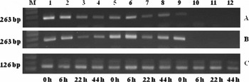
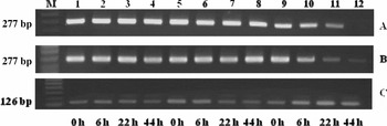
Denuded oocyte samples and their respective cumulus cells from immature and mature COCs of the control and treatment 4 groups were washed once in PBS, and total RNA was extracted using the RNeasy total RNA Kit (Qiagen) according to the manufacturer's instructions, and quantified by a GeneQuant Pro spectrophotometer (Biochrom Ltd). Another sample from the added DO used in co-culture of treatment 4 was also processed with the same RNA extraction protocol as above. cDNA was produced from 5 μg of total RNA from porcine cumulus cells and oocyte samples, using the SuperScript II reverse transcriptase kit (Gibco BRL) primed with oligonucleotide-dT (18-mer) and followed by RNase H digestion of RNA, in a total volume of 20 μl as per manufacturer's instructions. Polymerase chain reaction (PCR) was performed as described previously (Zhu et al., Reference Zhu, Guo, Pan, Mu and Feng2008), using the following oligonucleotide primers. GDF9 primer sequences were: forward strand 5′-CAGTCAGCTGAAGTGGGACA-3′ and reverse strand 5′-ACGACACGTGCACTTTGTTG-3′; BMP15 primer sequences were forward strand 5′-GGTCCTCCTCAGCATCATTAG-3′ and reverse strand 5′-TGCGGTTCTCCCTAGGGTG-3′. These primers were expected to generate 277- and 263-base pair (bp) cDNA fragments for GDF9 and BMP15, respectively. For PGK1, a housekeeping gene used as control, RT-PCR was performed as described previously (Kuijk et al., Reference Kuijk, du Puy, van Tol, Haagsman, Colenbrander and Roelen2007) using primers (forward 5′-AGATAACGAACAACCAGAGG-3′ and reverse 5′-TGTCAGGCATAGGGATACC-3′) that generated a 126-bp fragment. All samples were placed in a thermocycler (Biometra T1 Thermocycler). PCR reactions were performed as follows: 28 cycles of denaturation at 94 °C for 30 s; annealing at 60 °C for 20 s; extension at 72 °C for 20 s for both BMP15 and GDF9. For PGK1, initial denaturation at 94 °C for 3 min; 40 cycles of denaturation at 94 °C for 30 s, annealing at 56 °C for 30 s, extension at 72 °C for 30 s, and final extension of 94 °C for 1 min.
Scanning electron microscopy (SEM) observations on cumulus cells
Samples for SEM observations were submitted to and analysed at the National Instrumentation Center of Environmental Management (NICEM) of Seoul National University, Korea. Briefly, the representative COCs from the control and treatment 4 groups at 44 h culture were fixed in modified Karnovsky's fixative, consisting of 2% paraformaldehyde and 2% glutaraldehyde in 0.05 M sodium cacodylate buffer (pH 7.2) for 2 h. Next, the cells were washed and attached to small glass coverslips. The samples on the coverslips were post-fixed in 1% osmium tetroxide in 0.05 M sodium cacodylate buffer for 2 h. The fixed specimens were conductivity stained with 2% uranyl acetate and Reynolds’ lead citrate and then dehydrated in a series of increasing concentrations of ethanol, critical-point dried, and sputter-coated with gold. Observations were made with a JSM-5410LV SEM (JEOL).
Cumulus cell viability assessment
Cumulus cell samples generated from immature and mature COCs from the control and treatment 4 groups were used for the assessment of cumulus cell viability. Ten microlitres of each sample was mixed with 10 μl of 0.4% trypan blue stain. Next, 10 μl of the mixture was loaded onto a Countess cell counting chamber slide (Invitrogen), which was then inserted into the Countess Automated Cell Counter (Invitrogen). Live and dead cells were counted and cell viability was assessed.
Statistical analysis
Cell viability, maturation rate, cleavage rate, blastocyst rate and total cell number were analysed by one-way analysis of variance (ANOVA). Comparisons of mean values among treatments were performed using Tukey's post hoc tests using the statistical program for social sciences (SPSS) tool (SPSS Inc.). Total cell, live cell and dead cell data were analysed by Student's t-test. Experiments were performed in replicates and results were expressed as mean ± SD. Significance was established at the p < 0.05 level.
Experimental design
Experiment 1: Effect of co-culture of intact COCs with DOs during IVM on cumulus expansion
This experiment was carried out to determine the effect of DO on the regulation of cumulus expansion. Intact COCs were randomly allocated into control group or five treatment groups during IVM: control group, 25 COCs were cultured without DO in a 500 μl IVM media for 44 h; treatment 1, 25 COCs were co-cultured with 25 DOs which yields a ratio of 1 COC to 1 DO; treatment 2, 25 COCs were co-cultured with 50 DOs (ratio of 1 COC to 2 DOs); treatment 3, 25 COCs were co-cultured with 75 DOs (ratio of 1 COC to 3 DOs); treatment 4, 25 COCs were co-cultured with 100 DOs (ratio of 1 COC to 4 DOs); treatment 5, 25 COCs were co-cultured with 125 DOs (ratio of 1 COC to 5 DOs). For treatment groups 1 to 5, COCs were co-cultured with DO from 0 to 44 h and each group was cultured in 500 μl of IVM medium. IVM medium contained dbcAMP and GnRH for the first 22 h of IVM culture only. Expansion of COCs was assessed at 6, 22 and 44 h of IVM. At least three replicates in each treatment group were assessed.
Experiment 2: Effect of co-culture with DOs during IVM on cumulus cell viability
This experiment was carried out to determine if the presence of DOs affected the viability of the cumulus cells in COC. As treatment group 4 showed altered cumulus expansion morphology at 44 h IVM, we examined both live and dead cumulus cells and compared these with mature COC from the control group using an automated cell counter (Invitrogen). Five replicates were performed.
Experiment 3: Effect of DOs during IVM on the extracellular matrix
This experiment was conducted to examine the influence of DOs on the extracellular matrix, as effects on cumulus expansion might be due to changes in the extracellular matrix. Scanning electron microscopy was carried out to compare extracellular matrix formation in the control and treatment 4 groups after 44 h of IVM.
Experiment 4: Effect of DOs during IVM on maturation, cleavage and blastocyst rates and total cell number
This experiment was performed to assess if co-culture of COCs with DOs in the IVM medium improved the developmental competence of electrically activated porcine parthenogenetic embryos. Maturation rate was assessed by the presence of the first polar body after 44 h of IVM culture, while cleavage rate was examined on day 2. Blastocyst formation rate and total cell numbers in blastocyst were monitored on day 7.
Experiment 5: Expression of GDF9 and BMP15 genes in cumulus cells and oocytes from COC from the control and treatment 4 groups and in the added DO groups
This experiment was designed to determine the activity of GDF9 and BMP15 genes in the cumulus cells and oocytes from COCs in the control and treatment 4 groups and in the added DO groups at 0, 6, 22 and 44 h of IVM.
Results
Effect of co-culture of intact COCs with DOs during IVM on cumulus expansion
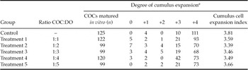
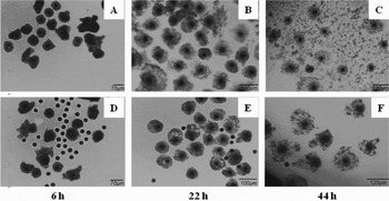
To examine the effect of DOs on cumulus expansion, COCs were cultured alone or were treated with increasing numbers of DO. COCs were considered to be expanded when the CEI was at least +2. After 44 h of IVM, COCs cultured alone expanded more (CEI 3.81). Treatment groups 1 to 5 had CEIs of 3.59, 3.39, 3.46, 3.49 and 3.66, respectively. The CEIs of each treatment group were not significantly different from each other but were lower than the control group (Table 1). Interestingly however, morphologically, in treatment groups 4 and 5, there was a noticeable difference in the outermost layer of the cumulus cells at 44 h of IVM culture compared with other treatment and control groups. There was expansion of all layers including the cells most proximal to the oocyte (corona radiata), but the outermost layer of cumulus cells did not fully disperse and intersperse with neighbouring COC in treatment group 4, as a representative sample (Fig. 1F), unlike the control group in which the cumulus cells interspersed with neighbouring COC (Fig. 1C). At 6 and 24 h of IVM, there was no morphological difference in the cumulus expansion between the control (Fig. 1A,B) and treatment 4 groups (Fig. 1D,E).
Effect of co-culture of intact cumulus–oocyte complexes (COCs) with denuded oocytes (DOs) on cumulus cell expansion at 44 h IVM.

aDegree of cumulus expansion as described by Vanderhyden et al. (Reference Vanderhyden, Caron, Buccione and Eppig1990).
(A) Pig cumulus–oocyte complexes (COC) from the control group during in vitro maturation (IVM) at 6 h; and (B) 22 h. (C) Expanded cumulus cells at 44 h IVM with full dispersion of the outer layer of COC, which interspersed with neighbouring COCs. (D) Pig cumulus–oocyte complexes from the 1 COC:4 denuded oocyte (DO) group at 6 h; and (E) 22 h. (F) Cumulus expansion at 44 h IVM. Note the outermost layer of cumulus cells is not fully dispersed and do not intersperse with the neighbouring COCs.

Effect of co-culture with DOs during IVM on cumulus cell viability
Cell viability was assessed using an automated cell counter to determine if the effect of DOs on cumulus expansion in treatment 4 group affects the viability of cumulus cells. There was no significant difference in cell viability and cell death between the control and treatment 4 groups (Table 2).
Cumulus cell numbers and cell viability in cumulus–oocyte complexes (COCs) at 44 ha.

aData are shown as mean ± standard error (SE) of total cells, live cells and dead cells from cumulus cells from COCs taken from the control group and the treatment 4 group.
Effect of DOs during IVM on extracellular matrix
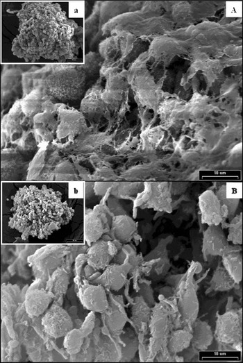
Co-culture with DO produced a lesser cumulus expansion in the treatment 4 and 5 groups. To determine if this situation was associated with changes in the extracellular matrix, scanning electron microscopy was performed for matured COC from the control and treatment 4 groups. Cumulus cells were covered completely with abundant extracellular matrix, showing mucification inside the cumulus mass in the control group (Fig. 2A,a). In the treatment 4 group, however, cumulus cells were oblong shaped with scanty extracellular matrix and elongated cellular projections between the cumulus cells (Fig. 2B,b).
Scanning electron micrographs of porcine cumulus–oocyte complexes (COCs) at 44 h in vitro maturation (IVM). (a,A) Control: cumulus cells are completely covered with abundant extracellular matrix, showing mucification inside of cumulus mass. (b,B) Treatment 4: note the oblong-shaped cumulus cells with scanty extracellular matrix and elongated cellular projections in the cumulus cells.

Effect of DOs during IVM on maturation, cleavage and blastocyst rates and total cell number
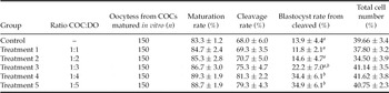
There was no significant difference in the maturation and cleavage rates and in total cell numbers between the control and treatment groups (Table 3). There was a significant difference in the blastocyst rate in treatment groups 4 and 5 (p < 0.05, 34.4% and 34.9% respectively) compared with the control (13.9%) and treatment groups 1, 2 and 3 (11.8%, 14.6% and 22.2%, respectively) (Table 3).
Effect of denuded oocytes (DO) during in vitro maturation (IVM) on oocyte developmental competence.

a ,b Values with no common superscripts within the same column are significantly different (p < 0.05). Values are expressed as mean ± SE. COC, cumulus–oocyte complexes.
Expression of GDF9 and BMP15 genes in cumulus cells and oocytes from COCs from the control and treatment 4 groups and in the added DO groups
Gel electrophoresis of RT-PCR products to assess BMP15 gene expression is shown in Fig. 3A,B with PGK1 as internal control in Fig. 3C. The BMP15 gene was expressed: (i) when DOs were added; (ii) when DOs from COCs from control and treatment 4 groups were added and (iii) when cumulus cells from COCs from control and treatment group 4 were added at the 0 h time-point. The BMP15 gene was not expressed in cumulus cells from COCs in the control and treatment 4 groups at the 6, 22 and 44 h time points (Fig. 3A,B). Gel electrophoresis of RT-PCR products representing GDF9 gene expression is shown in Fig. 4A,B with PGK1 as internal control in Fig. 4C. The GDF9 gene was expressed: (i) in DOs; and (ii) in DOs from COCs from the control and treatment 4 groups (Fig. 4A,B). In the cumulus cells from the COCs in the control group, GDF9 was expressed at 0, 6 and 22 h time points but not at the 44 h time point (Fig. 4A). In the cumulus cells from COCs in treatment group 4, GDF9 was expressed at the 0, 6, 22 and 44 h time points, which clearly demonstrated up-regulation in the 44 h cumulus cells from treatment group 4 (Fig. 4B).
mRNA expression of BMP15 in the control group (A) and in the 1 cumulus–oocyte complex (COCs):4 denuded oocyte (DO) group (B); in porcine COCs during in vitro maturation (IVM) culture, detected by reverse transcriptase-polymerase chain reaction (RT-PCR) assay. (C) PGK1 is used as the internal control. Representative photographs of RT-PCR products for specific genes. Lane M: 100-bp DNA marker; lanes 1–4: added oocyte; lanes 5–8: oocyte from COC; lanes 9–12: cumulus cells from COCs.

mRNA expression of the GDF9 in the control group (A); and in the treatment 4 group (B); in porcine cumulus–oocyte complexes (COCs) during in vitro maturation (IVM) cultivation detected by the reverse transcriptase-polymerase chain reaction (RT-PCR) assay. (C) PGK1 is used as the internal control. Representative photographs of RT-PCR products for specific genes. Lane M: 100-bp DNA marker; lanes 1–4: added oocyte; lanes 5–8: oocytes from COCs; lanes 9–12: cumulus cells from COCs.

Discussion
Recent studies in cattle and mice have demonstrated the concept and the validity of OSFs as IVM medium additives to improve oocyte quality and subsequent embryo and fetal developmental potential (Hussein et al., Reference Hussein, Thompson and Gilchrist2006; Yeo et al., Reference Yeo, Gilchrist, Thompson and Lane2008). The oocyte has a crucial function in its secretion of growth factors and its regulation of cumulus cell function (Gilchrist et al., Reference Gilchrist, Lane and Thompson2008) through their bidirectional communication. OSFs activate SMAD 2/3, which enables cumulus expansion (Dragovic et al., Reference Dragovic, Ritter, Schulz, Amato, Thompson, Armstrong and Gilchrist2007). However, one previous study (Vanderhyden et al., Reference Vanderhyden, Cohen and Morley1993) has demonstrated that the regulation of cumulus expansion in pig COCs is different from that of mice. Other studies have demonstrated that the expansion of porcine COCs in vitro is not dependent upon the oocyte (Prochazka et al., Reference Prochazka, Nagyova, Rimkevicova, Nagai, Kikuchi and Motlik1991; Vanderhyden, Reference Vanderhyden1993). Therefore, we know that addition of DO is not essential for porcine cumulus expansion of COCs that have been matured in vitro. However, our study has shown that addition of DOs at a certain ratio does alter the expansion of cumulus cells. We have shown here for the first time that the co-culture of COC with DO at a ratio of 1 COC to 4 DOs (treatment 4) and 1 COC to 5 DOs (treatment 5) resulted in an altered morphology of cumulus expansion that was different and had lower CEI scores than when COC were cultured alone. We noted that at 44 h of IVM, treatment groups 4 and 5 showed expansion of the outermost layers of cumulus cells, but did not have full dispersion to intersperse with the neighbouring COC. We investigated cumulus cell viability and found no significant difference with and without added DOs. These results demonstrated that alteration of cumulus expansion has no overt adverse effect on cell viability. Furthermore, scanning microscopy was performed to determine if DOs have an influence on the extracellular matrix. Results revealed scanty extracellular matrix in treatment group 4 that explained the changes seen in the morphology of cumulus cell expansion. Vanderhyden et al. (Reference Vanderhyden, Cohen and Morley1993) demonstrated that the pig oocyte is not necessary for cumulus expansion, but that pig oocytes have the cumulus expansion enabling factors (CEEF), as do the mouse oocyte. In that study, it was shown that co-culture of mouse oocytectomized complex (OOX) with pig oocytes enabled mouse cumulus expansion. It is probable that a different mechanism that needs to be described is involved in porcine CEEF. However, this study suggested that the porcine oocyte secretes factors that alter the morphology of the cumulus expansion.
GDF9 and BMP15 are two members of TGF-β superfamily (Chang et al., Reference Chang, Brown and Matzuk2002) that are only produced by growing oocytes in mice (McGrath et al., Reference McGrath, Esquela and Lee1995; Dube et al., Reference Dube, Wang, Elvin, Lyons, Celeste and Matzuk1998), rats (Fitzpatrick et al., Reference Fitzpatrick, Sindoni, Shughrue, Lane, Merchenthaler and Frail1998; Otsuka et al., Reference Otsuka, Yao, Lee, Yamamoto, Erickson and Shimasaki2000), and humans (Fitzpatrick et al., Reference Fitzpatrick, Sindoni, Shughrue, Lane, Merchenthaler and Frail1998; Aaltonen et al., Reference Aaltonen, Laitinen, Vuojolainen, Jaatinen, Horelli-Kuitunen, Seppa, Louhio, Tuuri, Sjoberg, Butzow, Hovata, Dale and Ritvos1999). In pig oocytes, it has been shown that BMP6, BMP15 and GDF9 expression decrease during IVM (Zhu et al., Reference Zhu, Guo, Pan, Mu and Feng2008). This finding is in agreement with the present study in which the control group showed no expression of GDF9 at 44 h. However, the addition of denuded oocytes to the IVM medium resulted in the detection of GDF9 at 44 h in treatment group 4. This up-regulation of GDF9 expression could be associated with the alteration of cumulus expansion in treatment group 4.
In this study, parthenogenesis was used to evaluate the developmental competence of the oocytes in vitro without confounding factors from the sperm (Hsieh et al., Reference Hsieh, Tang, Chang, Weng, Sha, Tseng, Chang and Ju2006). Parthenogenesis still provides a valuable measure of oocyte competence to initiate the developmental programme as progression of parthenogenetic embryos to the blastocyst stage is not affected by epigenetic imprinting (Latham et al., Reference Latham, Doherty, Scott and Schultz1994) and their developmental characteristics resemble those of in vitro fertilized embryos, as also reported by others (Kure-bayashi et al., Reference Kure-bayashi, Miyake, Okada and Kato2000). Our results showed that the maturation and cleavage rates of the treatment groups were not significantly different from that of the control group. However, the blastocyst formation rate was significantly difference in the control group compares with treatment groups 4 and 5. These two groups had higher rates of blastocyst formation when compared with the control and other treated groups, even though their cumulus expansion properties were altered. This finding is in agreement with the study of Yeo et al. (Reference Yeo, Gilchrist and Lane2009), which reported that when COC were matured with a SMAD 2/3 inhibitor (SB-431542) in the absence of FSH/epidermal growth factor (EGF), cumulus expansion was reduced, but fertilization, embryo development and embryo quality were not.
In conclusion, we have studied the effect of addition of porcine oocytes on the outcome of IVM. Our findings indicated, for the first time, the following: (i) porcine DOs alter the morphology of cumulus expansion and mucification; (ii) even though the appearance of cumulus expansion was altered, the developmental competence of porcine parthenotes was improved and could be attributed to the up-regulation of GDF9 expression; and (iii) with a surplus of porcine DOs available, it is feasible to use these cells as IVM additives to improve oocyte maturation and subsequent embryo development. Further studies should be performed to investigate the nature of factors secreted by the oocyte, their interactions, and their signalling mechanisms.
Acknowledgements
We acknowledge Dr Barry D. Bavister for his valuable assistance in editing this manuscript. This study was supported financially by Korean MKE (Grant no. 10033805, 10033839), the Research Institute for Veterinary Science, and the BK21 Programme for Veterinary Science.