Introduction
Breast cancer is the most common cancer in women worldwide and India as well.Reference Ferlay, Colombet and Soerjomataram1,Reference Malvia, Bagadi, Dubey and Saxena2 Treatment of breast cancer includes multiple modalities such as surgery (SX), chemotherapy, hormone therapy and radiotherapy (RT) depending on the disease stage. The role of RT in the adjuvant setting after mastectomy [radiation to the chest wall (CW)] or breast conservation surgery [radiation to the whole breast (WB)] is well established.3,Reference McGale, Taylor and Correa4 The benefit of adjuvant RT is more pronounced for node-positive cases as compared to node-negative.Reference McGale, Taylor and Correa4,Reference Zeidan, Habib and Ameye5 Most of the studies included in the early breast cancer trialists collaborative group meta-analysis irradiated the regional lymphatics as well. Axilla is generally treated by single modality either with axillary dissection or radiation to avoid an increased risk of lymphedema. The role of internal mammary nodal irradiation is still debatable as large randomised trials have failed to show overall survival benefit.Reference Whelan, Olivotto and Parulekar6,Reference Poortmans, Collette and Kirkove7 Hence, by and large, patients receive supraclavicular fossa (SCF) irradiation when axillary nodes are positive as it is considered the next echelon for cancer spread.
Radiation to either CW (post-mastectomy) or WB (post-lumpectomy) with ipsilateral (I/L) SCF is a common occurrence in clinical practice as per standard indications. Breast cancer generally accounts for at least one-fourth to one-third of the departmental load. The length of the target volume poses a radiation risk to various organs at risk (OARs) namely bilateral lungs, heart in left-sided tumours, contralateral breast, brachial plexus, thyroid gland and oesophagus. Meticulous radiation planning is vital as these patients are long-term survivors. They can present not only with acute radiation toxicities such as radiation dermatitis, dysphagia but also with chronic toxicities such as radiation pneumonitis, brachial plexopathy and radiation-induced cardiac morbidities which in turn can affect the quality of life.Reference Sloan and Alcorn8–Reference Olsen, Pfeiffer, Johannsen, Schrøder and Rose12
Radiation can be delivered in various ways. The conventional technique which uses bitangential portals for the WB or CW and anterior portal for SCF with separate isocentre for each is known as the dual isocentric technique (DIT). A gap between the two plans is maintained to avoid overlap between the inferior border of SCF and superior border of tangential portals. The policy of a gap is variable ranging from no gap to a few millimetres depending upon the use of energy, multi-leaf collimators and also institutional practice.Reference Dobbs13 The other common technique called the monoisocenteric technique (MIT) uses a single isocentre for bitangential and SCF portals by blocking the upper and lower portions of the portals, respectively.Reference Marshall14,Reference Klein, Taylor, Michaletz-Lorenz, Zoeller and Umfleet15 Each technique has its own advantage and disadvantages. Various dosimetric comparisons between two techniques to date have shown no significant differences.Reference Banaei, Hashemi and Bakhshandeh16,Reference Jooladi, Mahdavi and Nedaei17
The purpose of this study is to compare these two RT techniques in terms of dosimetry, ease of set-up and the reproducibility of set-up in treatment of breast cancer (either CW or WB patients). Both the techniques are used at our institute for breast radiation. This study was undertaken to understand the difference between the two techniques if any and allow uniform adoption of a particular technique for routine clinical practice.
Materials & Methods
Patient selection
Twenty consecutive patients diagnosed with carcinoma breast, in whom adjuvant RT to the WB or CW and I/L SCF was indicated, were studied. The study being of retrospective nature, local ethics committee approval was not mandatory for the study conduct as per the local standards. Of these, ten patients had received treatment with DIT and remaining ten with MIT. The patients treated with DIT had a second plan made using MIT, and the MIT patients had a second plan made with DIT retrospectively for dosimetric comparison. Overall, a total of 40 plans were evaluated. The planning process is briefly described below.
Simulation
All patients underwent computerised tomography (CT) simulation scan using 3–5 mm slice thickness without intravenous contrast. The patients were positioned supine with both hands above the head and neck turned to the opposite side, lying on an inclined breast board. The entire breast and any surgical scar was wired. Fiducials were kept to mark out the extent of the target volume: superiorly at the inferior border of clavicle, inferiorly 2 cm margin to infra-mammary fold or opposite breast, medially at midline or across to cover the surgical scar completely and laterally at the mid-axillary line or 2 cm beyond the palpable breast tissue. Care was taken to ensure that the patient was straight and the sternum was horizontal. The scanned volume extended from the level of the mandible to the level of T12 (upper abdomen).
Treatment planning
In all patients, OARs were contoured before treatment planning as per Radiation Therapy Oncology Group guidelines which included heart, bilateral lungs, contralateral breast, spinal cord, oesophagus, larynx and thyroid.Reference White, Arthur and Buchholz18 RT plans (both MIT and DIT for each patient) were generated with a dose prescription of 40 Gy in 15 # to planning target volume (PTV) comprising of WB or CW and I/L SCF. WB patients also received tumour bed boost as per standard practice, after completion of WB RT. No formal contouring for the target volumes was done as it is not a standard institutional practice. RT contouring and planning was done using Eclipse version 13 (Varian medical systems, Palo Alto, USA) treatment planning system, with energy ranging from 6 to 15 MV photons. A single physicist was involved in generating plans for both techniques in all patients. The dose calculation was done using Anisotropic Analytical Algorithm (AAA). All patients received treatment using Truebeam machine (Varian medical systems, Palo Alto, USA) at ACTREC Navi Mumbai.
Monoisocentric technique
For the MIT plans, the isocentre was placed on a horizontal line passing through the lower border of the clavicle, as it represents the junction between SCF portal and tangents and the position would be reproducible for daily implementation too. Both SCF and WB/CW plans were made keeping the same isocentre in both the plans with SAD (constant source to axis distance) technique. The dose was prescribed to two separate reference points for each plan. For the primary, bitangential portals were used, and for I/L SCF, a single anterior portal was used with the depth of prescription as per patient’s anatomy. Dynamic wedges and field-in-field technique were used to get adequate dose coverage such that 95% of PTV will receive 95% of the prescribed dose and homogenous dose distribution.
Dual isocentric technique
For the DIT, separate plans for SCF and WB/CW were generated independently using separate isocentres. Technique and portals for both SCF and WB/CW were placed similar to the MIT except for utilisation of SSD (constant source to surface distance) technique for SCF plan and use of 5 mm gap between the tangents and lower border of SCF portal. After the generation of optimal plans, fulfilling the dosimetric criterion of adequate and homogenous dose distribution, the respective sum plans were made and evaluated by the treating physician.
Treatment delivery
Patients were set up in treatment position. Set-up verification was done by obtaining single-exposure electronic portal images (EPIs) in anterior–posterior (AP) and medial tangent views. EPI was acquired on days 1, 2, 5, 8, 9, 10 and 12. The EPI was compared with digital reconstruction radiograph generated from the planning CT, and bony anatomy (clavicle, trachea, sternum and thoracic wall) was used for matching. The AP, right–left and superior–inferior (SI) shifts were recorded. Both clinician and radiation technologist verified the set-up errors. An online set-up error correction strategy was used and after set-up error correction planned treatment delivered. The time taken for setting up the patients was recorded before imaging in every patient for each fraction.
Variables for analysis
Various planning parameters, as shown in Table 1, were obtained from the plans for the two techniques. Estimation of dose to target volumes (WB, CW and SCF) as well as various OARs such as I/L lung, contralateral lung, contralateral breast, heart, spinal cord, oesophagus, larynx and thyroid gland was done and dose-volume histograms were used to obtain the dose-volume parameters. The parameters analysed were overdose volumes, that is, V107% (volume of the PTV receiving 107% of the prescribed dose) in the field junction, mean dose for I/L lung, contralateral lung, contralateral breast, and heart, max dose for spinal cord and mean dose to thyroid, larynx and oesophagus. The evaluation of the treatment reproducibility was performed in terms of mean displacements, systematic error (Σ) and random error (σ) calculated for both the techniques. The total error was calculated using the van Herk formula.Reference van Herk19,Reference Van Herk, Remeijer, Rasch and Lebesque20 The set-up time was compared between the two methods.
Treatment plan characteristics

* Posterior beam edge of lateral tangential portal beyond clinical reference (i.e., mid-axillary line).
# Ref point: point of normalisation and dose prescription.
Statistics
This data were analysed using Statistical Package for Social Sciences (SPSS) version 21. The statistical test used for the analysis was independent student t-test or Mann–Whitney U-test depending upon the normality distribution of each variable. Mean values are indicated with standard deviation. A p-value of less than 0.05 was considered significant.
Results
Twenty patients were included in this study of which 11 had right- and 9 had left-sided tumours. Ten patients each received WB and CW RT as well as were treated with DIT or MIT. However, for each patient, two plans (DIT and MIT) were produced to compare the dosimetry. The treatment plan characteristics are shown in Table 1. All plan characteristics were similar between MIT and DIT. The mean central lung distance was 1.8 cm. The mean central heart distance in 18 left-sided plans was 1.1 cm. In both techniques, the dose prescription reference point was at half of the depth (2 cm flash to skin + distance from skin to posterior beam edge) in a majority of plans. Ten megavolt was the most commonly used beam energy in both techniques especially for CW cases. Bolus was used only in CW cases. There was no difference in the gantry angle and wedge (degree) used in both techniques. The depth of prescription for SCF ranged from 2.7 to 3.1 cm.
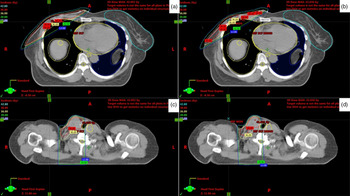
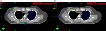
The dosimetric variables are compared between the two techniques and shown in Table 2. The dose distributions of the two techniques are shown in Figure 1. The dose distributions were very similar between the two techniques. There was no difference in mean dose to I/L lung, contralateral lung and contralateral breast between DIT and MIT. Overall, the mean heart dose was significantly less with the MIT (0.4 versus 0.79, p = < 0.001) as well as in left-sided tumours (0.37 versus 0.98, p = 0.003). Although a difference was noted in the dose to the heart, both the techniques achieved clinically acceptable cardiac doses. The maximum dose at the field junction was significantly higher with DIT (43 Gy versus 41 Gy, p = 0.003). Dose distribution at the junction for two techniques is shown in Figure 2. Spinal cord, larynx, oesophagus and thyroid dose were similar between the two techniques.
Dosimetric characteristics

Dose distribution of chest wall and supraclavicular fossa in axial for (a, c) dual isocentric technique and (b, d) monoisocentric technique.

(a) Dose distribution at the field junction for dual isocentric technique and (b) monoisocentric technique.

The average set-up time for each field, excluding the treatment time for MIT and DIT, was 8.83 and 8.86 minutes, respectively. There was no statistically significant difference between the two groups (p = 0.95). EPI was acquired on a minimum of 6 days for every patient. A total of 240 images (one each for Medial tangent (MT) and SCF/day) were analysed, 120 each for WB and CW. The set-up errors which include population systematic and random errors (in mm) for SI and AP and total error (in mm) calculated by van Herk formula for both techniques are shown in Table 3. The maximum total error was 1 cm (10–11 mm) in lateral for SCF and 8 mm in SI for tangents for both techniques. There was no statistically significant difference in set-up errors between the two techniques.
Set-up errors with MIT and DIT

Discussion
In the present study, we report the comparison of treatment planning parameters, dosimetric parameters, set-up time and set-up errors of MIT and DIT. The mean heart and maximum junction doses were less with MIT. The set-up times were similar between the two techniques. The random error for SCF was higher with the MIT.
DIT and MIT are both standard techniques for planning adjuvant treatment in breast cancer. MIT has the advantage of easier set-up, avoids overdosing the junctions and reduces doses to OAR. The downsides are that MIT planning has a learning curve, requires more physicist’s inputs and takes more time compared to DIT planning in the initial phase of adoption, only up to 20 cm can be treated with this technique (due to limitations of jaws) and no standard point of dose prescription. The conventional off-axis ratios and wedge factors may not be applicable for MIT, and detailed quality assurance should be carried out before adoption of monoisocentric planning. In contrast, DIT can overdose the junctions due to uncertainty in the implementation of the gap daily. Moreover, the technologist needs to set up a patient twice, once for tangents and the second time for the SCF field. Thus, it is cumbersome to implement DIT daily. The different techniques of matching tangents and SCF and advantages of MIT are highlighted in the review article by Kagkiouzis et al.Reference Kagkiouzis, Platoni and Kantzou21
The MIT has been described and implemented in various institutions. Urbancyzk et al. describe a ‘one isocentre quarter beam technique’ in 68 breast cancer patients using orfit for immobilisation in the supine position and show acceptable in vivo dosimetry.Reference Urbańczyk, Hawrylewicz and Ganowicz22 Galecki et al. describe the implementation of MIT in 18 breast cancer patients.Reference Gałecki, Grudzień-Kowalska and Kawczyńska23 The treatment set-up time was short (actual times not given), and there were no recurrences in 10-month follow-up period.
The dosimetric gains of MIT over conventional techniques have been confirmed with quantitative measurements using a phantom. Jooladi et al. compared the MIT with two other techniques dosimetrically using Gafchromic films in a phantom.Reference Jooladi, Mahdavi and Nedaei17 There was no difference in dose distributions with hot spots at field junction less with MIT. Chaikh et al. compared MIT with SSD technique quantitatively using polymethylmethacrylate phantom dosimetric measurements.Reference Chaikh, Fayolle and Gabelle-Flandin24 Although plan quality was similar in both techniques, dose distribution was more homogenous with MIT and resulted in a lower number of monitor units for treatment delivery.
Few studies have compared MIT with DIT dosimetrically. Assaoui et al. compared DIT and MIT in WB patients and showed that target volume coverage was similar with both techniques, but lung and heart mean doses were significantly less with MIT.Reference Assaoui, Toulba and Nouh25 Banaei et al. compared MIT and DIT in CW patients and showed that DIT resulted in higher max doses and 105% dose in target and mean dose to level II axillary lymph nodal regions.Reference Banaei, Hashemi and Bakhshandeh16 Dose to OAR was similar between the techniques. In the present study, the findings were identical to the literature, with dose to OAR, especially the heart reduced with MIT and DIT resulting in higher field junction doses.
This is the first study assessing and comparing set-up times and set-up errors of DIT with MIT. Urbancyzk et al. compared the daily treatment times and treatment planning time between DIT and MIT and found a lower treatment time with MIT (8.3 versus 16.8 minutes).Reference Urbańczyk, Hawrylewicz and Ganowicz22 Planning time, however, was higher (74.7 versus 59.7 minutes). In comparison, in the present study, the planning time was not estimated. The set-up time was estimated and was similar in both techniques (around 8 minutes). During the set-up, the systematic errors were within 3 mm in the present study. The random errors were higher in the ML direction, particularly for MIT. Hence, it seems prudent to verify the set-up after treating the tangential portals as the patient may slip inferiorly over the breast board or get fatigued due to prolonged overhead abduction of their arms, especially on the days when image verification is also performed.
This study has impacted the current practice in our hospital. It gave us the confidence to shift completely from DIT to MIT. A comprehensive quality assurance check was conducted prior to adoption of MIT. All the physics calculations, including off-axis ratios and wedge factors, were updated in the planning systems. The physicians, physicists and technologists are comfortable with using MIT in routine practice.
There are a few limitations to the study. Patients were not randomly assigned to MIT or DIT. The planning time, preferences of physicists and technologists were not assessed. Volumetric imaging for three-dimensional errors was not performed; hence PTV margin could not be proposed. We intend to compare the set-up errors of the two techniques on volumetric imaging in future.
Conclusion
MIT resulted in better dose homogeneity at the field junctions and reduced mean heart dose as compared to DIT. Otherwise, MIT and DIT were comparable in terms of doses to other OARs, treatment time and reproducibility. MIT can be safely implemented in clinical practice for breast cancer treatment.
Acknowledgements
None.
Financial support
None.
Conflict of interest
None.