I. INTRODUCTION
Nitinol (alloy of nickel-titanium) has numerous practical applications in biomedical field because of its exclusive characteristics like shape memory performance and super-elasticity. Its shape memory effect offers a marked advantage over other existing materials in manufacturing self-expanding cardiovascular stents, activated at body temperature.Reference Duerig, Pelton and Stokel1 Nitinol is also being tried as orthopedic implants, ocular implants, intramedullary nails, periosteal implants, and clamps because of its unique features to withstand fatigue and kinking activity, and good biomechanical and magnetic resonance compatibility.Reference Duerig, Pelton and Stokel1–Reference Shabalovskaya6 These characteristics are utilized in different surgical appliances and implants.Reference Duerig, Pelton and Stokel1,Reference Morgan7,Reference Shabalovskaya8
Nitinol has an inherent property to produce oxide layer on its surface. This oxide layer makes nitinol prevents leaching, but for long-term applications, the problem of leaching persists and cannot be overlooked particularly in physiological environments. It can be really detrimental to the biological tissues and organs.Reference Bernard, Balla, Davies, Bose and Bandyopadhyay9 Nickel, an essential element in the human body, should be present within a limit for daily digestion up to 200–300 µg.Reference Barrett, Bishara and Quinn10 Nickel releases from different grades of nitinol in different simulating biological environments have been studied in detail.Reference Katić, Metikoš-Huković, Babić and Marciuš11–Reference Trepanier, Venugopalan, Pelton and Yahia13 Keeping it in view, some systematic in vitro laboratory tests and clinical trials have also been conducted in last two decades. These studies focus toward the necessity of developing coating on nitinol and triggered series of further studies on surface modification.Reference Gill, Musaramthota, Munroe, Datye, Dua, Haider, McGoron and Rokicki14–Reference Tang, Wang, Shen, Wan, Xiao, Zhang, Yu, Liu and Wen23
Classically, surface modification of a material is necessary to alter different characteristics like roughness, hydrophilicity, surface charge and energy, biocompatibility, and reactivity. For nitinol too, various methods like mechanical and electrochemical treatments, chemical etching, heat treatments,Reference Tolomeo, Slater, Wu, Russell and Pelton24 conventional and plasma ion immersion implantation,Reference Yeung, Poon, Liu, Chu, Chung, Liu, Chan, Lu, Chan and Luk25 laser and electron-beam irradiation, design of bioactive surfaces,Reference Chrzanowski, Walke, Armitage and Knowles2 and coatingReference Bakhshi, Darbyshire, Evans, You, Lu and Seifalian26 are in use to modify the surface, so as to reduce nickel release without compromising its otherwise advantageous properties. Many people had also done biofunctionalization of the nitinol surface.Reference Dubruel, Vanderleyden, Bergada, De Paepe, Chen, Kuypers, Luyten, Schrooten, Van Hoorebeke and Schacht27 In most of the cases, the step preceding such biofunctionalization was silanization in which the surface is covered with organofunctional alkoxysilane molecules. From the literature, it is evident that this primary step is followed by a variety of biofunctionalization mechanisms adding gelatin, cytosan, carboxy methyl cellulose, silk fibroin, etc.Reference Bakhsheshi-Rad, Hamzah, Daroonparvar, Yajid, Kasiri-Asgarani, Abdul-Kadir and Medraj28–Reference Yu, Yan, Ma, Zeng, Liu and Zhao30 In vitro performance of these biofunctionalized nitinol is assessed in recent past to study their effect on Ni release and cytotoxity.Reference Haider, Munroe, Tek, Gill, Tang and McGoron31–Reference Pulletikurthi, Gill, Pandya, Persaud, Haider, Iyer and McGoron34 In most of the cases, biofunctionalization yielded better results. But none of these studies focused on the effect of silanization alone on in vitro and in vivo performance. Silanization is a well-known process of surface modification of nitinol using polymeric materials. Generally, silane (trialkoxysilanes) is used as the coupling agent, which forms a covalent bond between metal and the polymer. Silane forms strong a chemical bond with the oxide layer which is durable.Reference Bakhshi, Darbyshire, Evans, You, Lu and Seifalian26 In this study, we performed surface modification of the nitinol surface using the silanization technique without using any polymer coating and the purpose of this study is to evaluate the behavior of silanized nitinol in terms of nickel release and hemocompatibility. Also, we studied the in vivo performance of silanized nitinol and checked whether silane helps in new bone formation in vivo.
This study focuses on this so far unexplored domain and compares systematically the biological performance of silanized nitinol as compared to bare nitinol using in vitro and in vivo methodologies.
II. MATERIALS AND METHODS
A. Materials used
The material for the experiments was a commercially available nitinol shape memory wire. 1.5 mm diameter nitinol alloy wire consists of 54.5 wt% Ni and 45.5 wt% Ti (Manufacturer: Nitinol Devices & Components, NDC, California; Supplier: VRAS traders, India). As per the specification of the supplier, the composition conforms well to ASTM F-2063.
Required materials are acetone (Merck, Mumbai, India), ethanol (Merck), (3-aminopropyl)triethoxy saline (APTES) (Sigma-Aldrich, St. Louis, Missouri), toluene (Merck), sodium chloride (Merck), potassium chloride (Merck), sodium hydrogen phosphate (Merck), and potassium dihydrogen phosphate (Merck).
B. Methods
The NiTi wire of 1.5 mm diameter was cut into number of pieces with length 1 cm each followed by washing thoroughly for 15 min with acetone (70% by volume), ethanol, and deionized water, respectively, to remove residual surface impurities and finally dried to use for various studies.
1. Silanization process
Nitinol samples were silanized by immersing into 2% v/v solution of APTES ((3-aminopropyl)triethoxy saline) in toluene for 10 h to generate an amino-silane surface. After that, the silanized nitinol surface was washed three times with pure toluene to remove the excess APTES. Then the samples were washed two times with ethanol to remove toluene from the silanized nitinol samples and finally the samples were rinsed two times using deionized water to remove ethanol.
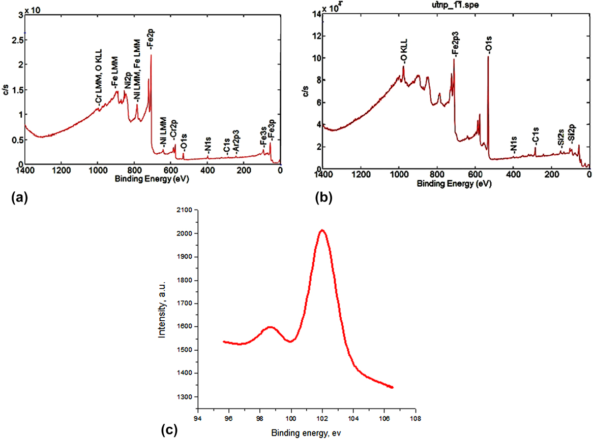
The X-ray photoelectron spectroscopy (XPS) study (PHI5000 Versa Probe II, Ulvac-Phi Inc., Chigasaki, Japan) was done on silanized samples so as to confirm the presence of Si.
2. Resorption study
The rate of bioresorption of bare nitinol and silanized nitinol was determined in relation to loss or gain as a function of time using protein-free simulated body fluid (SBF). It was prepared using the standard protocol, details of which is available in the literature.Reference Oyane, Kim, Furuya, Kokubo, Miyazaki and Nakamura35 Specimens (with dimension mentioned above) of bare nitinol and silanized nitinol were dipped in freshly prepared SBF under static condition in an incubator at 37 °C for four weeks. Over the whole course of the study, the SBF was changed once in every three days. Surface microstructures were observed using SEM to study the extent of mineralization at the top surface layer.
3. Leaching study
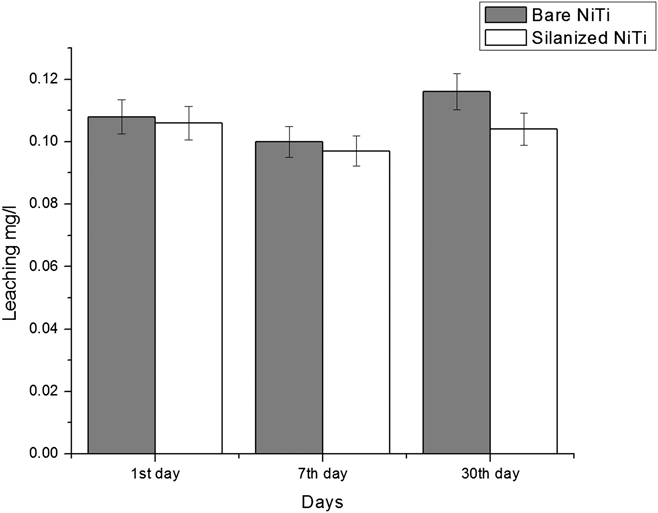
Leaching study of bare nitinol and silanized nitinol was done in a freshly prepared phosphate buffered saline (PBS)Reference Green36 for a span of one month by using atomic absorption spectroscopy (PerkinElmer, Model No. Pinaacle 900F, Waltham, Massachusetts). Data were taken intermittently on 1st day, 7th day, and 30th day.
4. Hemocompatibility study
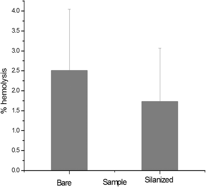
Hemocompatibility is one of the key issues as far as biological performance of the implant material is concerned. It can be defined as the property of a material that allows it to function in contact with the blood without inducing any adverse reaction. In the present study, human blood kept in citrate solution (3.8%) to prevent coagulation has been used. Blood was diluted using PBS (PBS-9.5 mL and blood-0.5 mL). Nitinol samples of definite length and weight were kept in the diluted blood suspension. 0.5 mL of 0.1 N HCl was taken as the positive control. 0.5 mL of PBS was used as the negative control. All the samples were kept inside the incubator at 37 °C for an hour and then centrifuged at 3000 rpm for 10 min. Optical density (OD) of the supernatant was taken using spectrophotometer (UV-Vis spectrophotometer Model-M1.177, Elico Ltd., Hyderabad, India) at 545 nm keeping PBS as the reference. The percentage hemolysis was calculated by using the following formula:
 $$\% \;{\rm{Hemolysis}} = {{{\rm{O}}{{\rm{D}}_{{\rm{test}}}} - {\rm{O}}{{\rm{D}}_{{\rm{nagative}}}}} \over {{\rm{O}}{{\rm{D}}_{{\rm{positive}}}} - {\rm{O}}{{\rm{D}}_{{\rm{nagative}}}}}} \times 100\quad .$$
C. In vivo study
Animal experimentation was performed as per the standard procedures of the Institutions’ Animal Ethical Committee of the West Bengal University of Animal and Fishery Sciences, Kolkata, India (Permit No. Pharma/188(ix) dated 31.07.2015). Twelve healthy New Zealand white rabbits of both genders, weighing 2–2.5 kg were randomly divided into two groups: control group I (6 animals for bare nitinol implants were inserted within the created bone defects) and the test animals, group II (6 animals in which silanized nitinol were inserted within the created bone defects).
The specimen was implanted transversely within the created defects in the distal epiphysis of femur. Prior to surgery, the rabbits kept individually in separate cages with alternating 12 h cycles of light and dark in temperature and humidity-controlled rooms, given water ad libitum and were without restriction of movement. Anesthesia was achieved by injecting a dose of 1 mg/kg body weight of xylazine hydrochloride (XYLAXIN®, Indian Immunologists Ltd., Ahmedabad, India) and ketamine hydrochloride (Ketalar®, Parke-Davis, Hyderabad, India) at a dose of 25 mg/kg body weight intramuscularly. A bone defect (5 × 1.5 mm) in each animal was created in the medial aspect of the distal epiphysis of femur bone with the help of a motorized dental drill. The implant was inserted in a press fit manner within the created bone defects and secured in position by suturing muscle, subcutaneous tissue, and skin in layers.
1. Local inflammatory reaction and healing of wound
All the animals were closely observed for lameness and weight bearing capacity, swelling of the surgical area and related signs of local inflammatory reactions were observed from the day of operation postoperatively, and changes were evaluated by visual and manual examinations. All the treated animals were administered using injection, cefotaxime sodium (Mapra India, Kolkata, India) at a dose rate 65 mg per animal intramuscularly, at 12 h interval daily for 5 days and meloxicam (MELONEX®, Intas Pharmaceuticals, Ahmedabad, India) at 0.2 mL once daily for 5 days. Dressing of surgical wounds was carried out using povidone iodine lotion and antibiotic ointment for 10 days postoperatively. Radiographs were taken just after the operation, 30th day, and on 90th day postoperatively of the operated distal epiphysis femur bone for studying the position of the implant and host bone reaction to the implant.
2. Histology study
The nitinol implanted distal femoral epiphysis was collected on the day of sacrifice for histological analysis to check the cellular response of host bone to the implants. The implanted nitinol implants along with the surrounding bones were collected from the animals after sacrificing the animal. The bone pieces (3–4 mm thick) taken from implanted area were cut (3–4 mm thick) using a hacksaw. These bone specimens from adjacent bone at the side and at the bottom of the original bone defect were collected and washed thoroughly with normal saline and were fixed in 10% formalin for 7 days. All the specimens of bone tissue were decalcified using Goodling and Stewart’s fluid (formic acid 15 mL, formalin 5 mL, and distilled water 80 mL solution), followed by fixation with 4% paraformaldehyde. Finally, the decalcified bone tissues were embedded in paraffin wax for preparation of 4 µm thick sections and stained with hematoxylin and eosin (HE). The histological scoring method was adopted to calculate bone formation semiquantitatively. A scale of 0–4 was taken to compare the cellular events with “0” indicative of absence, while “1–4” was for mild, moderate, marked, and severe, respectively. From each slide, three sites were examined by an assessor, and cross-examination was carried out by a blind observer.
3. Microstructural studies
The interface study of the bone and implant was verified by the detailed SEM study (JEOL JSM 5200 model, Tokyo, Japan). After removing the soft tissue, the implanted bone specimens were fixed in 5% glutaraldehyde phosphate solution followed by washing for 30 min with PBS (pH 7.4) and distilled water. The samples were then dehydrated in a series of graded alcohol solutions. Finally, the samples were dried with hexamethyldisilizane. A gold conductive coating was carried out by ion sputtering (JEOL ion sputter, Tokyo, Japan, model JFC 1100, Japan) at 7–10 mA and 1–2 kV for 5 min for the SEM study. The resin-mounted sample surfaces were then examined under SEM after proper alignment to assess the direction and orientation of newly formed bony tissues and distribution/absorption of materials at the defect site.
4. Oxytetracycline labeling study
The oxytetracycline labeling study was done to assess new bone formation. Fluorochrome (oxytetracycline dehydrate; Pfizer India, Mumbai, India), at a dose of 25 mg/kg body weight, was given 3 weeks before the sacrifice of the samples of 30 day and 90 days in the 2–6–2 pattern, postoperatively for double-toning of new bone. The implanted segments of the bone were collected and transverse section (2–3 mm) thickness including the implanted area was cut with the help of a hacksaw. Undecalcified ground sections were prepared as described in Ref. Reference Nandi, Ghosh, Kundu, De and Basu37 and the sections were ground to 20 µm thickness using different grades of sand paper. The specimens were kept wet by dipping in water during the entire procedure. The specimens were observed under UV light with Leica DM 2000 Bright, Leica Microsystems, Weztlar, Germany, phase contrast and a fluorescence microscope including Leica qwain software to find out the amount and source of newly formed bone. The amount of new bone formation was calculated using ImageJ software. Using this software, we measured the area that was covered by golden yellow color. The investigation was performed by taking data from three images of each group.
5. Histology of organs
To evaluate the toxic effects of these implant materials, the histology of three vital organs, viz., heart, liver, and kidney, where end products are supposed to get deposited, was studied. At the end of the study, heart, liver, and kidney of the animals were collected and preserved in 10% buffered formalin solution. The sections were treated and stained with HE solution for microscopic examination.
Statistical analysis was done on histological scores and fluorochrome labeling data between the two groups. One way analysis of variance (ANNOVA) with a Tukey’s post hoc test was carried out using ORIGIN software, National Institute of Health, Bethesda, Maryland. Absolute mean values and standard deviations were calculated. The data were taken from two samples (n = 2). An attempt has been made to compare comprehensively the in vivo performance of bare and silanized samples.
III. RESULTS AND DISCUSSION
A. In vitro studies
1. XPS study
The surface chemistry of bare and silanized nitinol has been studied by XPS. Figures 1(a) and 1(b) show the XPS spectra of bare and silanized nitinol. Bare nitinol surface mainly consists of relatively thick layer of carbon and oxygen. The survey scan reveals that surface also consists of N, Cr, Fe, and Ni. The silanized surface shows the presence of Si2p which indicates the APTES immobilization on the surface of nitinol [Fig. 1(c)]. This element was absent in the case of bare nitinol. The amount of C1s and O1s has been substantially increased, whereas the N1s is reduced in silanized surface compared to the bare nitinol.
(a) XPS of bare nitinol; (b) XPS of silanized nitinol; (c) Si2p spectra from the silanized sample.

2. Leaching study
Figure 2 shows the leaching characteristics over a span of one month. It is clearly evident that silanisation had a positive effect on controlling nickel release. With time, the superiority of the silanized sample became more evident. It hints toward the fact that silanization provides some sort of incremental barrier to nickel release, but the exact mechanism behind this gradual increment in superiority requires further in depth analysis on surface morphology and associated chemistry, to be reported in a subsequent article.
Ni+ leaching study over one month.

3. Hemocompatibility study
It is already a known fact that bare nitinol is a good hemocompatible material.Reference Roy, Choi, Yi, Moon, Lee, Han, Shin, Kamijo and Hasebe38–Reference Sinha, Pramanik, Begam and Chanda40 Figure 3 shows the hemocompatibility data of bare and silanized samples. Both samples showed high hemocompatibility. There is no significant difference between the two samples as checked by one way ANOVA analysis.
Hemocompatibility study of bare and silanized nitinol.

4. Resorption study
Apatite layer formation on both bare and silanized nitinol was clearly revealed in the present study. In both the cases, apatite formed in clusters. Apparently no difference in the quantum of apatite formation neither in its distribution pattern was evident from the figures.
B. In vivo studies
The control and experimental groups showed no inflammatory reactions postoperatively throughout the study period. In initial few days, mild lameness was observed, which is due to surgical stress. In this current study, no foreign body reaction or toxicity was revealed.
1. Histology of bone
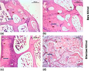
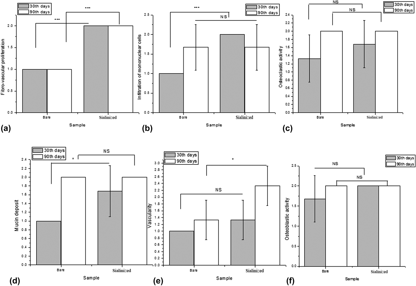
In Figs. 4(a)–4(d), histological images showed that the section taken from the defect part which was treated with bare nitinol (group-I) showed trabecular pattern of bone with few haversian systems and adjoining medullary position. The intramedullary spaces were filled up with fat cells, few mononuclear cells, and mild RBC and few osteoblasts. The vascularity inside the medullary structure was prominent in some places. But in group II samples, i.e., silanized nitinol samples, the osseous parenchyma showed the well cellular pattern of the osteogenic plate containing numerous blood vessels and phagocytic osteoclasts. The medullary structure showed fat cells, RBC, mononuclear cells, and scanty mucinious deposits. Fibrovascular proliferation was normal. After 90 days in bare nitinol samples, the bony plate showed matured cortical portion of the bony structure with few areas of remodeling and calcification. The medullary portion contained scanty amount of mononuclear cells, osteoblast, and fat cells. The haversian canal was intact in most of the places. Foreign body proliferation around the peripheral region was seen. Angiogenesis was normal. In the case of group II samples, the section depicted a well-plated bony structure consisting of numerous trabeculae along with few haversian canals and the adjoining cartilaginous matrix. The haversian canal was well marked and contained adequate amount of osteoblasts, and the bony plate formation was quite normal in the region of bony trabeculae. The medullary region contained fat cells, RBC, mononuclear cells, and scanty osteoclast. Figures 5(a)–5(f) depict clearly that in a number of terms silanized samples performed differently, sometimes better than the control group.
HE stained histological images of the (a) bare nitinol sample section after one month, (b) bare nitinol sample section after three months, (c) silanized nitinol sample section after one month, and (d) silanized nitinol sample section after three months (HC—haversian canal; OB—osteoblast; OC—osteoclast; BV—blood vessel).

(a) Bar chart of fibrovascular proliferation on the bare and silanized nitinol implant with statistical analysis; (b) Bar chart of infiltration of mononuclear cells on the bare and silanized nitinol implant with statistical analysis; (c) Bar chart of osteoclastic activity on the bare and silanized nitinol implant with statistical analysis; (d) Bar chart of mucin deposition on the bare and silanized nitinol implant with statistical analysis; (e) Bar chart of vascularity on the bare and silanized nitinol implant with statistical analysis; (f) Bar chart of osteoblastic activity on the bare and silanized nitinol implant with statistical analysis.

2. Radiology
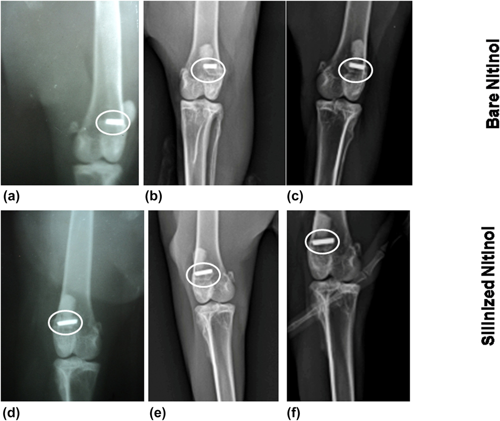
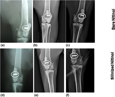
Figures 6(a)–6(f) show the postoperative radiological study. At “0” day of animal experimentation, the control group showed the presence of the radiodense implant in the cylindrical-shaped bone defect of distal epiphysis of femur of rabbit extending up to the opposite mid-cortex. The implant was more radiodenser than the bone. On 30th day, the group I implant material was observed in the similar position and the gap between the implant and bone has been reduced. Subsequently, at 90th day, the radiograph showed that the occupied implant material further reduced in diameter indicating initiation of new osseous tissue formation toward the implant. At “0” day of the experiment, the silanized nitinol implant was looked cylindrical-shaped radiodense implant in the distal epiphysis of femur extending up to mid-opposite cortex. On 30th day, the bony defect was observed with close association of the implant and host bone with slight reduction of radiodensity than what it had during “0” day. The radiograph taken on 90 days showed considerable diminution of bone–implant gap with more or less radiodensity of bone indicating firm attachment with host bone.
Radiological image of (a) bare nitinol sample on 0th day, (b) bare nitinol sample on one month, (c) bare nitinol sample on three months, (d) silanized nitinol sample on 0th day, (e) silanized nitinol sample on one month, and (f) silanized nitinol sample on three months.

3. Oxytetracycline labeling study
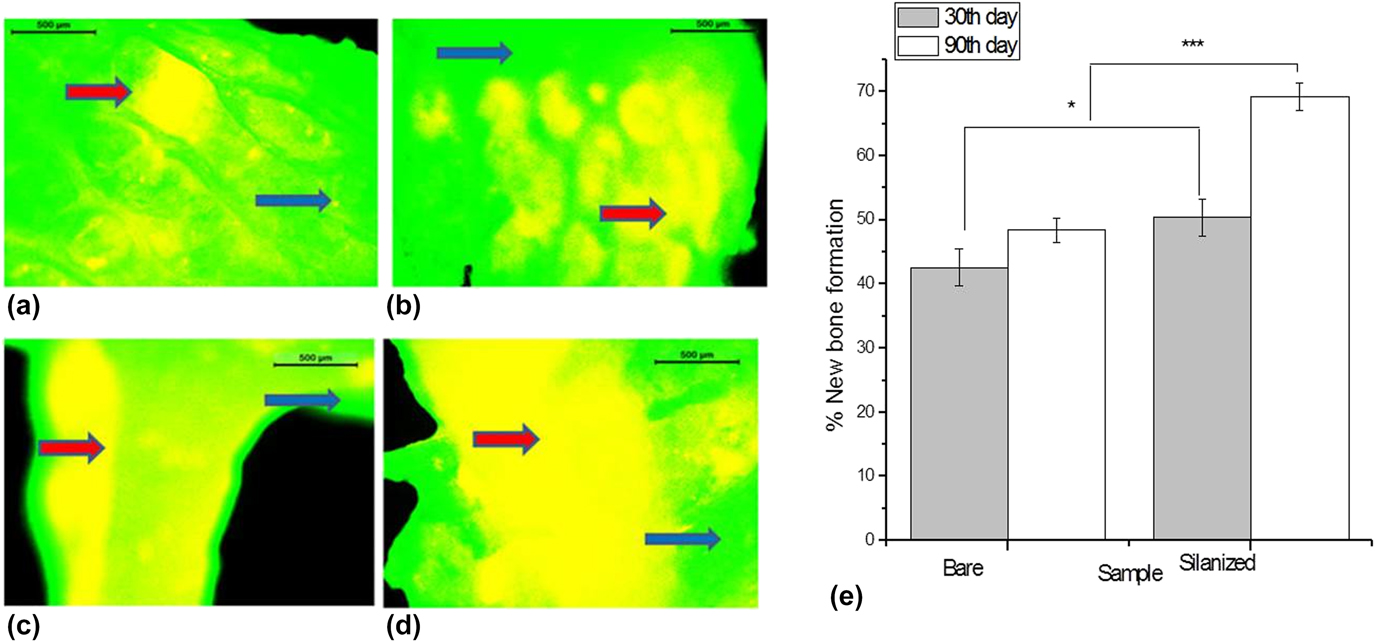
The oxytetracycline marker study revealed the new bone formation of both the groups of samples. Labeled fluorochrome images [Figs. 7(a)–7(d)] with golden yellow fluorescence imply new bone formation and sea green appearances signify host bone. At 30 day microphotographs in the bare nitinol implanted bone sample showed the presence of double tone golden yellow fluorescence in a narrow zone region in the defect site centrally and the majority of host bones depicted dark sea green homogenous color. More new osseous tissue formation (yellow fluorescence) was observed in both endosteal and periosteal side of silanized implants at this time point. On 90 days, bare nitinol implanted bone showed additional new bone formation in comparison to earlier days (30 day) of the same group. Wide and intense golden fluorescence were observed in the silanized implant at this time point indicating rapid bone formation at this time point. Apart from visual inspection and image analysis, statistical analysis [Fig. 7(e)] further confirmed the difference in the performance of bare and silanized samples.
(a) Fluorochrome labeling image of the bare nitinol sample after 1 month, (b) bare nitinol after 3 months, (c) silanized nitinol sample after one month, (d) silanized nitinol after three months; host/old bone (arrow blue) and new bone (arrow red); (e) Bar chart of new bone formation on the bare and silanized nitinol implant with statistical analysis.

4. Histology of organ
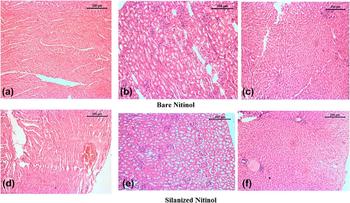
Vital organs like heart, kidney, and liver are reported to be susceptible to damage owing to nickel release. The present study took this possibility into account and did a detailed histology of all three vital organs after sacrificing the animals. Figures 8(a)–8(f) show that after 90 days of the experiment, defects treated with the bare nitinol sample showed formation of the normal network of myofibrils and adjoining matrices of heart muscles. Infiltrating cells between two cardiac muscles were very scanty. Some of the parenchyma showed mild mucinious degeneration. The other vascular structures were normal. In the case of silanized nitinol samples, the cardiac muscle fibers and surrounding stromal structure were in normal shape and size. A portion of cardiac vessels between myofibrils underwent congestion. The surrounding tissue reaction was very minimal with little infiltration of mononuclear cells.
Histological section after 3 months of the two types of implanted samples: (a) Heart sample of bare nitinol; (b) Kidney sample of bare nitinol; (c) Liver sample of bare nitinol; (d) Heart sample of silanized nitinol; (e) Kidney sample of the silanized sample; (f) Liver sample of the silanized nitinol sample.

In the section of kidney from the defects treated with bare nitinol showed that the renal structure depicted well-organised glomeruli and renal tubules of normal architecture and shape. Few tubules underwent mild degenerative changes and intratubular space was occupied by mononuclear cells. Blood vessels of the surrounding structure were normal. The section taken from the silanized nitinol sample after 90 days depicted mainly the cortical portion of kidney tissue containing sufficient glomeruli with adjoining proximal convoluted tubules and distal convoluted tubules. The medullary collecting tubule showed normal structure with mild focal infiltration of mononuclear cells.
The hepatic parenchyma from the bare nitinol sample of 90 days showed mostly normal hepatocytes with central vein and portal triads. The Kupffer cells among the hepatic spaces were minimum, although the adjoining tubules showed mucinious secretion and infiltration of mononuclear cells. But the hepatic parenchyma from the silanized sample of 90 days showed a number of central veins with numerous lobules characterized by hyaline degeneration and congestion in few central veins. The perivascular structure showed cuffing of mononuclear cell with normal angiogenesis.
5. Microstructural study
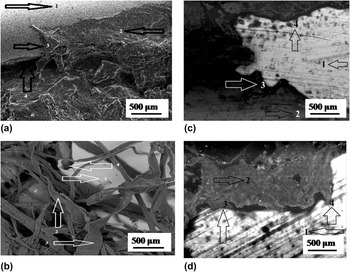
Figures 9(a)–9(d) show the SEM micrographs of the host bone–implant interface region after one and three months postoperatively. New tissue formation over the implant was observed for all specimens. But it was clearly evident that new bone tissues bridged the interface of the bone–implant more readily and more effectively in the silanized sample. Even after 30 days, there was still some gap between the bare nitinol implant and the host bone, which was not there in the case of silanized specimen. In fact the figures clearly denote how the new tissue formation gradually filled the gap and continued to spread and grow over the implant surface.
SEM micrographs of (a and b) bare nitinol sample of one and three months and (c and d) silanized nitinol of one and three months (1 denotes the nitinol implant, 2 denotes tissue, 3 denotes tissue growth over the implant, and 4 denotes the shortened gap between the implant and bone tissue).

One of the possible reasons behind the high growth rate of new bone formation in silanized nitinol, as evident from almost all the studies mentioned above, is the incorporation of silicon thorough the silanization process. From the literature, it is evident that silicon is involved in the collagen matrix formation as well as bone mineralization. Silicon is not a part of these tissues but instead acts as a catalyst in the processes. Researchers have established that silicon plays a crucial role in inter- and intracellular signaling pathways and hinder osteoclast formation vis-a-vis bone resorption interacts with inter- and intracellular signaling pathways and directly inhibits osteoclast formation and bone resorption. Silicon has the property to bind with glycosaminoglycans and helps in the formation of crosslinks between collagen and proteoglycans. From in vitro studies, it has been found that silicon stimulates type 1 collagen synthesis and osteoblast differentiation. The exact progression of mineralization is not known, but silicon plays a vital role to make the bone matrix more calcifiable. To maintain the bone strength, silica and calcium work together. Silica has the unique ability to “make the most” of available calcium, almost amplifying its effects in building bone. Consequently, it triggers the calcification process for new osseous tissue deposition. It also improves bone matrix quality and facilitates bone mineralization, so it is not surprising that fracture healing rates increase in the presence of silica, even if calcium is relatively low.Reference Beck, Ha, Camalier, Yamaguchi, Li, Lee and Weitzmann41–Reference Schwarz49
The marked superiority of the silanized samples may be attributed to this; however, further study with detailed cell–material interaction is needed before final conclusion. However, one thing is clearly evident that mere silanization of nitinol can definitely offer some specific advantages in terms of reducing leaching or promoting osseous bonding.
IV. CONCLUSION
Detailed in vivo performance of bare nitinol and silanized nitinol for their prospective use as orthopedic (intramedullary) pins has been conducted in this study. XPS study of the samples confirmed the addition of silicon through the silanization process. Both the samples’ hemocompatibility was appreciably good. Based on the microstructural, histological, radiological, and fluorochrome labeling study, as compared to bare nitinol, the silanized nitinol implant showed clear evidence of faster bone formation. Silanization increased the rate of the early stage in vivo osseointegration and bone remodeling due to the presence of silicon, a known stimulant for osseous growth. It is also evident that the silanization process can reduce the scope of leaching. The result also suggests that the silanization process can reduce the scope of leaching. Thus, this study opens up a new vista exploring the opportunity of an exclusive biomedical implant material, for better fixation at the fracture site.
ACKNOWLEDGMENTS
One of the authors (Sarmita Sinha) would like to acknowledge Council of Scientific and Industrial Research for financial support. The authors also acknowledge support from Mechanical Engineering Department and School of Bio Science & Engineering, Jadavpur University, Kolkata. The kind support from the Honorable Vice Chancellor, West Bengal University of Animal and Fishery Sciences, is gratefully acknowledged.