




No CrossRef data available.
Article contents
Effect of side of lesion on neuropsychological performance in childhood stroke
Published online by Cambridge University Press: 01 September 2004
Abstract
The purpose of the current study was to examine the effect of side of lesion on neuropsychological performance in childhood stroke. While laterality effects have been shown fairly consistently in adults who have experienced stroke, results from studies on children who have experienced childhood stroke are not as clear. Numerous methodological differences between previous studies on laterality effects in childhood stroke make it difficult to draw overall conclusions regarding laterality findings. The current study aimed to study a single group of children who experienced stroke in childhood across a number of cognitive domains. The participants were 13 children/adolescents with left hemisphere lesions and 16 children/adolescents with right hemisphere lesions, with a range of onset from prenatal to 13 years. All participants were administered a broad battery of neuropsychological tests including tests of intelligence, achievement, language skills, visuospatial skills, memory, and executive functioning. No significant differences were found between the groups on any of the measures and the calculated effect sizes were small for all but one of the measures examined. These results have implications for a greater understanding of the ability of the young brain to reorganize after childhood stroke. (JINS, 2004, 10, 698–708.)
Keywords
Information
- Type
- Research Article
- Information
- Journal of the International Neuropsychological Society , Volume 10 , Issue 5 , September 2004 , pp. 698 - 708
- Copyright
- 2004 The International Neuropsychological Society
INTRODUCTION
The study of brain–behavior relationships has long been an interest of psychologists, neurologists, psychiatrists, and cognitive scientists. Research on adults who have experienced stroke has yielded information regarding particular patterns of deficits associated with certain areas of brain damage. These studies have led to the idea of hemispheric specialization, the notion that each hemisphere of the brain is responsible for certain functions (Kolb & Whishaw, 1996).
Hemispheric specialization has been examined in terms of motor, sensory/perceptual, cognitive, and behavioral processes. The responsibility of each hemisphere for contralateral motor functioning is one of the clearest examples of brain lateralization. Adults and children who have right hemisphere or left hemisphere damage to the relevant motor tracks, are consistently left with contralateral motor deficits, while the severity of the deficit can vary.
With regard to cognitive functions, hemispheric specialization is not as clear-cut. Early thoughts regarding hemispheric specialization suggested that the left hemisphere was responsible for language while the right hemisphere was responsible for emotional and visuoperceptual functioning (Harrington, 1995). More recent research has defined the specialization of each hemisphere in a more detailed manner and has uncovered that lateralization of function in the brain is more complicated than previously believed. Studies on adults with stoke have found both hemispheres to be important for different aspects of language and visuospatial functioning. Regarding visuospatial functioning, it has been shown that the right hemisphere is more important in global processing (e.g., the gestalt of the information), while the left hemisphere seems to be important in local processing (i.e., the details of the visual stimulus) (Delis et al., 1986, 1988). Also, a dissociation in visuospatial processing has been hypothesized such that the left hemisphere is dominant for categorical spatial relations (e.g., prepositions such as in, on, behind, between, or simple phrases such as to the right of, next to) whereas the right hemisphere is specialized for coordinate relationships (i.e., precise location of objects and actual distance between objects) (Laeng, 1994). In terms of language, the left hemisphere seems to be responsible for speech production and comprehension (Bates et al., 2001; Blank et al., 2002; Damasio & Damasio, 2000), while the right hemisphere seems specialized for understanding the emotional aspects of language such as judging and expressing prosody (Ross, 1985, 2000), metaphor (Brownell et al., 1990) and humor (Brownell et al., 1983).
Research on children who have experienced focal brain damage allows for the study of brain-behavior relationships in the developing brain. This research can help answer questions regarding the nature of hemispheric specialization. Are the different hemispheres specialized for different functions from birth, as proposed by the invariant lateralization model (Kinsbourne, 1975)? Or are the hemispheres equally capable of performing different functions at birth and that after time and experience the hemispheres develop their specialization, as described by the theory of equipotentiality (Lenneberg, 1967)?
The answers to these questions regarding laterality have broader implications for a greater understanding of brain plasticity. The literature on brain plasticity suggests that the young brain is capable to some degree of reorganization after early damage. It appears that pre- or perinatal focal brain damage results in early diminished performances on numerous cognitive abilities, such as language acquisition, spatial cognition, and affect (Stiles, 2000a). However, some cognitive functions (e.g., language) show different patterns of lesion site/cognitive symptoms than that which is seen in adults and the cognitive deficits significantly improve over time (Bates, 1999). This suggests a compensatory mechanism in the brain to recruit other brain areas to take on these functions. Other cognitive abilities (e.g., spatial) mimic deficit patterns seen in adults and continue to show persistent process deficits over time, suggesting a more inflexible system (Akshoomoff et al., 2002; Stiles, 1995).
There are a number of ideas as to the mechanism behind the brain's ability to partially reorganize after early damage. Some have suggested a critical period during which time the brain is quite plastic (Bates, 1999; e.g., during the critical period for acquiring language, children can easily learn a second language; Bialystok & Hakuta, 1994; Johnson & Newport, 1989). If a lesion occurs before or during the critical period, the young brain is capable of reorganizing to compensate for the damage, resulting in less severe deficits, than if the damage occurs after the critical period. However, there is evidence accumulating that language acquisition does not appear to have a critical period, but instead follows a U-shaped curve, with better language outcome if the lesions occur congenitally or between 5 and 12 years of age and worse outcome if the lesions are acquired between 1 and 5 years of age (Goodman & Yude, 1996).
Another hypothesis is based on the idea of the brain reorganizing in such a way as to spare certain functions while crowding other cognitive functions out. This hypothesis suggests that when one area of the brain is damaged, that function is taken over by an analogous area (perhaps on the other side of the brain). Thus the original function is spared, however, the abilities that used to be performed by this newly recruited region become crowded out and the individual may be left with some diminished cognitive capacity (Stiles, 2000a; Teuber, 1974). For example, in some cases, early left hemisphere damage has led to the right hemisphere taking over language functions, but the individual was left with impaired visuospatial abilities (a more traditionally right hemisphere mediated task; Teuber, 1974). Although the questions that may be answered by research on the impact of stroke on the developing brain are important, the studies are limited due to the rarity of childhood stroke.
In general, children who suffer unilateral focal brain damage are left with milder deficits than those experienced by adults (Bates et al., 1999b, 2001; Trauner et al., 1993; Trauner & Mannino, 1986; Wulfeck et al., 1991). However, the pattern of deficits with regard to lateralization remains unanswered. Some studies have found that unilateral focal brain damage to either hemisphere is likely to result in a similar pattern of results on cognitive testing. A review by Hogan et al. (2000) on intelligence after childhood stroke concludes that there are no differences in IQ scores for children who experience early left hemisphere or right hemisphere stroke. This conclusion is based largely on two studies of children with early stroke. A study by Vargha-Kadem and Van Der Werf (1992) examined 82 children with congential or perinatal hemispheric cerebral palsy and found no differences between the left hemisphere and right hemisphere groups in Verbal IQ, Performance IQ, or Full Scale IQ (excluding children who were experiencing seizures). Another study by Goodman and Yude (1996) studied 149 children with pre- or perinatal (up to age 5 years) hemiplegia and failed to find any differences between the right-sided and left-sided groups. A study of pediatric ischemic stroke by Block et al. (1999) found that individuals in their left lesion and right lesion group exhibited a similar pattern of performance on verbal memory, functional memory, and speed of processing (range of age at lesion: 0.50 years–15.08 years). Another study by Ballantyne et al. (1994) also found no significant differences between children who experienced early (prenatal or perinatal) right hemisphere or left hemisphere focal brain damage in VIQ, PIQ, or FSIQ.
In contrast, other studies have found that damage to either the left or right hemisphere results in a pattern of results similar to adults (i.e., right hemisphere damage leads to visuospatial deficits and left hemisphere damage leads to language deficits). A study by Kolk and Talvik (2000) studied 37 children with congential hemiparesis on the NEPSY: A Developmental, Neuropsychological Assessment (Korkman et al., 1998). Their results suggest that the side of damage is important in predicting the profile of cognitive disorders with left hemisphere damage causing impairments in language and phonology whereas right hemisphere damage causes greater impairments in attention and visuospatial skills (Kolk & Talvik, 2000). Another study on language development found that prenatal or perinatal focal brain lesions in the left hemisphere resulted in greater deficits in the development of expressive lexicon and grammar when compared to children with right hemisphere damage (Chilosi et al., 2001). A detailed study on preterm children with unilateral intracranial hemorrhage detected during the neonatal period (first 5 days of life), found a pattern of cognitive asymmetry similar to adults, i.e., left hemisphere damage decreased Verbal IQ relative to Performance IQ whereas right hemisphere damage decreased Performance IQ relative to Verbal IQ (Raz et al., 1994).
Why is there so much discrepancy in the results of studies on children with early brain damage? The answer may lie in the many methodological differences between the studies themselves. For example, inclusion/exclusion criteria for participation as a stroke subject vary greatly. Some studies only use observation of a hemiparesis and medical history to presume a prenatal or perinatal ischemic event, while other studies verify the presence of a focal, unilateral lesion with a neuroimaging technique (i.e., CT or MRI). Also there were differences in the inclusion of strokes of differing etiologies (e.g., cardiac) and age at time of stroke (prenatal, perinatal, later). There are also differences in the types of tests used to measure cognitive functions. Some studies use standardized, more general clinical neuropsychological measures while others use very detailed, experimental tests that are designed to dissect specific areas of cognition. Also, there were differences in the types of comparisons made; for example, some studies compared the performance of the lesion subjects to a control group, some studies compare left hemisphere lesion and right hemisphere lesion group directly, and others use asymmetry scores within individual participants. Given the complicated nature of plasticity and reorganization of the developing brain, these methodological differences between studies may have dramatic effects on the results and conclusions that are drawn from them.
Additionally, most of the studies that have examined brain lateralization in children after stroke have focused on performance on one test (e.g., WISC performance) or small battery of tests covering a specific cognitive domain (e.g., language or visuospatial functioning). Since many of these studies are examining a theoretically “different” group of children (e.g., based on inclusion/exclusion criteria) and using different types of neuropsychological measures, it stands to reason that the results and conclusions drawn from those studies are quite disparate and at times contradictory.
The purpose of the current study was to examine the presence or absence of laterality effects across different cognitive domains using a single sample of children who experienced unilateral focal brain lesions. This study aimed to comprehensively examine hemispheric specialization in children who have experienced an early cerebral insult using a wide range of neuropsychological tests that measure different cognitive functions. This research has implications for a greater understanding of brain plasticity and the ability of the brain to reorganize after focal damage during childhood.
METHODS
Research Participants
The neuropsychological data used in this study were collected as part of a larger study examining psychiatric outcomes, neuropsychological functioning, family functioning, and adaptive functioning in children with strokes. Stroke subjects were included in the study if they experienced stroke prenatally or during childhood. Early lesions were classified as occurring prenatally or up to 12 months of postnatal life and lesions were categorized as late if they were acquired at age 12 months or later.
Inclusion criteria for stroke cases were (1) neuroimaging documentation of a focal, non-recurrent and non-progressive supratentorial brain parenchymal lesion caused by a stroke before age 14; (2) subjects aged 5–19 years at the time of the assessment; (3) 1 year or more since stroke; and (4) English as first language. The following exclusions were applied: (1) neonatal bleeds (e.g., intraventricular hemorrhages, germinal matrix hemorrhages) potentially associated with prematurity; (2) neonatal watershed infarcts associated with hypoxia; (3) hemoglobinopathies; (4) progressive neurometabolic disorders; (5) Down's syndrome and other chromosomal abnormalities; (6) malignancy; (7) congenital hydrocephalus; (8) shunts; (9) congenital and acquired CNS infections; (10) clotting factor deficiency; (11) stroke in a pregnant minor; (12) transplant status; (13) cerebral cysts; (14) trauma; (15) transient ischemic attack; (16) moya-moya; (17) severe and profound mental retardation; (18) quadriplegia, triplegia, or diplegia diagnoses; (19) syndromatic vascular malformations (excluding A–V aneurysm ruptures); (20) systemic lupus erythematosus; and (21) multiple lesions (unless in close proximity).
A pediatric neurologist supervised a record review guided by the ICD 9 codes for stroke, and congenital cerebral palsy. These procedures yielded 49 apparently eligible subjects. One male with a stroke at age 7 months was found upon screening to have severe mental retardation and was therefore ineligible to participate. We were able to locate 32 of the remaining 48 subjects and we studied 30 subjects. The research MRI revealed subtle bilateral lesions in 1 subject who had to be excluded. The parents of 2 prenatal stroke female subjects aged 9 and 11 years declined participation. The 16 children not located were demographically comparable to the sample (e.g., age, race, gender, timing of stroke) and did not differ from the participant group on lesion variables including laterality, etiology, mechanism (occlusive vs. hemorrhagic), or location (cortical vs. subcortical).
In all, 29 participants with stroke, including 13 with left hemisphere lesions and 16 with right hemisphere lesions, were evaluated. Twenty-seven participants were Caucasian, 1 participant in the left hemisphere lesion group was White–Asian and 1 participant in the right hemisphere lesion group was White–Hispanic. Of the 29 individuals, 17 experienced early lesions and 12 had late lesions. The mechanisms of stroke were occlusive in 21 cases and hemorrhagic in 8 cases. Etiology included 15 idiopathic occlusive cases, 2 idiopathic hemorrhagic cases, 4 cases related to congenital heart disease (3 after cardiac surgery or catheterization and 1 after varicella zoster infection), 5 cases of arterio-venous malformation rupture, 1 case of ruptured angioma, 1 case possibly linked to comorbid ulcerative colitis, and 1 case followed a varicella infection. The distribution of the brain lesions included 7 cases of predominantly putamen lesions, 9 large middle cerebral artery (MCA) distribution infarcts including deep gray structures, 10 smaller MCA distribution frontotemporal or temporoparietal lesions sparing the deep gray, and 3 cases of parietal or parieto–occipital strokes. All individuals were recruited from one university hospital.
Neuroimaging
Protocol magnetic resonance imaging (MRI) scans were obtained (T1-weighted volumetric mode, SPGR/400, TR = 26, TE = 7, matrix 256 × 192, NEX = 2, 1.5 mm thickness with no skip; T2-weighted multi-echo, FSE/V, TR = 2350, TE = 17/102, matrix 256 × 192, NEX = 1, 5 mm skip 1 mm). Twenty-six of 29 stroke subjects underwent research scans that were the basis of their lesion location analyses. The other 3 subjects who could not have a research MRI (due to refusal, concern about intracerebral metallic clips, and equipment failure respectively) had lesion location determined from previous clinical CT scans (2) or MRI scan (1).
Lesion volume was computed in absolute units (cm3) before and after normalization for intersubject differences in brain size (Lancaster et al., 1995; Max et al., 2002a).
Measures
Intelligence assessment
Wechsler Intelligence Scale For Children–Third Edition (Wechsler, 1991). IQ was measured using the Wechsler Intelligence Scale for Children–Third Edition (WISC–III). Two verbal and 2 performance subtests were administered (Information, Similarities, Block Design, and Picture Arrangement) from which estimated Verbal IQ and estimated Performance IQ were computed.
Academic Achievement
Wide Range Achievement Test–Revised (Jastak & Wilkinson, 1984). Academic achievement was measured using the Wide Range Achievement Test–Revised (WRAT–R; Jastak & Wilkinson, 1984). This test yields age adjusted standard scores in the areas of Reading, Spelling, and Arithmetic. For the Reading subtest, participants are required to recognize letters and read single words. The Spelling subtest required participants to copy simple geometric shapes, write their name, and spell single words. Participants were asked to complete mathematical problems of increasing complexity (e.g., counting, addition, subtraction, multiplication, division, fractions, decimals, etc.) for the Arithmetic subtest.
Language Skills
Multilingual Aphasia Examination (MAE) Sentence Repetition (Benton et al., 1994). MAE Sentence Repetition was administered to assess immediate verbal memory. Participants were read 14 sentences of increasing length and grammatical complexity and asked to repeat the sentence immediately after the examiner. For the purposes of the current study, this percentile was transformed into an estimated Standard Score using a standard conversion chart. In cases where a percentile corresponded to more than one standard score (e.g., near the upper and lower tails of the distribution), the highest standard score was assigned to the subject.
MAE Token Test (Benton et al., 1994). The MAE Token Test was administered to evaluate verbal comprehension and ability to carry out oral commands. Twenty blocks of varying colors, sizes, and shapes were presented. The participant was given commands of increasing complexity that require him/her to perform certain tasks with the blocks. See MAE Sentence Repetition for description of procedure used to obtain estimated standard scores.
Test of Written Language–Third Edition (Hammill & Larsen, 1996). The Test of Written Language–Third Edition (TOWL–3) was used to assess written language abilities. Participants were required to spontaneously write a story about a picture that is presented to them. Three aspects of the written story were then scored: Contextual Convention (spelling, punctuation, capitalization), Contextual Language (vocabulary, grammar, syntax), and Story Construction (composition of story, such as plot and organization). These three scaled scores were combined as described in the manual to obtain a Spontaneous Writing Quotient Standard Score.
Visuospatial skills
Developmental Test of Visual–Motor Integration (Beery, 1989). The Developmental Test of Visual–Motor Integration (VMI) was administered to assess visual-motor integration. The participant's task during the VMI was to copy geometric figures of increasing complexity. The figures were rated and a total score was computed which was then transformed into an age-adjusted Standard Score.
Memory
California Verbal Learning Test–Children's Version (Delis et al., 1994). The California Verbal Learning Test (CVLT–C) is a standardized measure of verbal learning. This test evaluates an individual's ability to learn a list of 15 words in three categories (toys, fruits, and clothing) over five learning trials. A second set of 15 words (a distractor list) is then presented for one trial, immediately followed by free and category cued recall of the first list. After a 20-min delay, free and cued recall and recognition of the first list is tested. The T score for the total number of words learned on the five learning trials was calculated and used as a measure of overall verbal learning ability.
Rey-Osterrieth Complex Figure Test (Meyers & Meyers, 1995). The Rey-Osterrieth Complex Figure Test (REY-O) was administered as a measure of visual memory. Participants were shown the Rey Complex Figure and asked to copy the figure while it remained in view. After the figure was removed, they were asked to draw it immediately from memory and again after a 20-min delay. The 20-min delay figure was scored using the 36-point scoring system and the raw score was transformed into an age-adjusted T score.
Executive functioning
Design Fluency (Jones-Gotman & Milner, 1977). The Design Fluency task was administered to assess numerous executive functions such as cognitive flexibility, creativity, constructional abilities, and working memory (Spreen & Strauss, 1998). Participants were required to make up as many different drawings as possible which were not real objects, geometric figures, or scribbles. There was a free condition (i.e., draw as many figures as possible in three minutes) and a fixed four line condition, in which the figures had to be comprised of four lines. An age-adjusted T Score was derived based on the participants overall performance.
Multilingual Aphasia Examination (MAE) Controlled Oral Word Association (COWA;Benton et al., 1994). Phonemic fluency was assessed by the MAE Controlled Oral Word Association (COWA) which required participants to generate as many words that begin with the letters C, F, and L. The executive functions tapped by phonemic fluency are “initiation, simultaneous processing, and systematic retrieval of phonemically similar lexical items” (Delis et al., 2001). Using the MAE manual (Benton et al., 1994), a performance percentile was assigned to the raw score based on the child's grade level. Estimated Standard Scores were obtained in the manner described for MAE Sentence Repetition.
Wisconsin Card Sorting Test (Heaton et al., 1993). The Wisconsin Card Sorting Test (WCST) was administered as a measure of problem solving ability. It requires many aspects of executive functioning including cognitive set-shifting, planning, the ability to use feedback to modify behavior, and inhibiting impulsive responding (Spreen & Strauss, 1998). The test consists of four stimulus cards that vary along the dimensions of color, geometric shape, and number. The participant was required to match the cards from a stack in front of him/her to the stimulus card that he/she believed it matches. The examiner does not tell the subject what the rule is (e.g., match on color, shape, or number). After the participant placed a card below a stimulus card, the examiner gave feedback (i.e., either correct or incorrect). The participant was required to use the examiner's feedback to determine the rule. After the subject successfully acquired the rule, the examiner switched the rule and the participant was required to learn the new rule based on the examiner's feedback. Age adjusted standard scores were calculated for Perseverative Errors.
Scoring
Since participants in this study ranged in age from 5.92 years to 19.75 years, some tests were given to individuals who were younger or older than the test age range. Consistent scoring rules were applied such that data from children who fell below the lowest normative age range was scored using the youngest normative age range available for the test and data from individuals who fell above the highest normative age range was scored using the oldest normative age range available.
Statistical analyses
A power analysis was conducted to estimate the sample size necessary to have adequate power to find differences between the groups. Upon review of the literature, there was a wide range of effect sizes from small (under 0.35) to large (over 1.0; Cohen, 1988). Using the estimates from the literature, as well as the largest effect size we found in our data set (.67) expected effect sizes between the left hemisphere and right hemisphere groups were estimated to be within the medium range (approximately .65). For a medium effect size, the current study is underpowered (would need 30 subjects per group), although there is ample power to find large effects, which have been found in some studies on childhood stroke. Since the current study may be underpowered, effect sizes have been provided which may be the best estimate of the size of the differences between the left hemisphere and right hemisphere groups on the measures included in this study.
Univariate ANOVAs were also used to statistically examine the effect of side of brain damage (left hemisphere or right hemisphere) on each measure included in the study. Age at lesion onset (in years) was used as a covariate to control for any differential effects of earlier versus later onset of stroke. Although numerous statistical analyses were run, alpha was set at .05 to allow the greatest sensitivity for the detection of a difference between groups. Demographic information was analyzed using Fisher's Exact tests and lesion volume was analyzed using a Mann-Whitney U test because the data are non-parametric.
RESULTS
Demographics
Table 1 shows demographic, lesion, and imaging characteristics of the left hemisphere lesion (LH) and right hemisphere lesion (RH) groups. The groups were not significantly different across these domains.
Participant demographic and lesion information and significance values for the left hemisphere and right hemisphere lesion groups

Neuropsychological Test Performance
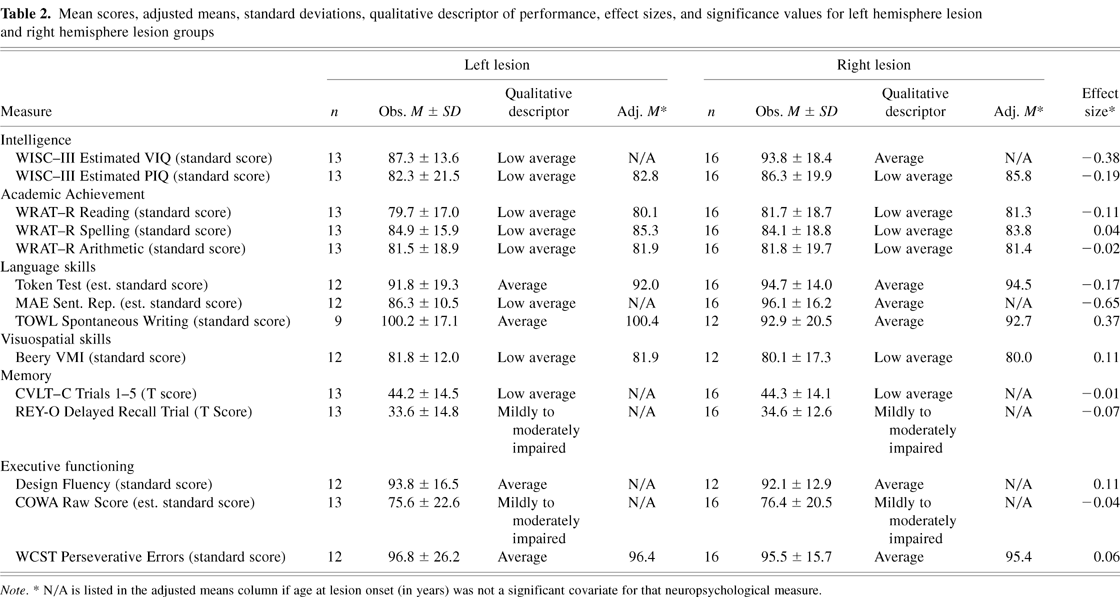
There were no significant differences found between the performance of the LH and RH groups on any of the measures administered. See Table 2 for mean scores, adjusted means (for measures in which age at lesion onset was a significant covariate), effect sizes, and qualitative descriptors of performance for each group. Not only were no significance differences found between the groups, but the effect sizes are all very small, with the exception of one medium effect size (Sentence Repetition; mean effect size = .17, range = .01–.67).
Mean scores, adjusted means, standard deviations, qualitative descriptor of performance, effect sizes, and significance values for left hemisphere lesion and right hemisphere lesion groups

DISCUSSION
The current study illustrates a relative lack of laterality effects in a group of children who experienced childhood stroke. There were no significant differences on any of the tests administered between children with left hemisphere and right hemisphere brain damage who had similar imaging and demographic characteristics. These results are quite different from the pattern of performance that would be expected in adults with stroke, that is, individuals with right hemisphere damage may perform more poorly on visuospatial tasks and individuals with left hemisphere damage may perform more poorly on language tasks.
The lack of performance differences in different cognitive domains between individuals with LH and RH damage during childhood, yield evidence for some degree of plasticity in the developing brain. These results suggest that if focal brain damage occurs during childhood, the brain is able to reorganize in such a way that the individual is not left with the striking lateralized deficits that are often seen in adults who experience stroke (e.g., aphasia, agnosia).
Interestingly, while no pattern of performance associated with either LH or RH childhood stroke was observed, both groups performed in the low average range on the majority of tests administered in the current study. In our effort to focus on lesion laterality and constrain the number of statistical tests, we have not shown the comparisons between stroke patients (combined LH and RH) and controls that were recruited as part of the larger study (Max et al., 2002b). However, as we shall show in future manuscripts, the stroke group performed more poorly than the controls at a statistically significant or trend level in 12 of the 14 measures studied here. This suggests that while the young brain is capable of some degree of reorganization, the resulting neurocognitive capacity of the brain is decreased by the damage. Our results suggest that these children are not left with the severe focal cognitive deficits with the sparing of other cognitive abilities that is seen in adults. Instead, it appears from the current findings that children who suffer focal brain damage in childhood are left with milder but more diffuse level of deficits that span a number of cognitive domains (i.e., general cognitive functioning, language, visuospatial skills, executive functioning, and academic achievement; Bates & Roe, 2001).
These results therefore do not support either the “invariant lateralization” theory or the “equipotentiality” theory, but rather lend evidence towards the newer “emergenist view” of brain reorganization after early focal damage (Bates & Roe, 2001). Invariant lateralization refers to the idea that the hemispheres are specialized from birth (or earlier) to perform different cognitive tasks. If the invariant lateralization theory were to hold true in this sample, we would expect to see a pattern of results similar to those seen in adults, with a pattern of certain preserved functions along with striking deficits. The equipotentiality theory suggests that either hemisphere is capable of performing different cognitive tasks and the hemispheres become specialized through time and experience. If our data were to support this theory, we would expect average performance across the cognitive domains, since lesions sustained in childhood allow much time for the brain to reorganize and compensate for the damaged area.
Instead, our data support the emergentist view which suggests that, at birth, the hemispheres have innate biases toward processing certain types of information but that neural and behavioral reorganization can occur across the course of development (Bates & Roe, 2001). Data supporting the emergentist view has been found in studies of children who have experienced early stroke, in particular in the area of language. Studies on children who have experienced a single, unilateral pre- or perinatal stroke found that these children perform lower than age-matched controls on IQ and language measures but found no differences in performance between the LH and RH lesion groups (Bates & Roe, 2001; Bates et al., 1999a, 1999b; Dick et al., 1999; Kempler et al., 1999). The results of the present study are consistent with the IQ and language results of the above-mentioned research, however, our study covers a wider range of cognitive functions, including intelligence, language skills, visuospatial skills, memory, and executive functioning. Interestingly, the rigorous inclusion/exclusion criteria implemented in the current study and the studies mentioned above are quite similar and thus these samples may be fairly comparable, eliminating some of the difficulty in generalization between other studies on childhood stroke.
One of the strengths of the current study is that it focuses on examining patterns of performance between children with LH and RH focal brain damage on a single group of children over a wide range of cognitive domains. Past studies that have examined the effect of childhood stroke on a single cognitive domain (e.g., IQ: Ballantyne et al., 1994; Goodman & Yude, 1996; Hogan et al., 2000), language skills (Bates & Roe, 2001; Bates et al., 1997b, 1999b; Reilly et al., 1998), visuospatial skills (Akshoomoff et al., 2002; Schatz et al., 2000; Stiles, 2000b), and behavioral profiles (Goodman & Yude, 1997; Max et al., 2002a, 2002b; Trauner et al., 1996, 2001). The strength of the current study lies in its breadth. It might be expected that by chance alone, a difference between children with LH and RH stroke might have been observed. The fact that the no significant difference was observed on any measure speaks towards the plasticity of the young brain.
It is important to note that statistically, the null hypothesis can not be proven. While no differences between the groups were found on this study, we are unable to conclude that no differences exist. There are many factors that may account for the lack of significant findings. The groups have a small number of subjects across a wide age range; the lesions are in different locations and are of different etiologies. However the small magnitude of the effect sizes found in the current study may help alleviate some concerns regarding simply not finding a significant difference between our groups because of the small sample size. Due to the rarity of childhood stroke, these methodological concerns are inherent in studying this population. Therefore, it is important to view the results in the light of these limitations and be cautious in the interpretation. However, the consistent pattern that is seen across almost every measure in this study may reduce concerns regarding possible methodological flaws or chance occurrence as a sole explanation of these findings. It is also scientifically important to report null findings in the literature to reduce possible publication bias, that is, “selective publication, in which the decision to publish is influenced by the results of the study” (Begg, 1994). Although the results of the current study are not “statistically significant,” we believe they are clinically significant and important in furthering our understanding of focal brain damage during childhood.
Another strength of the current study is its use of clinical neuropsychological measures to assess performance in the different cognitive domains. These tests are often used to draw conclusions about a child's cognitive ability and make recommendations regarding school placement, special services, and vocational ability. Our results suggest that children who have experienced stroke are not left with a striking pattern of lateralized strengths and weaknesses on these measures. However, this is not to say that more subtle differences between the groups do not exist. It is quite possible that by using more specific experimental measures that are designed to tax certain cognitive abilities we may find a pattern of results that resembles, to a much milder degree, the pattern of hemispheric specialization seen in adults.
For example, a study by Schatz et al. (2000) on children with early focal brain damage found that while there were no differences on overall performance on the WISC–R Block Design subtest between children with right hemisphere or left hemisphere stroke, the types of errors made by each group followed the pattern observed in adults with stroke (i.e., left hemisphere damage led to more local errors, whereas right hemisphere damage led to more global errors). Detailed analyses of language development between the ages of 10 and 17 months in children who experienced early focal lesions found that RH damage was more likely to result in receptive language delays, whereas LH temporal lobe damage was associated with a delay in expressive vocabulary (Bates et al., 1997a). These findings on a detailed analysis of language acquisition illustrate a complex pattern of findings in which side of lesion is predictive of performance, although the pattern is quite different from what would be expected in adults.
One of the limitations of the current study is that it used a broad battery to answer a number of scientific questions. A follow-up study should include more exacting measures reflecting laterality features; for example, a dichotic listening test. Another limitation is that only side of lesion was examined rather than more specific lesion location (e.g., MCA, basal ganglia, frontal vs. posterior). These types of analyses were not included in the current study in order to avoid additional comparisons. More detailed analyses on cognitive outcome as related to specific lesion locations will be important in a future study on a larger sample of children with stroke. Also, participants in the study varied substantially in their age at testing. There is certainly a significant amount of brain development that occurs between the early school age years and early adulthood. There is no question that performances on any of the measures are quite different when comparing the actual performances of a 7-year-old and a 17-year-old. Using standard scores allows us to obtain a score that takes into consideration what would be a typical performance at each age. In a future study, it would be helpful to examine cognitive performance on a group of children in a smaller age range in order to examine more specific hypotheses regarding the effects of development on cognitive performance. For example, do individuals with childhood stroke catch up over time or fall further behind their same-age peers? Or do individuals with childhood stroke use different cognitive strategies than their same age peers without childhood stroke?
While this sample may appear quite heterogeneous, great care was taken to use stringent inclusion and exclusion criteria in order to obtain a relatively homogenous sample for this rare population. It is difficult to balance the need for statistical power, hence a large sample size, with the need for a homogeneous sample, so as to ease the interpretation and generalizability of the results. Since childhood stroke is relatively rare, we have attempted to balance the competing needs of having both a large and homogenous sample. The results from the current study should be examined more closely using a larger, more homogeneous group of children with stroke. The difficulty in recruiting an ample number of similar individuals argues for the need for a larger, multi-site study in which the recruitment efforts can be combined between a number of investigators. This may be one way of alleviating this problem in the future.
Future studies should focus on a more detailed analysis of cognitive functioning in childhood stroke. For example, increasing the complexity of the measures administered or expanding the scoring to include more detailed error analyses may allow for a more fine-grained analysis of any differences in the cognitive processes utilized by children with LH or RH lesions. Also, it would be interesting to include tests of lateralized motor functioning since these functions may be less subject to reorganization than other skills. Another step may be attempting to correlate more detailed lesion location, rather than LH or RH, with specific patterns of performance. Furthermore, functional neuroimaging studies will be invaluable in answering the question of how the young brain is able to reorganize and which parts of the brain, if any, take over functioning for damaged areas.
ACKNOWLEDGMENTS
We would like to acknowledge the support of Katherine Mathews for subject selection, Farcundo Manes for lesion identification, Peter Fox and Jack Lancaster for neuroimaging analysis, Amy Schatz and Amy Lansing for data management, and Damien Ihrig for data collection. This work was supported by a NARSAD Young Investigator Award and a Career Development Award (K-08) MH 01800-05 to Dr. Max.
References
REFERENCES
Akshoomoff, N.,
Feroleto, C.,
Doyle, R., &
Stiles, J.
(2002).
The impact of early unilateral brain injury on perceptual
organization and visual memory.
Neuropsychologia,
40,
539–561.CrossRefGoogle Scholar
Ballantyne, A.,
Scarvie, K., &
Trauner, D.
(1994).
Verbal and performance IQ patterns in children after perinatal
stroke.
Developmental Neuropsychology,
10,
39–50.CrossRefGoogle Scholar
Bates, E.
(1999).
Plasticity, localization, and language development. In
S.H. Broman &
J.M. Fletcher (Eds.),
The changing nervous system (pp.
214–253).
New York:
Oxford University Press.
Bates, E.,
Reilly, J.,
Wulfeck, B.,
Dronkers, N.,
Opie, M.,
Fenson, J.,
Kriz, S.,
Jefferies, R.,
Miller, L., &
Herbst, K.
(2001).
Differential effects of unilateral lesions on language production
in children and adults.
Brain and Language,
79,
223–265.CrossRefGoogle Scholar
Bates, E. &
Roe, K.
(2001).
Language development in children with unilateral brain damage.
Handbook of developmental cognitive neuroscience (pp.
281–307).
Cambridge, MA:
MIT Press.
Bates, E.,
Thal, D.,
Trauner, D.,
Fenson, J.,
Aram, D.,
Eisele, J., &
Nass, R.
(1997a).
From first words to grammar in children with focal brain injury.
Developmental Neuropsychology,
13,
447–476.Google Scholar
Bates, E.,
Vicari, E., &
Trauner, D.
(1999a).
Neural mediation of language development: Perspectives from lesion
studies of infants and newborns. In
H. Tager-Flushberg (Ed.),
Neurodevelopmental disorders (pp.
533–581).
Cambridge, MA:
MIT Press.
Bates, E.,
Vicari, S., &
Trauner, D.
(1997b).
Neural mediation of language development: Perspectives from lesion
studies of infants and children. In
H. Tager-Flushberg (Ed.),
Neurodevelopmental disorders: Contribution to a new framework from
the cognitive sciences (pp.
533–581).
Cambridge, MA:
MIT Press.
Bates, E.,
Wulfeck, B.,
Opie, M.,
Fenson, J.,
Kriz, S.,
Reilly, J.,
Dronkers, N.,
Miller, L.,
Jefferies, R., &
Herbst, K.
(1999b).
Comparing free speech in children and adults with
left-vs.right-hemisphere injury.
Brain and Language,
69,
377–379.Google Scholar
Beery, K.E.
(1989).
The VMI: Developmental Test of Visual–Motor Integration, 3rd
Revision.
Cleveland, OH:
Modern Curriculum Press.
Begg, C.B.
(1994).
Publication bias. In
H. Cooper &
L.V. Hedges (Ed.),
Handbook of research synthesis (pp.
399–409).
New York:
Russell Sage Foundation.
Benton, A.L.,
Hamsher, K. de S., &
Sivan, A.B.
(1994).
Multilingual Aphasia Examination, Third Edition.
Iowa City, IA:
AJA Associates.
Bialystok, E. &
Hakuta, K.
(1994).
In other words: The science and psychology of second-language
acquisition.
New York:
Basic Books.
Blank, S.C.,
Scott, S.K.,
Murphy, K.,
Warburton, E., &
Wise, R.
(2002).
Speech production: Wernicke, Broca and beyond.
Brain,
125,
1829–1838.CrossRefGoogle Scholar
Block, G.W.,
Nanson, J.L., &
Lowry, N.J.
(1999).
Attention, memory and language after pediatric ischemic stroke.
Child Neuropsychology,
5,
81–91.Google Scholar
Brownell, H.,
Simpson, T.,
Bihrle, A.,
Potter, H., &
Gardner, H.
(1990).
Appreciation of metaphoric alternative work meanings by left and
right brain-damaged patients.
Neuropsychologia,
28,
375–384.CrossRefGoogle Scholar
Brownell, H.,
Michel, D.,
Powelson, J., &
Gardner, H.
(1983).
Surprise but not coherence: Sensitivity to verbal humor in
right-hemisphere patients.
Brain and Language,
18,
20–27.CrossRefGoogle Scholar
Chilosi, A.M.,
Cipriani, P.,
Bertuccelli, B.,
Pfanner, L., &
Cioni, G.
(2001).
Early cognitive and communication development in children with
focal brain lesions.
Journal of Child Neurology,
16,
309–316.CrossRefGoogle Scholar
Cohen, J.
(1988).
Statistical power analysis for the behavioral sciences.
Mahwah, NJ:
Lawrence Erlbaum Associates.
Damasio, A. &
Damasio, H.
(2000).
Language and the brain. In
K. Emmorey &
H. Lane (Eds.),
The signs of language revisited: An anthology to honor Ursula
Bellugi and Edward Klima (pp.
477–491).
Mahwah, NJ:
Lawrence Erlbaum Associates.
Delis, D.,
Kaplan, E., &
Kramer, J.
(2001).
The Delis-Kaplan Executive Function System (D-KEFS) Examiner's
manual.
San Antonio, TX:
The Psychological Corporation.
Delis, D.,
Kiefner, M., &
Fridlund, A.
(1988).
Visuospatial dysfunction following unilateral brain damage:
Dissociations in hierarchical and hemispatial analysis.
Journal of Clinical and Experimental Neuropsychology,
10,
421–431.CrossRefGoogle Scholar
Delis, D.,
Kramer, J.,
Kaplan, E., &
Ober, B.
(1994).
CVLT–C: California Verbal Learning Test–Children's
Version.
San Antonio, TX:
The Psychological Corporation.
Delis, D.,
Robertson, C., &
Efron, R.
(1986).
Hemispheric specialization of memory for visual hierarchical
stimuli.
Neuropsychologia,
24,
205–214.CrossRefGoogle Scholar
Dick, F.,
Wulfeck, B.,
Bates, E.,
Saltzman, D.,
Naucler, N., &
Dronkers, N.
(1999).
Interpretation of complex syntax in aphasic adults and children
with focal lesions or specific language impairment
[Abstract].
Brain and Language,
69,
335–336.Google Scholar
Goodman, R. &
Yude, C.
(1996).
IQ and its predictors in childhood hemiplegia.
Developmental Medicine and Child Neurology,
38,
881–890.CrossRefGoogle Scholar
Goodman, R. &
Yude, C.
(1997).
Do unilateral lesions of the developing brain have side-specific
consequences in childhood? Laterality,
2,
103–115.Google Scholar
Hammill, D.D. &
Larsen, S.C.
(1996).
Test of Written Language–Third Edition.
Austin, TX:
Pro-ed.
Harrington, A.
(1995).
Unfinished business: Models of laterality in the nineteenth
century. In
R. Davidson &
K. Hugdahl (Eds.),
Brain asymmetry (pp.
3–27).
London:
MIT Press.
Heaton, R.K.,
Chelune, G.J.,
Talley, J.L.,
Kay, G.G., &
Curtis, G.
(1993).
Wisconsin Card Sorting Test manual revised and expanded.
Odessa, FL:
Psychological Assessment Resources.
Hogan, A.M.,
Kirkham, F.J., &
Isaacs, E.B.
(2000).
Intelligence after stroke in childhood: Review of the literature
and suggestions for future research.
Journal of Child Neurology,
15,
325–332.CrossRefGoogle Scholar
Jastak, S. &
Wilkinson, G.S.
(1984).
The Wide Range Achievement Test–Revised.
Wilmington, DE:
Jastak Associates, Inc.
Johnson, J. &
Newport, E.
(1989).
Critical period effects in second language learning: The influence
of maturational state on the acquisition of English as a second
language.
Cognitive Psychology,
21,
60–99.CrossRefGoogle Scholar
Jones-Gotman, M. &
Milner, B.
(1977).
Design fluency: The invention of nonsense drawings after focal
cortical lesions.
Neuropsychologia,
15,
653–674.CrossRefGoogle Scholar
Kempler, D.,
van Lancker, D.,
Marchman, V., &
Bates, E.
(1999).
Idiom comprehension in children and adults with unilateral brain
damage.
Developmental Neuropsychology,
15,
327–349.CrossRefGoogle Scholar
Kinsbourne, M.
(1975).
The ontogeny of cerebral dominance.
Annals of the New York Academy of Sciences,
263,
244–250.CrossRefGoogle Scholar
Kolk, A. &
Talvik, T.
(2000).
Cognitive outcome of children with early-onset hemiparesis.
Journal of Child Neurology,
15,
581–587.CrossRefGoogle Scholar
Kolb, B. &
Whishaw, I.
(1996).
Fundamentals of human neuropsychology
(4th ed.),
New York:
W.H. Freeman and Company.
Korkman, M.,
Kirk, U., &
Kemp, S.
(1998).
NEPSY: A developmental neuropsychological assessment.
San Antonio, TX:
The Psychological Corporation.
Laeng, B.
(1994).
Lateralization of categorical and coordinate spatial functions: A
study of unilateral stroke patients.
Journal of Cognitive Neuroscience,
6,
189–203.CrossRefGoogle Scholar
Lancaster, J.,
Glass, T.,
Lankipalli, B.,
Downs, H.,
Mayberg, H., &
Fox, P.
(1995).
A modality-independent approach to spatial normalization of
tomographic images of the human brain.
Human Brain Mapping,
3,
209–223.CrossRefGoogle Scholar
Lenneberg, E.H.
(1967).
Biological Foundations of Language.
New York:
Wiley.
Max, J.,
Fox, P.,
Lancaster, J.,
Kochunov, P.,
Mathews, K.,
Manes, F.,
Robertson, B.,
Arndt, S.,
Robin, D., &
Lansing, A.
(2002a).
Putamen lesions and the development of
attention-deficit/hyperactivity disorder.
Journal of the American Academy of Child and Adolescent Psychiatry,
41,
563–571.Google Scholar
Max, J.,
Mathews, K.,
Lansing, A.,
Robertson, B.,
Fox, P.,
Lancaster, J.,
Manes, F., &
Smith, J.
(2002b).
Psychiatric disorders after childhood stroke.
Journal of the American Academy of Child and Adolescent Psychiatry,
41,
555–562.Google Scholar
Meyers, J. &
Meyers, K.
(1995).
The Meyers Scoring System for the Rey Complex Figure and the
Recognition Trial: Professional manual.
Odessa, FL:
Psychological Assessment Resources.
Raz, S.,
Foster, M.S.,
Briggs, S.D., &
Shah, F.
(1994).
Lateralization of perinatal cerebral insult and cognitive
asymmetry: Evidence from neuroimaging.
Neuropsychology,
8,
160–170.CrossRefGoogle Scholar
Reilly, J.S.,
Bates, E.A., &
Marchman, V.A.
(1998).
Narrative discourse in children with early focal brain injury.
Brain and Language,
61,
335–375.CrossRefGoogle Scholar
Ross, E.
(1985).
Modulation of affect and non-verbal communication by the right
hemisphere. In
M. Mesulam (Ed.),
Principles of Behavioral Neurology (pp.
239–258).
Philadelphia:
Davis.
Ross, E.
(2000).
Affective prosody and the aprosodias. In
M. Mesulam (Ed.),
Principles of behavioral and cognitive neurology (pp.
316–331).
New York:
Oxford University Press.
Schatz, A.,
Ballantyne, A., &
Trauner, D.
(2000).
A hierarchical analysis of block design errors in children with
early focal brain damage.
Developmental Neuropsychology,
17,
75–83.CrossRefGoogle Scholar
Spreen, O. &
Strauss, E.
(1998).
A compendium of neuropsychological tests
(2nd ed.).
New York:
Oxford University Press.
Stiles, J.
(1995).
Plasticity and development: Evidence from children with early
focal brain injury. In
B. Julesz &
I. Kovacs (Eds.),
Maturational windows and adult cortical plasticity (pp.
217–237).
Reading, MA:
Addison-Wesley.
Stiles, J.
(2000a).
Neural plasticity and cognitive development.
Developmental Neuropsychology,
18,
237–272.Google Scholar
Stiles, J.
(2000b).
Spatial cognitive development following prenatal or perinatal
focal brain injury. In
H.S. Levin &
J. Grafman (Eds.),
Cerebral reorganization of function after brain damage (pp.
201–217).
New York:
Oxford University Press.
Teuber, H.L.
(1974).
Why two brains? In
F.O. Schmidt &
F.G. Worden (Eds.),
The neurosciences, third study program (pp.
71–74).
Cambridge, MA:
MIT Press.
Trauner, D.,
Chase, C.,
Walker, P., &
Wulfeck, B.
(1993).
Neurologic profiles of infants and children after perinatal stroke.
Pediatric Neurology,
9,
383–386.CrossRefGoogle Scholar
Trauner, D. &
Mannino, F.
(1986).
Neurodevelopmental outcome after neonatal cerebrovascular accident.
Journal of Pediatrics,
108,
459–461.CrossRefGoogle Scholar
Trauner, D.,
Nass, R., &
Ballantyne, A.
(2001).
Behavioural profiles of children and adolescents after pre- or
perinatal unilateral brain damage.
Brain,
124,
995–1002.CrossRefGoogle Scholar
Trauner, D.A.,
Panyard-Davis, J.L., &
Ballantyne, A.O.
(1996).
Behavioral differences in school age children after perinatal
stroke.
Assessment,
3,
265–276.CrossRefGoogle Scholar
Vargha-Khadem, F.,
Isaacs, E., &
Van Der Werf, S.
(1992).
Development of intelligence and memory in children with hemiplegic
cerebral palsy. The deleterious consequences of early seizures.
Brain,
115,
315–329.Google Scholar
Wechsler, D.
(1991).
Wechsler Intelligence Scale for Children–Third Edition.
San Antonio, TX:
The Psychological Corporation.
Wulfeck, B.,
Trauner, D., &
Tallal, P.
(1991).
Neurologic, cognitive, and linguistic features of infants after
early stroke.
Pediatric Neurology,
7,
266–269.CrossRefGoogle Scholar

Participant demographic and lesion information and significance values for the left hemisphere and right hemisphere lesion groups
Mean scores, adjusted means, standard deviations, qualitative descriptor of performance, effect sizes, and significance values for left hemisphere lesion and right hemisphere lesion groups
 You have
Access
- 34
- Cited by