Introduction
Onchocerca volvulus is a tissue-dwelling, vector-borne nematode parasite of humans and is the causative agent of onchocerciasis or river blindness (http://www.who.int/tdr/diseases-topics/onchocerciasis/en/). Onchocerciasis is the second leading cause of infectious blindness worldwide. It affects about 18 million people worldwide – 99% living in Africa – 270,000 of whom are blind and 500,000 visually impaired. Onchocerca volvulus is transmitted during blood meals of black flies of the Simulium damnosum complex and can persist in the host for 10 years or longer (Karam et al., Reference Karam, Schulz-Key and Remme1987; Plaiser et al., Reference Plaiser, Van Oortmarssen, Remme and Habbema1991). The adult worms dwell in subcutaneous nodules. The pathology associated with onchocerciasis is mediated mainly by migrating first-stage larvae called microfilariae (mf) in the skin and by immune responses against the mf which are continuously released by fertilized adult female worms (Kazura et al., Reference Kuzura, Nutman and Greene1993; Allen et al., Reference Allen, Adjei, Bain, Hoerauf, Hoffmann, Makepeace, Schulz-Key, Tanya, Trees, Wanji and Taylor2008). The migrating mf can survive more than 1 year and thus accumulate in the skin. They may invade the eyes, and their presence in both anatomical positions can lead to tissue destruction (Anderson et al., Reference Anderson, Fuglsang, Hamilton and de Marshall1974). The inflammatory response against excretory/secretory products (ESP) of the living and internal components of the dead and disintegrating mf results in dermatitis, of which there are two types: hyperreactive onchodermatitis (‘sowda’) and hypo-reactive onchodermatitis (Brattig, Reference Brattig2004; Korten et al., Reference Korten, Hoerauf, Kaifi and Buttner2011).
Studies on the biology of O. volvulus have been limited due to lack of appropriate animal models (Lok & Abraham, Reference Lok and Abraham1992). In the recent past, investigations on the immunology of onchocerciasis have been focused on two animal models, using mice and cattle (Allen et al., Reference Allen, Adjei, Bain, Hoerauf, Hoffmann, Makepeace, Schulz-Key, Tanya, Trees, Wanji and Taylor2008). The mouse model employs the nematode Litomosoides sigmodontis, which is a natural parasite of the cotton rat, Sigmodon hispidus. This filaria was found to undergo complete development in BALB/c mice and can produce patent infections with blood-circulating, sheathed mf within 55–60 days post infection (Petit et al., Reference Petit, Diagne, Marachal, Owen, Taylor and Bain1992). Furthermore, L. sigmodontis belongs to the family of onchocercidae, like the human filariae, and has several common features, such as patterns of larval migrations (Bain et al., Reference Bain, Wanji, Voung, Maréchal, Le Goff and Petit1994), and genomic and biochemical structures (Xie et al., Reference Xie, Bain and William1994; Zahner et al., Reference Zahner, Hobom and Stirm1995). Litomosoides sigmodontis also shows extensive immunological cross-reactivities with O. volvulus (Bain et al., Reference Bain, Wanji, Voung, Maréchal, Le Goff and Petit1994), Brugia species, Loa loa, Wuchereria bancrofti and other Onchocerca species (Harnett et al., Reference Harnett, Grainger, Worms and Parkhouse1989; Maréchal et al., Reference Maréchal, Petit, Diagne, Taylor and Bain1994).
Another adequate approach is to study the interaction of the nematode Onchocerca ochengi, with its natural bovine host, the Zebu cattle in Africa. The skin-dwelling mf are very similar to those of O. volvulus and are transmitted by the same black fly vector S. damnosum sensu lato (Wahl et al., Reference Wahl, Enyong, Ngosso, Schibel, Moyou, Tubbesing, Ekale and Renz1998). It is the closest known relative of O. volvulus and therefore regarded as an excellent model for onchocerciasis research (Renz et al., Reference Renz, Trees, Achukwi, Edwards and Wahl1995; Achukwi et al., Reference Achukwi, Harnett, Enyong and Renz2007). There is extensive antigen cross-reactivity between O. ochengi and O. volvulus antigens (Hock et al., Reference Hock, Wahl, Enyong, Lueder, Harnett and Renz1992; Graham et al., Reference Graham, Lustigman, Trees and Bianco2000). The immunological cross-protection suggested (Wahl et al., Reference Wahl, Enyong, Ngosso, Schibel, Moyou, Tubbesing, Ekale and Renz1998) was demonstrated following the vaccination of cattle with live O. volvulus L3 (Achukwi et al., Reference Achukwi, Harnett, Enyong and Renz2007). Infection of cattle with O. ochengi leads to the formation of nodules, which resemble those induced by O. volvulus and can be quantified and enumerated by palpation of the skin (Trees et al., Reference Trees, Wahl, Klager and Renz1992; Wahl et al., Reference Wahl, Achukwi, Mbah, Dawa and Renz1994; Achukwi et al., Reference Achukwi, Harnett, Bradley and Renz2004). In general, four Onchocerca species are common parasites of cattle in Africa: O. ochengi (Bwangamoi, 1969) and O. dukei (Bain et al., 1974), which form nodules in the ventral skin or on the fasciae of the thoracic muscles, respectively. The adult worms of O. gutturosa (Neumann, 1910) normally reside in the loose connective tissues of the nuchal ligament and other tendons, whereas O. armillata (Raillet & Henri, 1909) inhibits the intima of the aorta. All these species generally exhibit low pathogenicity in cattle. Microfilariae of the four species can be detected by examination of infected skin biopsies (Wahl et al., Reference Wahl, Achukwi, Mbah, Dawa and Renz1994).
In these two models, the interaction of filarial stages with their hosts can be investigated (Allen et al., Reference Allen, Adjei, Bain, Hoerauf, Hoffmann, Makepeace, Schulz-Key, Tanya, Trees, Wanji and Taylor2008).
Four excretory/secretory proteins which are expressed at various stages of the life cycle of O. volvulus were selected for the study. These are: abundant larval transcript-2 (OvALT-2, 18 kDa) which is secreted in the late L2 and L3 larvae during growth in the vector, and also via the pseudocoelom and cuticle in the definitive host. This protein plays an important role in parasite entry into the host, and immunization experiments yielded 74–76% protection in animal models (Gregory et al., Reference Gregory, Atmadjia, Allen and Maizels2000; Yang et al., Reference Yang, Egerton, Pappin, Harrison, Wilkinson, Underwood and Bianco2004; Madhumathi et al., Reference Madhumathi, Prince, Nageswara Rao and Kaliraj2010). Previous studies have suggested ALT-2 as a promising vaccine candidate antigen (Munirathinam et al., Reference Munirathinam, Kakatura, Yi-Xun, Pankaj, Nutman, Kaliraj and Ramaswamy2004; Ramachandran et al., Reference Ramachandran, Kumar, Rami, Chinnaiah, Nutman, Kaliraj and McCarthy2004). BALB/c mice immunized with DNA as well as recombinant ALT-2 provided partial (DNA) to 70% (protein) protection against challenge with Brugia malayi (Ramachandran et al., Reference Ramachandran, Kumar, Rami, Chinnaiah, Nutman, Kaliraj and McCarthy2004). Similar immunization experiments with ALT-2 in jirds gave the same level of protection (Munirathinam et al., Reference Munirathinam, Kakatura, Yi-Xun, Pankaj, Nutman, Kaliraj and Ramaswamy2004). Antibodies to antigens of low molecular mass are considered to be the most reliable serological indicators of infection in humans (Weiss & Karam, Reference Weiss and Karam1989), and molecules of 16, 17 and 20 kDa have been cloned and applied for immunodiagnosis of onchocerciasis (Ramachandran, Reference Ramanchandran1993).
Onchocystatin (Ov7, 17 kDa) is expressed in all developmental stages of the parasite except the mf. It is a cysteine protease inhibitor (Lustigman et al., Reference Lustigman, Brotman, Huima, Prince and McKerrow1992a) which inhibits human T-cell proliferative responses and induces interleukin (IL)-10 release by macrophages (Schonemeyer et al., Reference Schonemeyer, Lucius, Sonneburg, Brattig, Sabat, Schilling, Bradley and Hartmann2001).
Microfilariae surface-associated antigen (Ov103, 15 kDa), is expressed in mf and also found in the cuticle and hypodermis of female adult worms (Lustigman et al., Reference Lustigman, Brotman, Huima and Prince1991). This protein stimulates immune responses in chimpanzees and cattle (Johnson et al., Reference Johnson, Lustigman, Kass, Irvine, Browne and Prince1995; Graham et al., Reference Graham, Lustigman, Trees and Bianco2000).
Novel larval transcript 1 (OvNLT-1, 24 kDa) is expressed by the larvae and adult worms (Frank et al., Reference Frank, Wisnewski, Brandt, Carter, Jennings and Selkirk1999) and plays a role in evasion of host immune response and immunopathology (Grieve, Reference Grieve1990).
The kinetics of specific and cross-reactive immune responses during the natural course of infection was studied. The proteins were produced and mice experimentally infected with L. sigmodontis were examined to see if they produce antibodies that cross-react with these O. volvulus antigens. Likewise, studies were conducted to see whether calves naturally exposed to the transmission of O. ochengi, O. gutturosa and other filarial species will produce antibody responses against these heterologous proteins, and hence whether there is a relationship between the O. ochengi nodule loads, the microfilaria levels in the skin and the protein-specific immune responses over a 26-month period of natural exposure.
Materials and methods
Preparation of DNA and candidate antigens
Total RNA was extracted from adult O. volvulus using Trizol reagent (Invitrogen, Carlsbad, California, USA) according to the manufacturer's protocol. First strand c′-DNA was synthesized using 1.5 μg of total RNA as template following the manufacturer's recommendations.
The accession numbers of proteins supplied the information needed for primer design: OvNLT-1 (AAD11969.1), OvALT-2 (AAD27588), Ov103 (AAA63412) and Ov7 (P22085). Following amplification, the polymerase chain reaction (PCR) products and the expression vector pJC40 (Clos & Brandau, Reference Clos and Brandau1994) were digested using appropriate FastDigest® restriction enzymes (Thermo Fisher Scientific, Lafayette, Colorado, USA) and ligated using T4 DNA ligase (Invitrogen). Five microlitres (μl) of each ligation were transformed into XL10-gold ultra competent cells according to the supplier's protocol (Stratagene, California, USA). Positive clones were identified by test digestion and sequencing. Following transformation of the respective expression plasmids into Escherichia coli Rossetta gami DE3 cells (OvNLT-1) or E. coli BL21DE3 Star cells (OvALT-2, Ov103, Ov7) (Stratagene), expression of the tagged proteins was initiated by the addition of isopropyl-beta-d-thiogalactopyranoside (IPTG) (0.05 mm IPTG for OvNLT-1, 0.5 mm IPTG for OvALT-2 and 1 mm IPTG for Ov7) once the cultures had reached A600= 0.5 (Ov-ALT-2, Ov7) or A600= 0.2 (OvNLT-1). Following induction, cells were left to grow for additional 3 h at 37°C (Ov7) or overnight (OvNLT-1, OvALT-2). Expression of Ov103was carried out by autoinduction (Studier, Reference Studier2005).
Cells were harvested by centrifugation and the resulting bacterial pellets were stored at − 20°C until further use. Pellets were resuspended in 50 mm Tris buffer (pH 8.0) containing 500 mm NaCl, 10% glycerol, 0.1% (v/v) Triton X-100, 10 mm imidazole and 1 mm phenylmethylsulphonyl fluoride (Ov103, Ov7), with the addition of 5 mm dithiothreitol (DTT) to the lysis buffer (OvNLT-1 and OvALT-2), and sonicated using a digital sonifier set to 30 W and 30% amplitude (Branson Ultrasonic Corp., Chicago, Illinois, USA). Following sonication, the resulting lysate was clarified by centrifugation at 10,000 × g for 30 min.
The resulting supernatant was purified by affinity column chromatography with profinityTM IMAC Ni2+-nitrilotriacetic acid resin (Bio-Rad Laboratories, Germany). Incubation of the resin–lysate mixture was increased to 2 h at 4°C and washing was carried out using 25 bed volumes of washing buffer (lysis buffer containing 20 mm imidazole and 5 mm DTT). The recombinant proteins were eluted with lysis buffer containing 300 mm imidazole (OvNLT-1, OvALT-2, Ov7) or 250 mm imidazole (Ov103) and dialysed in 20 mm Tris buffer, pH 7.5, containing 150 mm NaCl, 0.2 mm DTT.
The purity of the dialysed proteins was checked by resolution on 12.5% sodium dodecyl sulphate-polyacrylamide gel electrophoresis (SDS–PAGE) and Coomassie blue staining or Western blot analysis using anti-His antibody (Invitrogen). The purified and dialysed proteins were concentrated using Millipore 10,000 MWCO (Amicon Ultra, EMD Millipore Corporation, Billerica, Massachusetts, USA) (6000–8000 MWCO for OvALT-2) and the concentration was determined by Bradford protein assay (Bradford, Reference Bradford1976). To reduce the bacterial lipopolysaccharide (LPS) in the recombinant protein preparations, 60 μg/ml of polymyxin B was added to all stages of purification and the purified proteins were dialysed in buffers prepared with LPS-free water (Aqua B, Braun, Melsungen AG, Germany). The recombinant proteins were tested for contamination with LPS using a commercially available LAL Chromogenic Kit (Limulus amoebocytes lysate; QCL-1000, Lonza, Walkersville, Maryland, USA). Endotoxin contamination was: OvALT-2, 0.03 EU/mg; Ov103, 2.4 EU/mg; OvNLT-1, 1.2 EU/mg; and Ov7, 0.95 EU/mg.
Infection of mice
Female BALB/c mice and cotton rats (S. hispidus) were purchased from Charles River (Sulzfeld, Germany). The animals were kept in individually ventilated cages. The life cycle of L. sigmodontis was maintained in cotton rats, and natural infections of 6- to 8-week-old mice were performed by exposure to infected mites, as described by Al-Qaoud et al. (Reference Al-Qaoud, Taubert, Zahner, Fleischer and Hoerauf1997). Litomosoides sigmodontis-infected mice were sacrificed at days 30 and 64 post infection (pi) to collect sera and spleen tissues for analysis. Successful infection was confirmed by counting L. sigmodontis adults in the pleural cavity.
Litomosoides sigmodontis antigen was prepared by homogenization of vital worms isolated from infected BALB/c mice, followed by centrifugation at 10,000 × g for 30 min at 4°C. The supernatant was passed through a 0.22 μm sterile filter and the protein concentration was determined by Bradford protein assay (Bradford, Reference Bradford1976) with subsequent storage at − 80°C until use.
Detection of proteins by immunoglobulins and proliferation of immune cells of mice
Litomosoides sigmodontis-specific immunoglobulins (Ig) in the serum were quantified by enzyme-linked immunosorbent assay (ELISA) as described by Maréchal et al. (Reference Maréchal, Le Goff, Hoffman, Rapp, Oswald, Ombrouck, Taylor, Bain and Petit1997). Briefly, L. sigmodontis lysate or OvNLT-1, Ov103, OvALT-2, Ov7 (2.5 μg/ml in phosphate-buffered saline (PBS), pH 7.2) was coated overnight at 4°C on Microlon ELISA plates (Greiner Bio-one, Frickenhausen, Germany). Plates were washed four times with PBS 0.5% Tween 20 and blocked by incubation with PBS 1% bovine serum albumin (BSA) for 2 h at room temperature (RT). Serial dilutions of sera derived from naïve or L. sigmodontis-infected mice in PBS 0.1% BSA were incubated in duplicate, adding 50 μl/well overnight at 4°C. Plates were washed five times and antigen-specific Ig was detected by incubation with 50 μl/well of horseradish peroxidase-conjugated anti-mouse IgM or IgG1, respectively (Invitrogen, Karlsruhe, Germany) for 1 h at RT. Plates were washed five times and developed by incubation with 100 μl/well tetramethylbenzidine (TMB) 0.1 mg/ml, 30% H2O2 in 100 mm NaH2PO4 (pH 5.5) for 2.5 min. The reaction was stopped by addition of 25 μl/well of 2 m H2SO4 and the optical density at 450 nm (OD 450) was measured. The titre was defined as the highest dilution of serum that led to an OD 450 above doubled background. Non-specific background detection was always below an OD 450 of 0.15.
In vitro splenocyte stimulation and Western blotting
A total of 2 × 10⁵ splenocytes derived from L. sigmodontis-infected or age-matched naïve BALB/c mice were cultured in 96-well round-bottomed plates in RPMI 1640 medium supplemented with 10% fetal calf serum, 20 mm HEPES, l-glutamine (2 mm) and gentamycin (50 μg/ml) at 37°C and 5% CO2. Cells were stimulated in triplicate with OvNLT-1, Ov103, OvALT-2, Ov7 (10 μg/ml), L. sigmodontis lysate (10 μg/ml), anti-CD3 (1 μg/ml) or left unstimulated for 72 h. Cell proliferation was measured by the uptake of [H3]thymidine during an additional 18 h.
Proteins (1–10 μg/ml), were separated on 12% SDS–PAGE, transferred on to a 0.45 μm nitrocellulose membrane (Hybond ECL) at a constant amperage of 60 mA, 120 V at RT for 1 h 30 min. After blocking with PBS in 4% (w/v) skimmed milk overnight, the membranes were washed three times with PBS with 0.05% Tween 20. Subsequently, they were exposed to sera (derived from naïve or L. sigmodontis-infected mice) diluted at 1:200 in PBS in 1% w/v skimmed milk at RT for 1 h 30 min. After washing with PBS and 0.05% Tween 20, the membranes were incubated with polyclonal goat anti-mouse immunoglobulin (1:2000 diluted) conjugated with horseradish peroxidase (Dako, Hamburg, Germany) for 1 h at RT. After repeated washings, the immobilized monoclonal antibodies were detected using the ECL-Western blotting detection system (GE Healthcare, Germany) according to the manufacturer's instructions.
Selection of calves for natural transmission
A total of 30 calves (Zebu Goudali breed), less than 1 month old, were purchased locally and kept on the project-owned fenced meadow along the ‘Vina du Sud’ river in a high-grass savanna area situated about 20 km south of Ngaoundere (Adamawa plateau, Northern Cameroon). Together with their dams, calves were introduced on to the large project site (approximately 15 ha) and exposed to natural transmission of the bovine filarial, O. ochengi, transmitted by the local population of S. damnosum s.l. In this hyperendemic area, the biting density of S. damnosum on cattle is high. The annual transmission potential (ATP) for O. ochengi has been measured by regular fly catches and dissections at a monitoring point located on the paddock at the ‘Vina du Sud’ close to the fly biting sites. Regular fly catches on cattle and human bait were carried out close to the S. damnosum breeding sites at the river bank and upland near the paddock, where the calves spent the majority of the day. Flies were caught between 07.00 and 18.00 hours and were screened for filarial infections, identified to species by morphological identification and verification by a PCR-based approach (Eisenbarth et al., Reference Eisenbarth, Ekale, Hildebrandt, Achukwi, Streit and Renz2013). During their 26 months of exposure, calves received an estimated total of 127,000 fly-bites per calf and 19,300 infective L3 larvae of O. ochengi. Calves were monitored at 2-monthly intervals (for 26 months), during which skin biopsies were taken for mf counts. Sera from blood were collected from the jugular vein, heat inactivated at 56°C and frozen at − 20°C. Calves were weaned after 1 year and their dams removed.
Nodule mapping and examination of microfilariae
At every time point, nodule and mf counts were made according to the method described by Renz et al. (Reference Renz, Trees, Achukwi, Edwards and Wahl1995). Briefly, each animal was restrained in a lateral recumbent position and the nodules counted by palpation of both lateral sites of the animal. The position of all known and newly acquired worm nodules were identified and marked on a sketch map (‘nodule map’).
After shaving, three superficial skin biopsies were collected with a sterile scalpel blade along the linea alba: one posterior to the umbilicus, one mid-way between the umbilicus and udder and the last one just anterior to the udder/scrotum. Skin biopsies (ranging from 15 to 40 mg for the total of three skin snips) were processed (Achukwi et al., Reference Achukwi, Daiber, Renz, Wahl and Wanji1994) and mf were identified morphologically and distinguished following the method described by Wahl et al. (Reference Wahl, Achukwi, Mbah, Dawa and Renz1994).
Recognition of Onchocerca proteins by immunoglobulins in sera from exposed cattle
Serum levels of antigen-specific IgM, IgG1 and IgG2 were measured by ELISA as described by Achukwi et al. (Reference Achukwi, Harnett, Bradley and Renz2004) with some modifications. Briefly, 50 μl of solution containing 2.5 μg/ml of either OvNLT-1, Ov103, OvALT-2, Ov7 or O. ochengi lysate (positive control) in PBS (pH 7.2) were coated in 96-well high-binding plates (Microlon, Greiner bio-one, Germany) overnight at 4°C. After washing with PBS/0.05% Tween 20, 100 μl of blocking buffer, containing 5% milk powder (Roth, Karlsruhe, Germany) in PBS, 0.05% Tween 20 (PBS/T), was added to each well and incubated for 2 h. Individual cattle sera (at initial dilutions of 1:100) in 0.2% milk in PBS/T were serially diluted, and incubated overnight at 4°C. Freshly collected sera from non-infected European cows (control) were pooled and diluted side by side with the test sera. After washing, rabbit anti-bovine IgM or sheep anti-bovine IgG1 or IgG2 (Bethyl laboratories Inc., Texas, USA) conjugated with horseradish peroxidase diluted at 1:4000 in 0.2% milk in PBS/T was added and incubated for at least 1 h. After 5–6 washings, the plates were incubated with 100 μl substrate buffer (0.1 m NaH2PO4, pH 5.5 in TMB, 30% H2O2) for 2.5 min and the reaction afterwards stopped by adding 25 μl 2 m H2SO4. Optical densities were read at 450 nm in a Dynatech MRX ELISA reader (MTX Lab. Systems Inc., Vienna, Virginia, USA). Titres were defined as the highest serum dilution that resulted in an OD 450 above twice the OD 450 level of the background for L. sigmodontis lysate-specific IgM, L. sigmodontis lysate-specific IgG1, protein-specific IgM, respectively, and the highest dilution that gave an OD 450 value above the mean of the background for protein-specific IgG1 and IgG2.
Withdrawal of nodules/sera from human hosts
The collection of sera and removal of nodules were performed as previously documented by Liebau et al. (Reference Liebau, Hoppner, Muhlmeister, Burmeister, Luersen, Perbandt, Schmetz, Buttner and Brattig2008). Onchocercomas were derived from studies conducted in Liberia and Ghana (Liebau et al., Reference Liebau, Hoppner, Muhlmeister, Burmeister, Luersen, Perbandt, Schmetz, Buttner and Brattig2008). The storage of adult worms was accomplished by freezing in liquid nitrogen. Production of total worm extract was carried out by appropriate homogenization and subsequent centrifugation steps. Cell-free protein supernatant was then adjusted to 1 mg/ml protein and stored at − 20°C until further use. Blood was collected from 24 individuals with onchocerciasis residing in Dungwa (Ghana), an area highly endemic for O. volvulus infection (Brattig et al., Reference Brattig, Lepping, Timmann, Buttner, Marfo, Hamelmann and Horstmann2002), as well as from eight healthy Europeans as controls. The diagnosis was confirmed by the detection of skin microfilariae and/or onchocercomas. Sixteen patients expressed a generalized form of onchocerciasis, while eight of the onchocerciasis patients showed the rare hyperreactive form (Mpagi et al., Reference Mpagi, Buttner, Tischendorf, Erttmann and Brattig2000; Brattig, Reference Brattig2004).
Recognition of onchocercal proteins in human sera
A semi-quantitative analysis of serum IgG antibody levels was performed by ELISA as described previously (Liebau et al., Reference Liebau, Hoppner, Muhlmeister, Burmeister, Luersen, Perbandt, Schmetz, Buttner and Brattig2008), with modifications. In brief, polystyrene microtitre plates (Maxi-Sorb, Nunc, Roskilde, Denmark) were coated with recombinant OvALT-2, OvNLT-1 and Ov7, as well as O. volvulus lysate, at a concentration of 200 ng per well in carbonate buffer (pH 9.6). After sealing with saran wrap, the plates were incubated overnight at 4°C. After removal of unbound protein by washing three times with PBS/T, the plates were blocked with 5% (w/v) BSA in PBS for 1 h at 37°C. Human sera were diluted 1:300, 1:600 and 1:1200 in PBS/0.5% BSA, and were then incubated at 37°C for 1 h. Unspecifically bound proteins were removed by three washing steps. For detection of bound IgG anti-human peroxidase-conjugated IgG (Jackson ImmunoResearch, Dianova, Hamburg, Germany) was applied at a final concentration of 1:5000 and TMB (Sigma, Deisenhofen, Germany) was used as the substrate. Data are expressed as endpoint titres derived from titration curves and presented as boxplots and whiskers (Mpagi et al., Reference Mpagi, Buttner, Tischendorf, Erttmann and Brattig2000).
Data analysis
Data analysis was performed with Graphpad prism software (San Diego, California, USA) using Student's unpaired t-test for the tested groups of mice and cattle. The data are represented as arithmetic means ± SD. A value of P< 0.05 was considered statistically significant. In addition, Mann–Whitney U-test was used for comparison of the titres obtained for the tested group of humans. Analysis by bivariate Pearson correlation coefficients for nodule loads, mf counts and protein-specific IgM and IgG1 responses were performed using SSPS 15.0 (SPSS Inc., Chicago, Illinois, USA).
Results
Cross-reactivity to recombinant proteins in mice
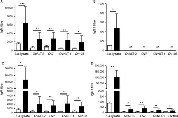
Using Western blots, three of the four recombinant proteins, namely OvALT-2, OvNLT-1 and Ov7, were specifically detected by immunoglobulin present in sera derived from L. sigmodontis-infected BALB/c mice but not by sera derived from naïve mice (figure not shown). Ov103 was neither detected by infected nor naïve animals. To identify the isotype of the antigen-specific immunoglobulins and to quantify the immunoglobulin responses, further ELISA studies were performed. Binding of sera derived from ten naïve and ten L. sigmodontis-infected BALB/c mice to the recombinant proteins and to complete L. sigmodontis antigen-lysate as a positive control were tested (fig. 1A–D). Sera derived from infected mice contained L. sigmodontis lysate-specific IgM and IgG1 at day 30 and day 64 post infection (pi). Furthermore, in infected mice, all recombinant proteins were specifically recognized by IgM (fig. 1A and C). Sera derived from naïve mice did not bind to the recombinant O. volvulus proteins, thus demonstrating antigen-specificity of this IgM. However, a higher immunoglobulin titre for complete L. sigmodontis-lysate was recorded compared to the individual recombinant proteins. This quantitative difference is most likely due to the high number of potential antigens in the lysate compared to single antigens in the purified proteins. Titres for protein-specific IgG1 at day 30 pi were not detected, although L. sigmodontis lysate-specific IgG1 was produced (fig. 1B). At a later time point, i.e. day 64 pi, IgG1 specific for the investigated O. volvulus proteins OvALT-2, OvNLT-1, Ov7 and Ov103 was detected in sera derived from L. sigmodontis-infected mice (fig. 1D). No antigen-specific IgG2a and IgG2b against the recombinant proteins or complete L. sigmodontis antigen lysate were detected (data not shown).
Mean titres (+ SD) of O. volvulus-derived proteins and L. sigmodontis lysate-reactive (A) IgM day 30, (B) IgG1 day 30, (C) IgM day 64 and (D) IgG1 day 64 post infection, derived from infected mice (black bars) or left naïve (white bars), with levels of significance: *P< 0.05; **P< 0.01; ***P< 0.001; nd, not detected.

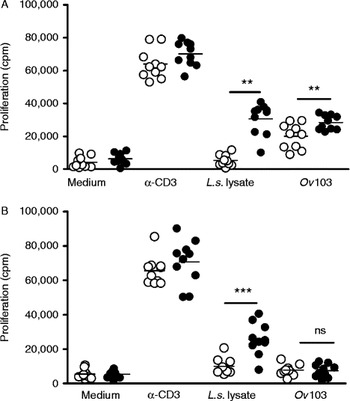
To test whether the recombinant O. volvulus proteins are targeted by cellular immune responses, splenocytes derived from naïve and L. sigmodontis-infected BALB/c mice were stimulated with OvALT-2, OvNLT-1, Ov7, Ov103, L. sigmodontis lysate or anti-CD3, respectively. Anti-CD3 stimulation readily induces proliferation of splenocytes, hence proving viability of T cells within the splenocyte culture. Litomosoides sigmodontis lysate antigens specifically induced proliferation of splenocytes derived from infected mice, but not from naïve mice, demonstrating antigen-specific proliferation (fig. 2A and B). Also, the purified recombinant protein Ov103 selectively induced a higher proliferation in splenocytes derived from infected mice at an early time point (day 30 pi, fig. 2A), but this proliferation was not recorded at a later time point (i.e. day 64 pi, fig. 2B). OvALT-2, OvNLT-1 and Ov7 did not induce any proliferation in splenocytes, neither from naïve nor from infected mice. These results strongly suggest that L. sigmodontis-specific IgM and IgG1 elicited during L. sigmodontis infection of BALB/c mice cross-reacted with the O. volvulus recombinant proteins. In addition, Ov103 was recognized by L. sigmodontis-specific T cells at an early time point of infection.
Cellular immune responses to recombinant proteins to show the proliferation of splenocyte cells from naïve (○) and infected (●) mice on (A) day 30 and (B) day 64 post infection, in the presence of medium, L. sigmodontis lysate, Ov103 or anti-CD3, with levels of significance **P< 0.01 or ***P< 0.001.

Immune responses to bovine filarial infections
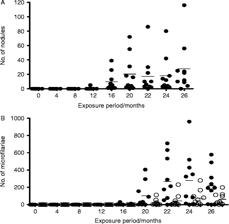
To study the recognition of O. volvulus recombinant antigens in the bovine model of onchocerciasis, sera were analysed for antigen-specific humoral responses, i.e. IgM, IgG1 and IgG2. To monitor parasite burden, the cumulative nodule loads during the first 26 months of exposure were counted (fig. 3A). Intradermal nodules were first detected after 12 months post exposure (pe) and subsequently the nodule loads increased steadily over time. Meanwhile, detection of mf in skin snips occurred from 16 months pe onwards, and mf counts increased steadily thereafter (fig. 3B). Onchocerca ochengi and O. gutturosa were the Onchocerca species detected from skin snips. The present results show that calves naturally exposed to Onchocerca transmission, develop patency after 12 months, with the appearance of palpable intradermal nodules and mf in the skin after 16 months pe.
Infection levels of calves naturally exposed to Onchocerca infections, to show (A) the number of nodules and (B) levels of microfilariae of O. ochengi (●) and O. gutturosa (○) in skin snips (n= 10).

Nodule and total microfilaria counts (O. ochengi and O. gutturosa) correlated positively with individual protein-specific immune responses (table 1). Significant positive correlations (P< 0.01) were found between nodule and total microfilaria loads, between total mf and OvNLT-1-specific IgM (P< 0.01), OvNLT-1-specific IgG1 and Ov103-specific IgG1 (P< 0.05) (table 1).
Pearson correlation coefficients between the number (N) of nodule loads, total microfilarial counts, including O. ochengi and O. gutturosa, and protein-specific immune titres; levels of significance **P<0.01 and *P<0.05.

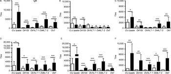
Next, sera were analysed for humoral immune responses during this period of natural infection. Figure 4A shows that all the recombinant proteins OvALT-2, OvNLT-1, Ov7 and Ov103 and adult worm lysate were specifically recognized by IgM in sera derived from cows (mothers), while sera derived from the control (uninfected European cattle) did not react with the recombinant proteins or with the O. ochengi lysate. The cows (mothers) were pre-exposed to the Onchocerca infections before they entered the study site. Similarly, analysis of the sera derived from calves before exposure ( < 1 month after birth) revealed the absence of IgM reactive with the recombinant proteins or O. ochengi lysate (fig. 4B). This was also observed after 4 months pe (figure not shown). After 8 months of exposure, the first antibodies of the IgM class were seen, reacting against both the O. ochengi lysate and the recombinant O. volvulus antigens (fig. 4C). After 12 months pe the sera from exposed calves showed IgM detection with all the proteins tested (fig. 4D). This finding is concomitant with the first appearance of palpable intradermal worm nodules (patency) in the calves (fig. 3A). We did not detect OvNLT-1-specific IgM in sera derived from calves at early time points but OvNLT-1-specific IgM was detected at the same time point when mf occurred (fig. 4D–F).
Mean titres (+SD) of the O. volvulus-derived proteins and O. ochengi lysate-reactive IgM: (A) mother cows, (B) pre-exposure calves, (C) 4 months pe, (D) 8 months pe, (E) 22 months pe and (F) 26 months pe cattle (black bars) or control sera (white bars), with levels of significance *P< 0.05; **P< 0.01 or ***P< 0.001 (n= 10).

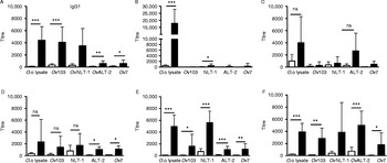
Cows introduced into the study site contained IgG1 specific for Ov-ALT-2, OvNLT-1, Ov7 and Ov103 in their sera at an early time point (fig. 5A). Likewise, analysis of sera of the calves at 1 month after birth for IgG1 isotype show O. ochengi lysate- and OvNLT-1-specific responses (fig. 5B), which most likely reflects a maternal diaplacenta and/or lactogene transfer of IgG1. At 4 months pe, a decline in O. ochengi lysate-specific titres for IgG1 (figure not shown) was observed, which in part recovered at 12 months pe (fig. 5D). There was no Ov103 and OvNLT-1 protein-specific recognition in exposed calves up to 12 months pe (fig. 5B–E). After 12 months exposure, a re-emergence of OvALT-2 and Ov7 protein-specific IgG1 responses was detected (fig. 5E), which was maintained throughout the rest of the exposure period (fig. 5E–F). In addition, this was evident for the other proteins tested (O. ochengi lysate, Ov103 and OvNLT-1; fig. 5E–F). This consistent protein-specific recognition of sera at the onset of 16 months exposure throughout the rest of the exposure period (fig. 5E–F) coincided with the appearance of worm nodules and mf in the skin. There was no observed protein or O. ochengi lysate-reactive recognition of IgG2 by sera in all the time points tested.
Mean titres (+SD) of O. volvulus-derived proteins and O. ochengi lysate-reactive IgG1: (A) mother cows, (B) pre-exposure calves, (C) 4 months pe, (D) 8 months pe, (E) 22 months pe and (F) 26 months pe cattle (black bars) or control sera (white bars), with levels of significance *P< 0.05; **P< 0.01 or ***P< 0.001 (n= 10).

The present results show that there is no maternal transfer of antigen-specific IgM from cow to calf, as expected. Protein-specific recognition of IgM was first detected after 8 months pe and antigens of O. ochengi lysate, OvALT-2, OvNLT-1, Ov7 and Ov103 were gradually recognized following prolonged exposure to the parasite. Moreover, antigen-specific IgG1 is maternally transferred, as expected, and declines after 8 months pe. A second re-emergence of antigen-specific IgG1 starts as from 12 months pe, thereby most likely reflecting the humoral immune response elicited by prepatent and early patent infections.
Immune recognition of onchocercal proteins in human sera
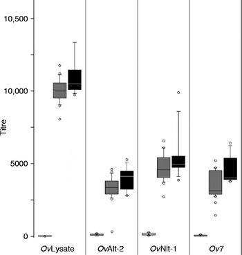
In order to examine the immunogenicity of three of the investigated onchocercal proteins, a possible IgG reactivity of sera from 24 onchocerciasis patients, eight of those expressing the rare hyperreactive form of the infection, towards OvALT-2, OvNLT-1 and Ov7, applying total O. volvulus lysate as the positive control, was investigated. The IgG reactivities of these sera were compared to the reaction found in sera from eight healthy European controls. IgG in sera from patients with generalized onchocerciasis compared to the control persons reacted strongly with the total protein extract of O. volvulus (median titre for patients versus controls: 10,380 versus 35; P< 0.001) and showed lower reactivities towards the single onchocercal proteins: OvALT-2 (median titre: 3913 versus 145; P< 0.001), OvNLT-1 (median titre: 4850 versus 195; P< 0.01) and Ov7 (median titre: 3770 versus 70; P< 0.01) (fig. 6). Interestingly, the eight sera from the rare hyperreactive form uniformly showed a trend of higher IgG reactivities for onchocercal extract (median titre: 10,550), OvALT-2, OvNLT-1 and Ov7 (median titres: 4175, 4975 and 4060, respectively).
Humoral immune responses to the mean titres of O. volvulus proteins compared to O. volvulus lysate-reactive IgG in human patients with a generalized common form (generalized onchocerciasis) (grey bars) or hyperreactive form (black bars) of onchocerciasis, compared with those in healthy Europeans (white bars).

Discussion
Using murine and bovine models for human O. volvulus infection, we show that recombinant O. volvulus antigens (OvALT-2, OvNLT-1, Ov103 and Ov7) are targets of the immune responses. In mice, specific IgM and, at a later stage of infection, also IgG1 exclusively in L. sigmodontis-infected but not in naïve animals, were detected. These experiments indicate that antigens generally function as targets for B-cell response during nematode infection. The detection of IgG1 at a later time point indicates a T-helper (Th)-dependent class-switch, thus suggesting that a preceding priming of specific T cells occurs. Also, the very early Ov103-specific proliferation at day 30 pi indicates the presence of Ov103-specific T cells. There were no observed OvALT-2-, OvNLT-1-, Ov103- and Ov7- specific IgG2a and IgG2b titres in mice in these studies. This is in line with the association of IgG2a or IgG2b with Th1-associated immune responses, whereas L. sigmodontis infection predominantly provokes Th2-associated responses in BALB/c mice (Torrero et al., Reference Torrero, Hübner, Larson, Karasuyama and Mitre2010).
Proliferation of spleen cells was recorded in response to Ov103 at 30 days, but not at 64 days post L. sigmodontis infection. Previous studies suggested a possible correlation between levels of mf and antibody response to Ov103 antigen (Lustigman et al., 1992b). They found that a significantly greater proportion of individuals with low levels of skin mf recognized the protein encoded by clone Ov103 than did individuals with higher levels of skin mf. Dynamics of response mirrors that observed in the current study, in which Ov103-protein-specific cellular response at an early time point (day 30) was recorded, but was absent at a later time point (day 64) in the L. sigmodontis model. In human onchocerciasis, Lustigman et al. (1992b) therefore proposed that the presence of specific anti-Ov103 antibodies before patency may reflect the existence of Ov103 epitopes in early developmental stages. A probable reason for a proliferation in response to Ov103 at the early time point of day 30, but not on day 64, may be that the protein is only transiently released on to the surface (or in solution) at an early course of infection and not continuously. Further studies, such as a longitudinal study of the cellular immune response to Ov103 in L. sigmodontis infection, could be used to test this hypothesis.
Calves (Goudali breed, Bos indicus), which were naturally exposed to the Simulium and other arthropod vectors at our study site, developed intradermal nodules of O. ochengi and harboured O. ochengi and O. gutturosa microfilariae (and other filariae) in the skin, as has already been shown in previous studies (Wahl et al., Reference Wahl, Achukwi, Mbah, Dawa and Renz1994; Achukwi et al., Reference Achukwi, Harnett and Renz2000). By introducing very young Goudali calves into an area with high parasite transmission, dynamics of nodule acquisition, microfilaria loads and protein-specific immune responses for more than 2 years have been studied. This time-frame correlates to a series of biological events occurring during the life cycle of bovine Onchocerca parasites.
First, the serological profiles (IgM and IgG1) of the cows (mothers) were established. Their protein-specific responses were an indication of ongoing infection with filarial parasites (figs 4A and 5A). The significant protein-specific IgM and IgG1 responses imply that the cows were already exposed to Onchocerca infection. In addition, intradermal palpation of the mother cows indicated that 7 out of 10 were already positive for Onchocerca nodules and skin mf of O. ochengi and O. gutturosa.
OvNLT-1- and O. ochengi lysate-reactivity of IgG1 of pre-exposed calves (fig. 5B) indicated a maternal transfer of specific antibodies through the colostrum. In cattle, IgG1 is the only (predominant) isotype transferred maternally in large amounts (Butler, Reference Butler and Ogra1999) and this transfer gradually declines after the calves start to build their own protective immunity. Hence, there was no protein-specific IgM and a reduced O. ochengi lysate-reactive IgG1 response in calves after 4 months pe. The predominant IgG1 responses observed in this study are in line with earlier reports of cattle experimentally infested with O. lienalis (Kuo & Bianco, Reference Kuo and Bianco1995) and cattle naturally exposed to O. ochengi (Achukwi et al., Reference Achukwi, Harnett, Bradley and Renz2004). The high anti-Onchocerca IgG1 levels at an early stage of a calf's life could have an inhibitory effect on the establishment of L3s and therefore delay the acquisition of nodules (Achukwi et al., Reference Achukwi, Harnett, Bradley and Renz2004).
In general, infections with helminths typically lead to a strong Th2-immune response (Allen & Maizels, Reference Allen and Maizels2011), more probably because generic invertebrates produce signals through host receptors in a way that amplifies Th2 responses and is antigen specific. Bovine IgG consists of two isotypes, IgG1 and IgG2 (with allotypes 2a and 2b), neither of which is a homologue of human or murine IgG isotypes. Production of IgG2 responses were stimulated by interferon (IFN)-γ (a Th1-associated cytokine) from bovine cells in vitro (Estes, Reference Estes1996) and in vivo (Graham et al., Reference Graham, Trees, Collins, Moore, Guy, Taylor and Bianco2001). Results show that protein-specific IgG1 antibody responses could be an indicator of infection with bovine onchocerciasis. However, in the current study, a Th2-associated IgG1 response in mice was observed, while in cattle there was no such correlation.
Previous work conducted on a ranch along the Vina du Sud river in Ngaoundere, Cameroon (Achukwi et al., Reference Achukwi, Harnett, Bradley and Renz2004) identified morphologically other species (O. gutturosa, O. armillata and Setaria species) with yet largely unknown transmission cycles. In the present study, O. ochengi and O. gutturosa were identified morphologically in the investigated cattle. Even though O. ochengi was the predominant filarial parasite identified in this study, the infection with O. gutturosa was closely related to that of O. ochengi (fig. 3) which is similar to earlier observations (Voung et al., Reference Voung, Wanji, Prod'Hon and Bain1994; Wahl et al., Reference Wahl, Achukwi, Mbah, Dawa and Renz1994; Achukwi et al., Reference Achukwi, Harnett, Bradley and Renz2004). Also, previous studies (Bradley et al., Reference Bradley, Gregory, Bianco and Maizels1989) have already indicated that surface antigens of O. gutturosa from bovines and O. volvulus from humans are similar in size and share antigenic cross-reactivities. Thus, it is very likely that the E/S protein-specific immune responses observed in this study are also a result of O. gutturosa infection, or of undetected filarial species, and not only of O. ochengi.
The immunogenicity and the exposure of the investigated proteins from O. volvulus to its natural host was examined in 24 individuals infected by O. volvulus. By ELISA analysis high IgG titres were demonstrated in the sera of the onchocerciasis patients for OvALT-2, OvNLT-1, Ov7 and much higher for O. volvulus lysate. The reaction of the sera of the more rare chronic hyperreactive form was uniformly more pronounced with all tested proteins and the total extract, representing the characteristic immune response pattern as demonstrated earlier (Mpagi et al., Reference Mpagi, Buttner, Tischendorf, Erttmann and Brattig2000; Brattig, Reference Brattig2004).
This study confirms cross-reactions to proteins expressed by the different developmental stages of Onchocerca and Litomosoides in filaria-infected cattle and mice. Mice display antigen-specific immunoglobulin levels in the sera 30 days pi, thus before patency is achieved, and a marked increase after production of mf 64 days pi. Calves naturally exposed to O. ochengi develop patency after 12 months of exposure. Protein-specific immune responses are proportional to the parasite loads and may function as an indicator of infection. This study suggests that the proteins are highly immunogenic and qualifies them as potential vaccine candidates for human onchocerciasis.
Acknowledgements
The herdsmen are acknowledged for maintenance of the cattle, and Achille Paguem, Nancy Ngwasiri, David Ekale and Guimbang Abanda for field and laboratory assistance. Mfopit Youssouf is equally acknowledged for assistance in data analysis.
Financial support
T.K.M. was supported by the DFG-funded project on ‘Analysis of host-parasite cross- talk based on the bovine model for human onchocerciasis, Onchocerca ochengi’ (grant number: PAK 296 BR 1020/4-1).
Conflict of interest
None.
Ethical standards
The in vivo experiments were carried out in the animal facility of the Bernhard Nocht Institute for Tropical Medicine, Hamburg, with permission of the federal health authorities of the state of Hamburg, Germany. The collection of human sera and removal of nodules was approved by the Ethics Commission of the Medical Board in Hamburg (Germany) and by ethic committees and medical authorities in the respective countries, in accordance with the principles of the Helsinki declaration of 1975, revised 2000.