

Introduction
Beekeeping is a long-standing agricultural practice and an important economic subsector in the rural communities of the Kingdom of Saudi Arabia. For a significant number of rural households, beekeeping is an important source of income. Additionally, because they are the major pollinators of many cultivated crops and wild plants, honey bees are of great ecological and economic importance (Free, Reference Free1993). The total annual global economic value of pollination is estimated to be USD 202 billion, representing 10% of the total value of global agricultural production (Gallai et al., Reference Gallai, Salles, Settele and Vaissière2009). Apis m. jemenitica, the smallest race of A. mellifera, is the only indigenous honey bee race of Saudi Arabia. Apis m. jemenitica is well adapted to the harsh local environmental conditions, including high temperatures and low rainfall, and can tolerate hunger for long periods of time. In contrast, A. m. carnica, which was imported to the Kingdom, cannot tolerate harsh environmental conditions (Alqarni, Reference Alqarni1995; Abou-Shaara et al., Reference Abou-Shaara, Al-Ghamdi and Mohamed2012). These conditions affect the health, performance and productivity of imported honey bee colonies. A decline in bee populations leads to a decline in pollination, crop yield and food supply (Potts et al., Reference Potts, Biesmeijer, Kremen, Neumann, Schweiger and Kunin2010). Hence, conducting research on these factors and diseases, including potential treatments and preventative measures, is beneficial to the agricultural industry and conservation strategies in general.
In the last few decades, significant losses have been observed in imported bee colonies in Saudi Arabia (Alattal and AlGhamdi, Reference Alattal and AlGhamdi2015). A mysterious decline in honey bee colonies has gained worldwide attention, including in Saudi Arabia. Much attention has been given to Colony Collapse Disorder (CCD), which is a syndrome defined as a dead colony with no adult bees and with no dead bee bodies but with a live queen, and usually honey and immature bees still present (vanEngesldorp et al., Reference vanEngesldorp, Evans, Saegerman, Mullin, Haubruge, Nguyen, Frazier, Frazier, Cox-Foster, Chen, Underwood, Tarpy and Pettis2009). Five major abiotic and biotic factors (parasites and pests, pathogens, poor nutrition, sublethal exposure to pesticides and harsh environmental conditions) threaten honey bee health on a global scale. In reality though, these factors tend to overlap and interact with one another, which complicates issues and synergistically results in the abrupt disappearance of worker bees from the colony. Abiotic factors include environmental stresses such as high summer and low winter temperatures, a lack of foraging capacity, and use of insecticides in agriculture (Naug, Reference Naug2009; Watanabe, Reference Watanabe2008). Biotic factors include infection by parasites such as P. larvae, Nosema spp., and more than 18 viruses (Genersch et al., Reference Genersch, von der Ohe, Kaatz, Schroeder, Otten, Büchler, Berg, Ritter, Mühlen, Gisder, Meixner, Liebig and Rosenkranz2010; Ansari et al., Reference Ansari, Al-Ghamdi, Nuru, Ahmed, Ayaad, Al-Qarni, Alattal and Al-Waili2016; Ansari et al., Reference Ansari, Al-Ghamdi, Nuru, Khan and Alattal2017).
One honey bee infection, American foulbrood (AFB) disease, is caused by the bacterium P. larvae (Genersch et al., Reference Genersch, Forsgren, Pentikäinen, Ashiralieva, Rauch, Kilwinski and Fries2006). This gram-positive, rod-shaped bacterium can form spores that are picked up by adult bees and brought back to the hive (Genersch, Reference Genersch2010). Adult bees are resistant to the bacterial disease; however, they become vectors and transmit the infectious endospores to young larvae, which are highly susceptible to it (Hitchcock et al., Reference Hitchcock, Stoner, Wilson and Menapace1979; Wilson, Reference Wilson1971). Spores are resistant to antibiotics and heat, (the spores of P. larvae are particularly resistant) and can persist for at least several decades (Genersch, Reference Genersch2010), making the eradication of this disease difficult. Woodrow (Reference Woodrow1942) and Woodrow and Holst (Reference Woodrow and Holst1942) report that as few as 10 spores can cause the disease in a larva. Once spores enter the gut of a larva, they germinate and become vegetative bacterial cells (Bamrick, Reference Bamrick1967) that proceed to grow and divide in the larva. However, in doing so, they compete with the larva for nutrients and propagate to the point of rupturing the midgut lumen (Yue et al., Reference Yue, Nordhoff, Wieler and Genersch2008). The larva disintegrates into a ropy mass that deteriorates into scales, which is a condition that is characteristic of larvae killed by this particular disease (Genersch, Reference Genersch2010). The death of one larva can produce millions of spores that are then released in the hive (Sturtevant, Reference Sturtevant1932). As the larvae of a hive succumb to the disease and fewer bees are able to reach adulthood, the hive collapses, because it can no longer maintain its population.
These findings have led to calls for research that explores the agents that cause disease in A. m. jemenitica and A. m. carnica in relation to honey bee health under the local environmental conditions in the Kingdom of Saudi Arabia. However, even though some studies have reported on disease-causing organisms of honey bees in Saudi Arabia (Al-Ghamdi, Reference Al-Ghamdi1990; Ellis and Munn, Reference Ellis and Munn2005; Alattal and AlGhamdi, Reference Alattal and AlGhamdi2015; Abdel-Baki et al., Reference Abdel-Baki, Mares, Dkhil and Al-Quraishy2016), there has been little research reported on the molecular characterization of honey bee pathogens in Saudi Arabia. Therefore, detailed studies of various honey bee pathogens, including their identification and characterization using various molecular and microbiological methods, are needed. In a previous study of Al-Ghamdi (Reference Al-Ghamdi1990), American foulbrood infection was reported in two apiaries out of 102 apiaries examined. The fundamental goal of the research described here was to characterize pathogenic agents from different geographical locations in Saudi Arabia that infect honey bees, particularly bacterial pathogens, such as P. larvae.
Materials and methods
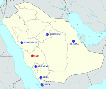
The presence of American foulbrood (AFB) in honey bee colonies was investigated in different beekeeping locations during the spring season (March to April 2015), which is the active season for honey bees in Saudi Arabia. Seven different geographical localities where beekeeping is common were included in this survey (Fig. 1): Al-Ahsa (25°25′46″N, 49°37′19″E), Abha (18°13′24″N, 42°30′26″E), Jazan (16°53′21″N, 42°33′40″E), Taif (21°16′0″N, 40°25′0″E), Al-Madinah (24°28′0″N, 39°36′0″E), Al-Bahah (20°0′0″N, 41°30′0″E) and Al-Qassim (25°49′19.72″N, 42°50′6.85″E). At least 10 apiaries were visited in each area, and 10 colonies in each apiary were inspected.
Map showing AFB inspection areas by region in Saudi Arabia. AFB has been detected only in the Taif Region (red solid circle).

Sampling
A total of 700 hives in seven targeted localities (100 hives each) of A. m. jemenitica and A. m. carnica were investigated in this study. Samples were collected from local (A. m. jemenitica) and imported bee races (A. m. carnica). Honey bee broods were visually inspected for any signs of abnormality and clinical disease status. The clinical signs of AFB are diverse and depend on the genotype involved, the stage of the disease and the strength of the bee colony (OIE, 2008). Visual signs of AFB infection include a sealed brood with a patchy and irregularly patterned appearance and sunken, dark and perforated wax cappings containing the dead larvae, which are brown in colour. Additionally, in the case of severe infection, the colony may emit a rotten odour. The ropy thread test has been employed as a key diagnostic test for AFB (Brødsgaard et al., Reference Brødsgaard, Hansen and Ritter2000; De Graaf, et al., Reference De Graaf, Alippi, Antúnez, Aronstein, Budge, De Koker, De Smet, Dingman, Evans, Foster, Fünfhaus, Garcia-Gonzalez, Gregorc, Human, Murray, Nguyen, Poppinga, Spivark, vanEngelsdorp, Wilkins and Genersch2013).
Biosafety practices as recommended (De Graaf et al., Reference De Graaf, Alippi, Brown, Evans, Feldlaufer, Gregorc, Hornitzky, Pernal, Schuch, Titĕra, Tomkies and Ritter2006; OIE, 2008) were followed during the sampling period, to avoid contamination between colonies during sampling. To initially confirm AFB, suspect larvae were removed from the combs and tested using an AFB diagnostic field test kit [Vita (Europe) Limited, Basingstoke, UK] according to the manufacturer's instructions. AFB-suspected honey bee broods were collected for further lab examination. A piece of brood comb (10 × 10 cm) containing suspect larvae was excised, wrapped in paper towels, packaged in a plastic bag, labelled and transported to the laboratory of the Bee Research Unit (BRU) at the Department of Plant Protection of the College of Food and Agricultural Sciences at King Saud University. The brood comb samples were subjected to microbiological examination and molecular analyses to test for the presence of the causative bacterium, P. larvae.
Data collection
The data collected from the survey included the following information for each inspected apiary: the date of inspection, the apiary location (to facilitate repeat visits), the name of the owner, the hive type (local or modern), the honey bee race (indigenous or imported), the number of honey bee colonies and the number of colonies with positive visual clinical symptoms of AFB.
Field diagnosis of Paenibacillus larvae
Bacterial strain isolation and identification
Pieces of comb of approximately 10 cm2 in size were ground in phosphate-buffered saline (PBS), and different serial dilutions prepared. A total of 50 µL of each dilution was spread on MYPGP agar [1% Mueller-Hinton broth (Difco) and 1.5% yeast extract supplemented with 0.1 mg/l of thiamine] containing 15% v/v glycerol in Petri dishes. The medium was also supplemented with 6-μg/mL nalidixic acid and 10-μg/mL pipemidic acid to inhibit the growth of Bacillus, Brevibacillus and other Paenibacillus species. The plates were then incubated at 36 ± 0.5 °C under micro-anaerobic conditions for 10 days, and the resulting bacterial colonies subjected to microscopic examination.
Colonies with a P. larvae-like morphology (whitish with a transparent and glistening appearance) were further analyzed for catalase activity and mannitol fermentation. For the catalase activity test, part of the colony was transferred to a microscope slide and mixed with a drop of 30% H2O2. The production of bubbles indicated catalase activity, while the absence of bubbles indicated a lack of activity. Strains were also tested for the production of acid from mannitol according to Gordon et al. (Reference Gordon, Haynes and Pang1973). One colony from each characterized bacterial isolate was subjected to molecular identification using the P. larvae-specific primers (AF1-f-5′- GCTCTGTTGCCAAGGAAGAA-3′; AF2-r-5′- AGGCGGAATGCTTACTGTGT-3′) described by Bakonyi et al. (Reference Bakonyi, Derakhshifar, Grabensteiner and Nowotny2003).
Microscopic examination of spores
The modified hanging-drop technique (Kostecki, Reference Kostecki1969) was used to observe the P. larvae spores in larval remains. Suspect larval material was mixed with water. This spore suspension was heat-shocked at 80 °C for 10 min to kill the vegetative forms of other microorganisms, including other spore-forming bacteria. A drop of this suspension was placed on a coverslip, dried, heat-fixed and stained with carbol fuchsin for 5 min at 37 °C. After staining, the samples were mounted on glass slides and observed with a high-resolution microscope (Carl Zeiss, North Ryde, NSW, Australia) by examining fields where pockets of water had formed in the emersion oil. The spores of P. larvae exhibited Brownian movement (Shimanuki and Knox, Reference Shimanuki and Knox2000).
Morphological, physiological and biochemical characterization
Biochemical confirmation of the isolates
Cell and colony morphologies of all isolates were described, and their biochemical profile determined according to Gordon et al. (Reference Gordon, Haynes and Pang1973) and/or Plagemann (Reference Plagemann1985), with the following tests: gram staining, subculturing in nutrient broth, haemolysis on CSA, catalase tests, nitrate reduction, casein hydrolysis, mannitol fermentation, salicin fermentation and acid- and alkaline-phosphatase reactions. All tests were carried out in triplicate.
Gram staining: A smear from the suspected larvae colony was taken on a slide and Gram stained. The slide was flooded with crystal violet for 60 sec, washed, then flooded again with iodine solution for 1 min. Ethanol was used as a decolorizing agent for 5 sec, and the final step involved applying safranin. After each step the slide was rinsed with water for 5 sec. The prepared slide was then examined under a microscope (Carl Zeiss, North Ryde, NSW, Australia) and photographed. The bacterium was identified as P. larvae with gram-positive rods (Shimanuki and Knox, Reference Shimanuki and Knox1988).
Catalase test: This involved a drop of 3% hydrogen peroxide (H2O2) placed on an actively growing culture on a solid medium. Most aerobic bacteria break down the peroxide to water and oxygen, and produce bubbly foam, but P. larvae responds negatively to this reaction (Haynes, Reference Haynes1972).
Hydrolysis of casein: The 24-h-old suspected culture of bacteria was inoculated on the sterilized medium containing 10% skimmed milk and 3% agar, and incubated for 7 days at 37 °C. The culture was identified as P. larvae and a clear zone around the growing culture due to casein hydrolysis was visible (Neuendorf et al., Reference Neuendorf, Hedtke, Tangen and Genersch2004).
Molecular characterization of bacterial isolates
Bacterial genomic DNA isolation
All the positively identified samples of isolates (by cultural methods) were subjected to PCR analysis. Bacterial pellets were incubated in 200 µl enzyme solution [20 mg lysozyme, 20 mM Tris-HCl (pH 8.0), 2 mM EDTA and 1.2% Triton] at 37 °C for 1 h. Then, 25 µl Proteinase K and 200 µl buffer AL (Qiagen) were added, and the lysates incubated, first at 56 °C for 30 min and then at 96 °C for 5 min. DNA was removed with 200 µl of elution buffer and stored at −20 °C. Bacterial DNA was isolated using the QIAamp® genomic DNA isolation mini kit for gram-positive bacteria (Qiagen Inc., Valencia, CA) according to the manufacturer's instructions. In addition, buffer controls were prepared in parallel during the DNA extraction to monitor for extraction contamination. Each DNA extract was tested for the presence of P. larvae 16S rDNA.
PCR amplification using AFB-specific primers
The PCR reactions were performed in a total volume of 50 µL containing 5µl 10xPZR buffer (100 mM Tris-HCl, pH 9.0, 500 mM KCl, 15mM MgCl2, 1% Triton X-100), 250 µM of each deoxynucleotide triphosphates, 2U Taq DNA polymerase enzyme (Promega, USA), 40 pmol of each primer and 5 µl target DNA. The surface of the mixture was covered with 100 µl mineral oil. The following PCR conditions were used: one cycle at 95 °C (15 min), followed by 30 cycles of 93 °C (1 min), 55 °C (30 sec) and 72 °C (1 min) and a final cycle at 72 °C (5 min). Samples of the amplicons were electrophoresed in 1.0% agarose gel. Approximate product size was determined using the 100-bp molecular size marker (Promega, USA). The PCR product was visualized and photographed using a Gel Doc EZ system (Bio-Rad, USA).
BLAST and phylogenetic analyses
16S rRNA gene sequencing and phylogenetic analysis
The 16S rRNA gene sequencing was performed using a Genetic Analyzer DNA Sequencer (Applied Biosystems, USA) according to the manufacturer's instructions. Sequences obtained were ‘cleaned up’ using MEGA4 software (Tamura et al., Reference Tamura, Dudley, Nei and Kumar2007). Partial 16S rRNA gene sequences of the isolates were compared with 16S rRNA gene sequences available by the BLAST search (Altschul et al., Reference Altschul, Gish, Miller, Myers and Lipman1990), in the National Centre for Biotechnology Information (NCBI) database (http://www.ncbi.nlm.nih.gov/). Multiple sequence alignments were performed using CLUSTAL W version 1.8 (Thompson et al., Reference Thompson, Higgins and Gibson1994). The method of Jukes and Cantor (Reference Jukes, Cantor and Munro1969) was used to calculate evolutionary distances. A phylogenetic tree was constructed by the neighbour-joining method (Saitou and Nei, Reference Saitou and Nei1987), and the reliability of the tree topology was evaluated by bootstrap analysis using MEGA 4.1 software (Tamura et al., Reference Tamura, Dudley, Nei and Kumar2007).
Results
Field diagnosis of Paenibacillus larvae
Bacterial strain isolation and identification
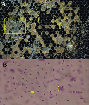
Infected honey bee combs were collected from the A. m. jemenitica colonies kept in the modern hives in the Taif region (Figs. 1 and 2A and B). No infection was reported in the imported honey bee race, A. m. carnica. The collected infected combs had a mottled appearance, including healthy capped brood, uncapped cells containing the remains of diseased larvae and empty cells. The capped cells containing infected larvae appeared moist, darkened and concave, and became perforated as the infection progressed (Fig. 2A). The larvae and pupae changed colour from dark brown to black. Some infected larvae became glutinous and were drawn out as a thread when a forceps was inserted into the infected cell. The whole comb emitted a foul odour. These field diagnostics all indicated an AFB infection in the colony.
(A) Irregular pattern of sealed broods with sunken, darker cells and perforated cell caps with the foul odour (arrow) that typifies AFB disease. (B) Spores of P. larvae isolates (arrow) at 1000x resolution.

Morphological, physiological and biochemical characterization
Biochemical confirmation of the isolates
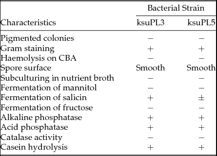
All the colonies of P. larvae isolates were typical of P. larvae subsp. larvae: small (3 mm in diameter), regular, mostly rough, raised and whitish, with a somewhat transparent and glistening appearance on MYPGP medium. Colonies grown on MYPGP agar under micro-anaerobic conditions for 10 days were creamy in colour, regular and disc-shaped. Cells were examined, and all isolates were gram-positive rods with a width of about 1 μm and a length of 3–5 μm. Bacteria appeared as single cells or pairs, and sometimes as short chains. Biochemical tests carried out on the plated bacterial isolates revealed that all were gram-positive and catalase- and oxidase-negative. Moreover, bacterial isolates also exhibited haemolysis on Columbia blood agar (CBA) with 5% sheep blood. When cultured on nutrient media, these isolates failed to grow. The isolates were unable to ferment mannitol. However, ksuPL3 exhibited fermented salicin well, whereas ksuPL5 exhibited fermented salicin poorly. Both isolates reacted with acid- and alkaline-phosphatases (Table 1). These results confirm and expand earlier reports of genotype-specific differences in the metabolism and biochemistry of P. larvae subsp. larvae (Genersch et al., Reference Genersch, Forsgren, Pentikäinen, Ashiralieva, Rauch, Kilwinski and Fries2006). Spores of both isolates displayed Brownian motion under high-resolution microscopy at 1000x when the microscope slide was prepared by the hanging drop method. The Brownian motion also confirmed that the spores belong to P. larvae (Fig. 2B), and therefore, the isolated strains were identified as P. larvae.
Phenotypic characteristics of isolated Paenibacillus larvae genotypes

Molecular characterization of bacterial isolates
Bacterial genomic DNA isolation
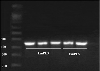
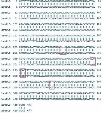
Amplification products from ksuPL3 and ksuPL5 were obtained, resulting in 451- and 453-bp DNA fragments, respectively, corresponding to the expected size and all AFB suspected isolates were positive in PCR. The sequence of ksuPL3 (GenBank accession number: KR780760) and ksuPL5 (GenBank accession number: KR780761), resulting in 451- and 453-bp DNA fragments, respectively, were deposited in the NCBI GenBank (Fig. 3).
Visualization of the 16S rRNA gene PCR amplification products from the two selected bacterial isolates (ksuPL3 and ksuPL5). Lane MM: molecular size marker (100-bp ladder); Lanes 1–3: isolate ksuPL3; Lanes 4–5: isolate ksuPL5.

BLAST and phylogenetic analyses
16S rRNA gene sequencing and phylogenetic analysis
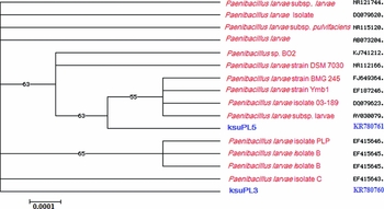
A similarity search using the Basic Local Alignment Search Tool (BLAST) (Altschul et al., Reference Altschul, Gish, Miller, Myers and Lipman1990) showed that the ksuPL3 and ksuPL5 sequences exhibited more than 98.9% and 98.8% sequence identity, respectively, with some P. larvae isolates (Fig. 4). The Nucleotide BLAST demonstrated that the ksuPL3 isolate DNA sequence (451 bp) showed 99% sequence identity with the 16S rDNA of P. larvae isolates (gb| NR_121744.1, gb| CP003355.1 and gb| AB680856.1). Similarly, the DNA ksuPL5 isolate sequence (453 bp) showed 99% sequence identity with the 16S rDNA of other P. larvae isolates (gb| FJ649363.1, gb| FJ649362.1 and gb| FJ649360.1).
Pairwise sequence alignment of representative sequences belonging to the ksuPL3 and ksuPL5 bacterial isolates. These sequences were aligned using the ClustalW pairwise alignment tool. The ranges of sequence identity and sequence similarity of the aligned sequences have been demonstrated.

The evolutionary relationship between the two isolates and previously reported isolates was constructed using MEGA4 software (Tamura et al., Reference Tamura, Dudley, Nei and Kumar2007). The results illustrate the degree of evolutionary relatedness between the two Saudi Arabian isolates and other previously reported isolates (Fig. 5). From our study, the ksuPL5 isolate showed greater relatedness to four previously reported P. larvae isolates (gb| FJ649364.1, gb| EF187246.1, gb| DQ079623.1 and gb| AY030079.1), while isolate ksuPL3 formed a separate clade by itself. This indicates that the genotypes of the two isolates differ.
Neighbor-joining phylogenetic tree of the two P. larvae isolates (ksuPL3 and ksuPL5) based on 16S rRNA gene sequence comparisons and closest NCBI (BLASTn) strains based on the 16S rRNA gene sequences (Neighbor-joining tree method). The scale bar indicates 0.0001 nucleotide substitutions per nucleotide position. The numbers at node show the bootstrap values obtained with 1000 resampling analyses.

Discussion
American foulbrood is the most dangerous and contagious of the infectious diseases in bees. Hornitzky and Clark (Reference Hornitzky and Clark1991) have reported that the smell in the infected hives and the empty, shrunken and perforated comb cells observed in the combs with larvae are among the specific symptoms of the disease. Al-Ghamdi (Reference Al-Ghamdi1990) reported AFB infection in some apiaries in Saudi Arabia on the basis of morphological characterization. In this study, a large survey was carried out and AFB caused by P. larvae reported in brood combs from Saudi Arabian hives. AFB disease was found to infect the honeycombs of local indigenous race of Saudi Arabia, A. m. jemenitica collected from Taif Region. Taif is a city in Mecca Province of Saudi Arabia on the slopes of the Sarawat Mountains. Taif has a hot summer and mild winter, and precipitation is low with more rain in spring and late autumn than in other months. The identification of this causative agent was based on symptomatology, morphological characteristics, biochemical reactions, microscopic analysis and molecular detection using PCR. Due to the low selectivity of P. larvae on different growth media, various PCR methods have been developed to detect P. larvae (Bakonyi et al., Reference Bakonyi, Derakhshifar, Grabensteiner and Nowotny2003; Alippi et al., Reference Alippi, Reynaldi, López, De Giusti and Aguilar2004; D'Alessandro et al., Reference D'Alessandro, Antúnez, Piccini and Zunino2007). Adult bees (Lindström et al., Reference Lindström, Korpela and Fries2008), brood larvae and pupae are efficient testing materials, because they are all susceptible to AFB (Bakonyi et al., Reference Bakonyi, Derakhshifar, Grabensteiner and Nowotny2003).
PCR is a reliable, rapid and widely used method in microbiological diagnostics, and the testing of pathogen DNA is an alternative to classic cultivation tests on agar. Many types of samples can be used as a source of infectious material for AFB testing. Interpretation of positive and negative PCR results can be challenging. Similar to traditional pathogen detection techniques, PCR results must be strictly interpreted in conjunction with the history, clinical signs and evidence of disease. A positive PCR result only indicates the detection of the target genetic sequence. It cannot differentiate between the incidental presence of an organism, colonization without disease, transient infection or active infection with disease.
AFB has recently been diagnosed by molecular methods in Pakistan by Anjum et al. (Reference Anjum, Shah, Kamran Azim, Yousuf, Khan and Khan2015) and also detected in several locations in Jordan (Nizar et al., Reference Nizar, Alaa, Noureddine, Fares and Quddoumi2015), which is on the northern border of Saudi Arabia. Masry et al. (Reference Masry, Kabeil and Hafez2014) studied AFB in honey bee colonies in Egypt in 2012−2013. We were unable to compare the sequences of our isolates with other isolates reported in Pakistan, Egypt and Jordan, because of lack of sequence information in the NCBI database for other isolates. We herein report, for the first time, the isolation and molecular characterization of P. larvae, the causative agent of AFB in honey bees in the kingdom of Saudi Arabia.
Fragments of the 16S rRNA gene of P. larvae subsp. larvae were amplified using PCR. Bakonyi et al. (Reference Bakonyi, Derakhshifar, Grabensteiner and Nowotny2003) developed AFB-specific primers based on 16S rDNA that amplify a 451-bp fragment of the target AFB-specific sequence. Govan et al. (Reference Govan, Allsopp and Davison1999) stated that the culture-dependent identification of P. larvae subsp. larvae is not a rapid confirmation method for identifying this pathogen. Therefore, the authors claim that a PCR-based method can be of greater utility to rapidly confirm the presence or absence of these bacteria.
Using the AFB primers developed by Bakonyi et al. (Reference Bakonyi, Derakhshifar, Grabensteiner and Nowotny2003), we amplified 451- and 453-bp DNA fragments from the ksuPL3 and ksuPL5 isolates, respectively. Masry et al. (Reference Masry, Kabeil and Hafez2014) succeeded in detecting two different P. larvae strains by amplifying a 359-bp fragment from the 16S rRNA gene of P. larvae, representing an important alternative for rapid AFB diagnosis. The results presented herein agree with those of Masry et al. (Reference Masry, Kabeil and Hafez2014), who suggested that partial 16S rDNA PCR may be an easier method for rapidly confirming the presence of P. larvae.
A nucleotide BLAST search indicated that the DNA sequence obtained from the ksuPL3 (451 bp) isolate showed a 99% sequence identity with the 16S rDNA of some P. larvae isolates (gb| NR_121744.1, gb| CP003355.1 and gb| AB680856.1). The first two close hits of ksuPL3 were closely related to the DNA sequence of P. larvae isolated from honey samples in Germany (Djukic et al., Reference Djukic, Brzuszkiewicz, Fünfhaus, Voss, Gollnow, Poppinga, Liesegang, Garcia-Gonzalez, Genersch and Daniel2014), and the third close hit belongs to the sequence of P. larvae isolated in Japan (Nakagawa, direct submission to NCBI). Similarly, the DNA sequence obtained from the ksuPL5 isolate (453 bp) showed a 99% sequence identity with the 16S rDNA of other P. larvae isolates (gb| FJ649363.1, gb| FJ649362.1 and gb| FJ649360.1). All three closely related hits of ksuPL5 belong to P. larvae isolates from Tunisian infected honey bee broods (Hamdi et al., Reference Hamdi, Essanaa, Sansonno, Crotti, Abdi, Barbouche, Balloi, Gonella, Alma, Daffonchio, Boudabous and Cherif2013). This indicated that Tunisia, a neighbouring country, has the same type of genotype as found in Saudi Arabia. Molecular diagnostic methods based on the comparative sequence analysis of the 16S rRNA gene are useful tools for the detection and identification of P. larvae (Bakonyi et al., Reference Bakonyi, Derakhshifar, Grabensteiner and Nowotny2003; Lauro et al., Reference Lauro, Favaretto, Covolo, Rassu and Bertoloni2003). When the DNA nucleotide sequences of both the isolates in this study (ksuPL3 and ksuPL5) were compared, the resulting alignment score was approximately 98.9% (identity: 449/454; similarity: 449/454; gaps: 4/454).
Drought conditions, short nectar flow and long summers are major drawbacks of the Saudi Arabian beekeeping industry (Al-Ghamdi et al., Reference Al-Ghamdi, Nuru, Khanbash and Smith2013; Alqarni et al., Reference Alqarni, Hannan, Owayss and Engel2011). As a result, Saudi Arabian beekeepers experience significant annual losses (Alattal and AlGhamdi, Reference Alattal and AlGhamdi2015). To compensate for these annual losses, beekeepers commonly import exotic honey bee packages, primarily from Egypt (Alattal et al., Reference Alattal, Al-Ghamdi and Alsharhi2014). In 2012, approximately 200,000 package bees were introduced into the Kingdom (MoEP, 2012). Generally, there is no quality control for these packages, which may be contaminated by disease-causing agents and parasites (Alattal et al., Reference Alattal, Al-Ghamdi and Alsharhi2014). Masry et al. (Reference Masry, Kabeil and Hafez2014) recently discovered AFB infections in Egypt. Because Saudi Arabia primarily imports bee packages from Egypt, cross-contamination from the imported bees to local bees may be a possible source of AFB infection.
Migratory beekeeping is a common practice in Saudi Arabia to avoid severe weather and food deficiency (Alqarni et al., Reference Alqarni, Hannan, Owayss and Engel2011). Limited nectar sources spur many seasonal migratory beekeepers to move thousands of honey bee colonies for honey production during the flowering period of the target bee's forage species (Adgaba et al., Reference Adgaba, Al-Ghamdi, Shenkute, Ismaiel, Al-Kahtani, Tadess, Ansari, Wodajo and Abdulaziz2014). Moving infected or healthy colonies into close proximity to infected apiaries may also spread the disease in the Kingdom. This infection may originate from feeding on contaminated honey/or pollen and robbing diseased hives.
These results illustrate the degree to which the two Saudi Arabian isolates are evolutionarily closer to other previously reported isolates (Fig. 5). Four different genotypes (ERIC I, II, III and IV) are differentiated (Genersch et al., Reference Genersch, Forsgren, Pentikäinen, Ashiralieva, Rauch, Kilwinski and Fries2006). Genotypes ERIC I and II correspond to the former subspecies P. l. larvae, while genotypes ERIC III and IV correspond to the former subspecies P. l. pulvifaciens (Genersch, Reference Genersch2010). All four genotypes differ in colony and spore morphology, haemolysis, metabolism of carbon sources and, most importantly, in virulence (Genersch et al., Reference Genersch, Ashiralieva and Fries2005; Genersch, Reference Genersch2010; Djukic et al., Reference Djukic, Brzuszkiewicz, Fünfhaus, Voss, Gollnow, Poppinga, Liesegang, Garcia-Gonzalez, Genersch and Daniel2014). Epidemiological studies showed that only ERIC I and II are common in AFB-diseased colonies. ERIC III and IV have not been identified in the field for decades, but a few isolates exist in culture collections (Genersch, Reference Genersch2010; De Graaf et al., Reference De Graaf, Alippi, Antúnez, Aronstein, Budge, De Koker, De Smet, Dingman, Evans, Foster, Fünfhaus, Garcia-Gonzalez, Gregorc, Human, Murray, Nguyen, Poppinga, Spivark, vanEngelsdorp, Wilkins and Genersch2013). With regard to phenotypic characterization of ERIC II genotype, it was able to ferment fructose while no strains of ERIC I genotype possessed this ability (Bassi et al., Reference Bassi, Formato, Milito, Trevisiol, Salogni and Carra2015). As per the results of Bassi et al. (Reference Bassi, Formato, Milito, Trevisiol, Salogni and Carra2015), both ksuPL3 and ksuPL5 isolates fall in the ERIC 1 genotype category of P. larvae, as they were unable to ferment fructose (Table 1).
A phylogenetic tree was constructed based on the two obtained DNA nucleotide sequences of the 16S rRNA genes and the sequences of 14 different Paenibacillus isolates. The results presented in Fig. 5 reveal that the 16 bacterial isolates could be grouped into eight sub-groups. Isolate ksuPL5 clusters into a major clade with six other bacterial isolates, whereas ksuPL3 is isolated in a separate clade (Fig. 5). The phylogenetic tree reflects the inferred evolutionary links between each bacterial isolate and the previously reported isolates. These results indicate that the two isolates and the other examined 14 isolates share a distant, common origin. Based on all previous analyses, we assume that the two bacterial isolates are new isolates.
This is the first report of molecular detection of American foulbrood in Saudi Arabia. In this article, we reported the infection in the indigenous bee from the Taif Region of Saudi Arabia. Further research and analysis of more colonies, with and without apparent AFB symptoms, are needed to determine the actual prevalence of this new agent in the country. Intensive survey and further research are thus necessary to determine the distribution, prevalence and ERIC type of P. larvae in the Kingdom of Saudi Arabia and their preventive measures. Because we reported infection in the indigenous race, it is a wake-up call to conserve and protect it from honey bee pathogens. Beekeepers must pay attention when moving their colonies in different seasons to avoid pathogens, including the AFB infective agent.
Conclusion
The honey bee colonies in only one apiary in one of seven locations (Taif) surveyed for AFB were infected with P. larvae. These test results emphasize the need to systematically monitor beekeeping locations in the Kingdom of Saudi Arabia for AFB. Such monitoring will lower the risk of the disease spreading without the need to destroy honey bee colonies in the Kingdom.
Acknowledgements
This project was funded by the National Plan for Science, Technology and Innovation (MAARIFAH), King Abdulaziz City for Science and Technology, Kingdom of Saudi Arabia, Award number (11-AGR2082-02). We are grateful to all the beekeepers who allowed us to sample their hives.