Charcot–Marie–Tooth (CMT) constitutes a large group of hereditary motor and sensory peripheral neuropathies characterised by phenotypic and genetic heterogeneity, which can be inherited as autosomal dominant, autosomal recessive and X-linked traits. Clinical, electrophysiological and pathological features allow us to distinguish three main subtypes: demyelinating (CMT1), axonal (CMT2) and intermediate (I-CMT) (Ref. Reference Shy, Dyck and Thomas1). Patients with demyelinating CMT1 are characterised by a marked slowing in motor nerve conduction velocities (NCVs) to less than 38 m/s in upper limb motor nerves. Myelin is the primary target of the disease, and the pathology is characterised by segmental demyelination, which results in demyelinated axons. Efficient remyelination is indicated by the presence of thin myelinated fibres, whereas ‘onion bulbs’ are a sign of abnormal Schwann cell proliferation and failure to remyelinate. Specific forms may also have signs of abnormal myelination such as tomacula, myelin folding and myelin uncompaction (Fig. 1). Secondary axon damage may be present and its severity can vary depending on the type and severity of the neuropathy. Axonal CMT2 is characterised by compound motor and sensory action potentials with small amplitude and normal or mild reduction of NCVs (usually >45 m/s). In this case, the primary target of the disease is the axon. Histological features include signs of degeneration, such as axon atrophy, shrinkage, vesicular degeneration and ovoids (myelin lipid droplets), which leads to a reduced number of myelinated and non-myelinated fibres. Regeneration is characterised by clusters of myelinated axons and large axons that are thinly myelinated. Patients with I-CMT normally have NCVs of 25–45 m/s in upper limb motor nerves, associated with reduced amplitudes. The nerve histology is intermediate between CMT1 and CMT2, showing signs of both demyelination and axonal damage.
Morphological characteristics of sural nerve in Charcot–Marie–Tooth neuropathies. (a, b, c) Semi-thin sections showing demyelinating neuropathy characterised by demyelinated fibres (arrowhead in a), and onion bulbs (arrows in b and c) and denervated onion bulbs (arrowheads in b). (d, e) Electron micrographs of demyelinating neuropathy showing a thinly remyelinated fibre with redundant basement membrane (d, arrows) and onion bulbs (e). (f, fI) Semi-thin section of demyelinating neuropathy characterised by focal myelin folding (tomaculum, arrow) and small onion bulb [black arrowhead in magnified (fI)]. (g, gI) Semi-thin section of axonal neuropathy showing clusters of regenerating fibres (arrows) and degenerating axon [arrowhead in magnified (gI)]. Bar, 24 µm (a, b, f, g); 16 µm (c, gI); 2 µm (d); 6 µm (e); 14 µm (fI).

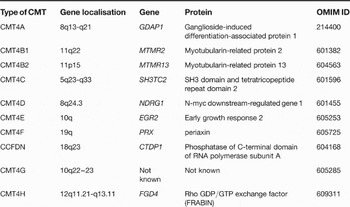
Autosomal recessive forms of Charcot–Marie–Tooth (ARCMT) are demyelinating (ARCMT1 or CMT4), axonal neuropathies (ARCMT2) or intermediate sensory motor neuropathies; however, they are clinically distinguished by an earlier onset and a more severe disease course. In the West, ARCMTs account for about 5% of the CMT neuropathies, and often appear as isolated cases because of the small kindred size. In communities with a high occurrence of consanguineous marriage, ARCMT may constitute up to 50% of all CMT cases (Ref. Reference Dubourg2). In the past few years, progress in molecular genetics has allowed a better classification of the different forms of ARCMT based on the underlying gene defect (Table 1). At present, demyelinating forms of ARCMT are more widely studied at the genetic level than the axonal forms are.
Classification of autosomal recessive Charcot–Marie–Tooth disease type 4

Abbreviations: CCFDN, congenital cataracts, facial dysmorphism and neuropathy; CTDP1, C-terminal domain of RNA polymerase II phosphatase; EGR2, early growth response; FRABIN, FGD1-related F-actin binding protein; GDAP1, ganglioside-induced differentiation-associated protein; NDRG1, N-myc downstream-regulated gene 1; PRX, periaxin; SH3TC2, SH3-domain tetratricopeptide repeats.
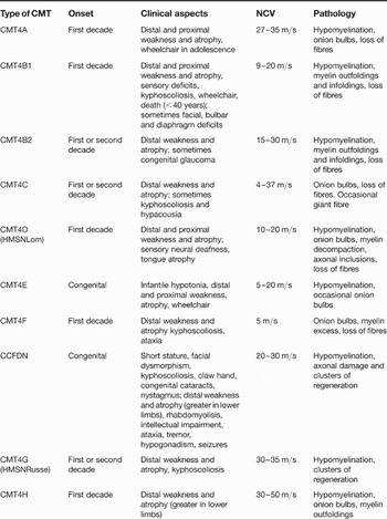
Demyelinating CMT4 subtypes are generally characterised by early onset and are sometimes congenital, with patients displaying delayed motor milestones. Muscle atrophy and weakness affects the distal muscles of all four limbs – although the lower limbs are usually more severely involved – and sometimes weakness may progress to proximal muscles. For example, the peripheral neuropathy may also involve bulbar and facial nerves, resulting in vocal cord paresis or diaphragm weakness. Sensorineural deafness may also be observed. Neurophysiological and morphological criteria are usually sufficient to classify CMT4 neuropathies in the demyelinating subtype to initiate adequate gene screening. However, secondary axonal loss may sometimes be severe enough to make diagnosis difficult. The main clinical characteristics of the various forms of CMT4 neuropathies are summarised in Table 2.
Main characteristics of the Charcot–Marie–Tooth type 4 neuropathiesa

aData obtained from Refs Reference Shy, Dyck and Thomas1, Reference Dubourg2, Reference Benstead73, Reference Vallat74, Reference Pareyson75.
Abbreviations: CMT, Charcot–Marie–Tooth; NCV, nerve conduction velocity.
CMT4B1 and CMT4B2: clinical characteristics
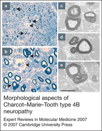
CMT4B is the first recognised neuropathy to be characterised by focally folded myelin (Fig. 2) and was initally described by Ohnishi in 1989 (Ref. Reference Ohnishi3), and in the subsequent year by Gabreels-Festen (Ref. Reference Gabreels-Festen4), in patients showing an autosomal recessive inheritance. The gene responsible for CMT4B1 neuropathy was identified as myotubularin-related 2 (MTMR2) in 2000 (Ref. Reference Bolino5).
Morphological aspects of Charcot–Marie–Tooth type 4B neuropathy. (a, b) Semi-thin sections of sural nerves showing a reduced number of myelinated fibres, myelin outfolding (arrows) and onion bulb (arrowhead). (c, d, e) Electron micrographs showing myelin outfoldings. Bar, 24 µm (a); 18 µm (b); 6 µm (c, d, e).

Disease onset is in early infancy, although early developmental milestones are normal. Only two cases of neuropathies with focally folded myelin, the pre-molecular hallmark of CMT4B, have been reported with congenital onset. In these cases, a lack of investigation at the molecular level makes it difficult to achieve a definitive diagnosis (Ref. Reference Vallat6). Usually, the disease begins between 2 and 7 years of age, when children show progressive atrophy and weakness of the distal muscles of all four limbs. Patients present with an unsteady gait – usually walking on tiptoe – and with an increasing difficulty in manipulating small objects. Weakness may be severe enough to result in footdrop (Ref. Reference Houlden7). Pes equinovarus deformity and hammertoes are present; tendon reflexes are absent, as are pathological reflexes. Moderate-to-severe sensory deficits are present in the distal parts of all four limbs, although vibratory sensation is more affected than touch and pain sensation. Muscle atrophy and weakness progressively proceed toward proximal muscles, resulting first in a clumsy and waddling gait, and then in the need for a wheelchair from late childhood into adolescence.
Facial, bulbar and diaphragmatic involvement have been reported in a few cases (Refs Reference Houlden7, Reference Quattrone8, Reference Verny9). Houlden used molecular analysis to confirm the original case described by Tyson (Ref. Reference Tyson10), which was characterised by vocal cord paralysis with stridor and subsequent difficulty with phonation, chewing and swallowing. Examination also showed facial and jaw weakness, tongue wasting and dysarthric speech. Scoliosis is usually present in adult patients. The lifespan can be shortened, with death occurring in the fourth to fifth decade, mostly as a result of respiratory failure (Ref. Reference Quattrone8). Intellectual function is not affected.
Neurophysiology is characterised by a marked reduction in NCVs, which can range from 9 to 20 m/s and have a prolonged distal latency. Amplitudes also are markedly reduced (0.7–1 mV) and may be undetectable in adult patients. Sensory action potentials and velocities are also severely reduced. Fibrillation potentials, positive sharp waves and a decrease in the recruitment pattern of motor unit potentials are found upon needle electromyography. Polyphasic motor unit potentials and giant potentials can be present in distal muscles. Increased protein (up to 280 mg/dl) may be found in lumbar cerebrospinal fluid, despite normal cell counts (Ref. Reference Ohnishi3).
CMT4B2 is the second type of AR-CMT with myelin outfoldings, and is due to mutations in MTMR13/SBF2. The phenotype of CMT4B2 patients is usually less severe than that seen in CMT4B1 (Refs Reference Azzedine11, Reference Senderek12, Reference Conforti13). The disease onset is between 4 and 13 years of age (mean age of 8) involving motor and sensory modalities distally in all four limbs. The proximal muscles are usually spared. Tendon reflexes are reduced or absent. Foot deformities, such as pes cavus and hammertoes are present. The cerebrospinal fluid may show increased protein (up to 114 mg/dl). In some families the neuropathy segregates with congenital glaucoma (Refs Reference Azzedine11, Reference Hirano14). The neurophysiology overlaps with the B1 subtype and mean NCV is around 22 m/s in CMT4B2, suggesting a relatively milder phenotype.
Pathology of CMT4B1 and CMT4B2
The histology of affected nerves was first described by Onhishi in 1989 (Ref. Reference Ohnishi3) and pathological findings were confirmed in subsequent descriptions (Refs Reference Houlden7, Reference Quattrone8, Reference Gabreels-Festen and Gabreels15, Reference Salih16). The histology is characterised by a severe loss of myelinated fibres, and the remaining fibres are generally hypomyelinated or non-myelinated. The pathological hallmark is the presence of fibres with focal regions where myelin is thrown into irregular protrusions from the outer myelin sheath, known as ‘myelin outfoldings’ (Fig. 2). Inward protrusions, which extend towards the axon, are also present and these are termed ‘myelin infoldings’. The protrusions may contain axons or exclusively Schwann cell cytoplasm. Supernumerary Schwann cell processes or basal lamina structures may encircle some fibres (‘onion bulbs’) indicating the occurrence of repeated episodes of demyelination and remyelination and abnormal proliferation. In CMT4B2, the number of onion bulbs is usually higher, and the complete loss of myelinated fibres delayed, indicating a milder phenotype compared with CMT4B1 (Refs Reference Azzedine11, Reference Hirano14, Reference Salih16). Muscle biopsy has been performed in a few patients and this has revealed only minor abnormalities compatible with neurogenic atrophy (Ref. Reference Quattrone8).
Genetic characterisation of CMT4B1 and CMT4B2
Loss-of-function mutations in either MTMR2 or MTMR13 cause CMT4B1 and CMT4B2, respectively. MTMRs represent a family of proteins sharing homology with protein tyrosine phosphatases (PTPs) (discussed in more detail below). This family is highly conserved and comprises 14 members in humans, of which eight are catalytically active proteins, whereas six are inactive. Despite their homology with PTPs, MTMRs dephosphorylate both PtdIns3P and PtdIns(3,5)P 2 phosphoinositides in the D3 position of the inositol ring (Refs Reference Robinson and Dixon17, Reference Clague and Lorenzo18, Reference Laporte19). MTMR2 and MTMR13 are catalytically active and inactive phosphatases, respectively, which can interact and form heterotetramers of homodimers, consisting of two MTMR2 and two MTMR13 molecules (Refs Reference Robinson and Dixon20, Reference Berger21). The catalytically inactive MTMR13 is thought to regulate both the phosphatase activity and the subcellular localisation of active MTMR2. MTMR2 was the first gene identified in CMT4 and thus CMT4B constitutes the first neuropathy known to be caused by loss of phosphoinositide phosphatases, which have a non-redundant biological role in the nerve, despite their ubiquitous expression.
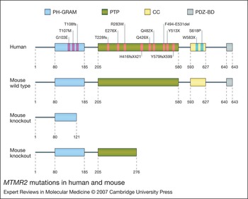
MTMR2 is a protein of 643 amino acids with a PH-GRAM (Pleckstrin homology, glucosyltransferase, Rab-like GTPase activator and myotubularin) domain, which binds phosphoinositides [mainly PtdIns5P or PtdIns(3,5)P 2]; a PTP domain; a coiled-coil domain for homo- and heterodimerisation with other members of the family, such as MTMR13; and a PSD-95–Dlg–ZO-1 binding domain (PDZ-BD) at the C- terminus (Ref. Reference Robinson and Dixon17) (Fig. 3).
MTMR2 mutations in human and mouse. The structures of the highly homologous human and mouse myotubularin-related 2 (MTMR2/Mtmr2) proteins are shown. PH-GRAM, pleckstrin-homology glucosyltransferase, Rab-like GTPase activators and myotubularin domain; PTP, tyrosine-phosphatase-like domain; CC, coiled-coil domain; PDZ-BD, PSD-95–Dlg–ZO-1 binding domain. The G103E, E276X, F494-E531del, Q426X and Q482X mutations have been demonstrated to abolish phosphatase activity in vitro towards both PtdIns3P and PtdIns(3,5)P 2 (Ref. 22). The Y579fsX599 and the G103E mutations also impair binding of the protein to membranes of vacuoles formed under hypo-osmotic stress (Ref. 64). The consensus motif for catalytic activity C-X5-R corresponds to amino acids 417–423 of the MTMR2 protein sequence. The upper mouse knockout structure is from Ref. 28 and the lower one is from Ref. 32

MTMR2 mutations are loss-of-function mutations, such as base-pair insertions or deletions leading to frameshifts, and missense mutations creating stop codons or leading to single amino acid changes. Most of the identified MTMR2 mutations affect the PTP domain, resulting in loss of enzymatic activity, as has been functionally demonstrated (Ref. Reference Berger22). Mutations in the PH-GRAM domain are predicted to affect the phosphatase activity and binding to the membrane, as shown for the G103E mutation. Other MTMR2 mutations, such as Y579fsX599 and W583X, remove part of the coiled-coil domain (residues 593-627) and the PDZ-BD (residues 640-643) (Refs Reference Bolino5, Reference Houlden7, Reference Parman23, Reference Nelis24) (Fig. 3). Four new mutations have been recently identified in patients with CMT4B1: T107M in the PH-GRAM domain; H416fsX421 and Y513X in the PTP domain; and the missense mutation S618P in the coiled-coil domain at the C-terminus. Homozygous T107M and S618P mutations have been found in the same family (E. Nelis and P. De Jonghe, University of Antwerp, Antwerp, Belgium, pers. commun.; S. Erdem, Atatürk Research and Training Hospital, Ankara, Turkey, pers. commun.).
MTMR13 is a catalytically inactive phosphatase of 1849 amino acids with a PH-GRAM domain, a PTP domain with substitutions in the Cys and Arg residues of the Cys-X5-Arg site for catalytic activity, and a coiled-coil domain and PDZ-BD as in MTMR2. MTMR13 also possesses a DENN (‘differentially expressed in normal versus neoplastic’) domain at the N-terminus and a canonical PH domain at the C-terminus. The first reported mutation is an in-frame deletion, which removes part of the DENN domain (Ref. Reference Senderek12). DENN–GEF–AEX-3 is a well-conserved motif in signalling molecules. Cytoplasmic proteins such as MTMR5/SBF1, which lack part of this domain, relocalise to the nucleus where they might activate signalling events leading to oncogenic transformation (Refs Reference Firestein and Cleary25, Reference Levivier26, Reference Cui27). Other MTMR13 mutations are classical loss-of-function mutations that remove the PH-GRAM, PTP and C-terminal PH domains (Refs Reference Azzedine11, Reference Senderek12, Reference Conforti13, Reference Hirano14).
Animal models of CMT4B
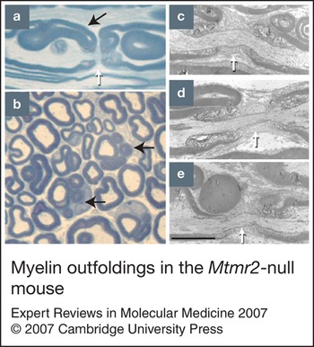
To demonstrate that CMT4B is caused by loss of MTMR2 in the nerve, two animal models have been generated. The first mouse model was the Mtmr2-null mouse, which was produced using Cre/loxP technology to remove Mtmr2 function (Ref. Reference Bolino28). Upon removal of exon 4, which encodes part of the PH-GRAM domain, a truncated product is produced, which lacks all the downstream functional domains such as PTP, coiled-coil and PDZ-BD. These mice are viable and do not show any functional impairment upon external examination. However, behavioural and electrophysiological tests suggest a neuromuscular defect. In both motor and sensory nerves of Mtmr2-null mice, myelin outfoldings were observed mainly at paranodal regions 3–4 weeks after birth – a time when myelinating Schwann cells are still elongating during postnatal development (Box 1 and Fig. 4). Notably, paranodes are known sites of membrane remodelling during myelin assembly in postnatal development (Ref. Reference Berthold and Nilsson29). At 12–15 months of age, myelin outfoldings also arise at Schmidt–Lanterman incisures, which are internodal channels that are enriched in the Schwann cell cytoplasm (Ref. Reference Bolis30).
Myelin outfoldings in the Mtmr2-null mouse. (a, b) Semi-thin longitudinal (a) and transverse (b) sections of sciatic nerves with myelin outfoldings (black arrows). White arrow in (a) indicates the node of Ranvier. (c, d, e) Electron micrograph of redundant myelin loops (outfoldings) in longitudinal sections at the node of Ranvier (white arrows). Bars, 4 µm (a); 14 µm (b); 2 µm (c, d, e).

Box 1 Nerve fibre architecture in the peripheral nervous system
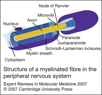
Impulse propagation by saltatory conduction in vertebrates is achieved using myelin, which wraps around axons with diameters greater than 1 µm in the peripheral nervous system. Here, myelin is formed from compacted layers of Schwann cell membrane ensheathing a single internode – the portion of the axon isolated by myelin – with lamellae, which are produced in proportion to the axon diameter. Both radial and longitudinal polarisation can be distinguished in a myelinated fibre. The radial polarisation extends from the basal lamina surrounding the outside of the Schwann cell – the compact myelin – to the periaxonal space. Longitudinal polarisation consists of a few subdomains centred on the node of Ranvier, such as paranodal and juxtaparanodal regions (Fig. 5) (Refs Reference Poliak and Peles36, Reference Salzer37). Compact myelin is produced by the compaction of lamellae of the Schwann cells membrane ensheathing an axon whereas the so-called ‘non-compact’ myelin comprises regions of Schwann cells where the cytoplasm is more abundant along the fibre, such as nodal–paranodal regions, inner and outer mesaxons and Schmidt–Lanterman incisures. These structures allow the radial diffusion of soluble and small molecules across different layers of membrane. Incisures, as well as paranodal Schwann cell membranes, are enriched with tight junctions, adherens junctions and gap junctions, which are referred to as ‘autotypic’, because they link membrane layers within the same cell.
Structure of a myelinated fibre in the peripheral nervous system. Schwann cell membrane layers are depicted in blue ensheathing the axon, which is shown in brown. Dots in the axon represent neurofilament clusters. Paranodal and juxtaparanodal regions flank the node of Ranvier, which is devoid of myelin. The cytoplasm of Schwann cells is restricted to the perinuclear region, paranodal loops and microvilli, finger-like extensions of Schwann cells facing the axolemma in the node of Ranvier.

Myelin compaction and the ultrastructure of the myelinated fibres from Mtmr2-null mice were both normal, suggesting that the gross molecular architecture of the myelinated fibre with redundant myelin is not altered (Ref. Reference Bolino28). Mtmr2-null mice also have defects in spermatogenesis, which become apparent from 3 to 4 weeks of age. Immature germ cells were found in the lumen of tubules of the seminiferous epithelium of these mice, suggesting that a loss of adhesion occurs between Sertoli cells and germ cells (Ref. Reference Bolino28). Consistent with this, one familial CMT4B1 case has been reported with azoospermia (Ref. Reference Laporte19). Moreover, when the inactive Mtmr5 phosphatase – which interacts in vitro with Mtmr2 – is ablated, a defect in spermatogenesis is also observed (Ref. Reference Firestein31). Thus Mtmr2, perhaps through interaction with Mtmr5, has a relevant role in the testis and its absence is not compensated by other MTMRs.
Another animal model has been produced by introducing the E276X mutation in exon 9 (Ref. Reference Bonneick32). This homozygous mutation has been found in one familial CMT4B1 case (Ref. Reference Bolino5). The nerve morphology in these mice was similar to that observed in Mtmr2-null mice, although a huge variability in the number of myelin outfoldings in individual animals was noted. Moreover, no electrophysiological or behavioural alterations were reported and the testis was grossly normal (Ref. Reference Bonneick32).
In Mtmr2-null mice and E276X mutants, the dysmyelinating phenotype was less severe than that observed in human CMT4B1. Axonal loss was observed in Mtmr2-null mice only at later stages (around 15 months of age) in more distal nerves. The variation in phenotype observed between mice and humans might be due to the shorter mouse lifespan and to their shorter nerve lengths. Alternatively, a partial compensation by other MTMRs in the mouse nerve might account for the less-severe pathology. Expression of Mtm1 and Mtmr1 proteins, which share the strongest homology with MTMR2 within the MTMR family, was investigated in nerve, and Mtm1 but not Mtmr1 was detected in myelin-forming Schwann cells. Using quantitative expression analysis, levels of Mtm1 mRNA were found to be significantly upregulated (1.32-fold) in mutant nerves, but not in muscle and brain, suggesting a possible compensation by Mtm1 in this tissue (Ref. Reference Bolis30).
MTMR2 mRNA is ubiquitously expressed in human and mouse tissues (Refs Reference Bolino5, Reference Berger22, Reference Bolino33). In the adult rat peripheral nervous system, the Mtmr2 protein is detected in vivo in the cytoplasm of myelin-forming and non-myelin-forming Schwann cells, and in axons in peripheral nerves (Ref. Reference Previtali34). To assess the cell-autonomous role of Mtmr2, two conditional-null mice with Mtmr2-specific ablation in either Schwann cells or motor neurons were also generated (Ref. Reference Bolis30). Only the Schwann-cell-null mouse displayed a dysmyelinating phenotype very similar to that observed in human CMT4B1 and in the Mtmr2-null mouse. Dysmyelination and axonopathy were not observed in the motor-neuron-null mouse. It is possible to conclude that loss of Mtmr2 phosphatase activity in Schwann cells is sufficient and necessary to cause CMT4B1 with myelin outfoldings. Mtmr2 might still have a function in motor neurons, which is redundant or compensated for by other MTMRs at E9.5, when Mtmr2 is inactivated in mice. It also has to be considered that axonal phenotypes have only rarely been reproduced in mice, owing to differences in nerve length and their shorter lifespan, as mentioned earlier (Ref. Reference Zu Horste and Nave35).
Myotubularin-related protein family
Myotubularin-related proteins constitute a family of phosphatases within the PTP superfamily (Ref. Reference Robinson and Dixon17). The founding member of this family, MTM1, was identified as the mutated gene responsible for X-linked myotubular myopathy (XLMTM) (Ref. Reference Laporte19). Despite their homology with PTP and dual-specificity phosphatase (DSP) proteins, MTMRs are 3-phosphatases, which act on PtdIns3P and PtdIns(3,5)P 2, both in vitro and in vivo (Refs Reference Berger22, Reference Schaletzky38, Reference Blondeau39, Reference Zhao40, Reference Walker41, Reference Kim42, Reference Laporte43, Reference Mochizuki and Majerus44, Reference Tronchere45, Reference Taylor, Maehama and Dixon46, Reference Begley47).
Polyphosphorylation of phosphatidylinositol (PtdIns) at the D3, D4 and D5 positions of the inositol ring generates polyphosphoinositides (PPIn), which account for less than 10% of the total amount of lipids in eukaryotic cells. Nevertheless, PPIn are important second messengers that control a variety of biological functions, such as membrane trafficking, cellular motility, survival, growth and cell division. A tightly controlled spatial distribution of PPIn within the cell is at the basis of their function as signalling mediators (Refs Reference Balla48, Reference De Matteis and Godi49, Reference Carlton and Cullen50, Reference Behnia and Munro51). PPIn are concentrated in different subcellular membranes, such as early endosomes, late endosomes, the Golgi and endoplasmic reticulum, by the concerted and opposed function of specific kinases and phosphatases. Moreover, PPIn bind to domains of ‘effector’ proteins that are concentrated on the same membranes, such as PH, PX (phox homology), ENTH (epsin N-terminal homology) and FYVE (Fab1p–YOTB–Vac1p–EEA1) domains. It is generally accepted that each organelle has a specific identity that is defined by macrodomains formed by PPIn, specific effectors that are recruited and GTPases. PtdIns3P for example, which is a substrate of MTMRs, is highly enriched in early endosomes and in the internal vesicles of multivesicular bodies (Refs Reference Schu52, Reference Zerial and McBride53). PtdIns3P is produced by a class III PtdIns 3-kinase in mammals, which corresponds to the Vps34 protein (vacuolar protein sorting 34) in yeast. On early endosomes, PtdIns3P recruits its effector protein, EEA1 (early endosomal antigen 1), which possesses a FYVE domain for lipid binding and associates with Rab5, Rabex, Rabenosin and other proteins forming a lipid–protein macrodomain in these membranes.
In yeast, PtdIns(3,5)P 2 – the other MTMR substrate – is required at the limiting membrane of vacuoles (Refs Reference Dove54, Reference Gary55, Reference Odorizzi, Babst and Emr56). In mammalian cells, PtdIns(3,5)P 2 localisation is not well defined, and a recent report indicates that it may localise at early endosomes rather than late endosomes (Refs Reference Dove54, Reference Gary55, Reference Odorizzi, Babst and Emr56, Reference Ikonomov, Sbrissa and Shisheva57, Reference Michell58, Reference Rutherford59). Perturbation of PtdIns(3,5)P 2 levels in both yeast and mammalian cells that overexpress the kinase or an inactive mutant, provokes swollen endosomes and vacuolisation, suggesting that this phosphoinositide is important for membrane homeostasis. PtdIns(3,5)P 2 effectors have been mainly identified in yeast, whereas only putative effectors have been demonstrated in mammalian cells (Refs Reference Robinson and Dixon17, Reference Michell58). The metabolic pathway and the roles of PtdIns5P – the product of the 3-phosphatase activity of MTMRs on PtdIns(3,5)P 2 – are poorly understood. However, recent findings define PtdIns5P as a potential new second messenger, not only in endosomal membranes but also within the nucleus (Refs Reference Coronas60, Reference Pendaries61).
MTMR phosphatase activity toward PtdIns3P and PtdIns(3,5)P 2 obviously points to its involvement in the regulation of membrane trafficking and homeostasis. However, MTMRs are mainly cytosolic or peripheral-membrane-associated proteins and they have not been reported to localise to early endosomes or late endosomes/lysosomes. Partial localisation of some MTMRs with intracellular membranes has been reported. For example, MTM1 might be partially located at the plasma membrane upon sustained overexpression or following co-transfection with dominant-negative forms of Rac1 that induce membrane ruffling (Refs Reference Laporte43, Reference Tsujita62). MTMR3 and its C413S mutant form have also been detected in Golgi membranes and in internal multivesicular structures resembling autophagosomes (Refs Reference Walker41, Reference Lorenzo, Urbe and Clague63). MTMR2 and MTMR13 may localise at membranes of vacuoles formed under hypo-osmotic stress, which increases levels of PtdIns(3,5)P 2 (Refs Reference Berger21, Reference Berger64). However, these studies are based mainly on overexpression under nonphysiological conditions. Endogenous localisation of MTMRs to endosomal compartments remains to be demonstrated. Expression studies at physiological levels have been hampered by (1) the difficulty in obtaining specific antibodies recognising a single protein; (2) the low level of expression of these proteins; and (3) the fact that MTMRs might be spatially targeted, enriched and controlled under specific physiological conditions depending on the cell type.
Several studies have suggested a role of myotubularins in regulating endocytosis and the trafficking of membranes and proteins. MTM1 overexpression in COS7 cells extensively treated with epidermal growth factor (EGF) induces large endosomal vacuoles and inhibits EGF receptor (EGFR) trafficking from the late endosome to lysosomes. By contrast, in myotubes, MTM1 overexpression leads to the displacement of early endosomal antigen 1 (EEA1) from early endosomes and also to decreased insulin-induced glucose uptake (Refs Reference Tsujita62, Reference Chaussade65). However, lack of endogenous MTM1 in XLMTM myoblasts does not significantly affect the pool of PtdIns3P or the localisation of EEA1 (Ref. Reference Tronchere45). In Caenorhabditis elegans, MTM-6 and MTM-9 (homologous to MTMR6–8 and MTMR9, respectively, in mammals) have been identified by genetic screening as proteins required for endocytosis in coelomocytes, with a dramatic alteration in PtdIns3P localisation observed in coelomocytes from mtm-6 and mtm-9 mutants (Ref. Reference Dang66).
Despite these results, the physiological role of myotubularins in mammalian cells is still not clear. Moreover, the finding that MTMR6 can inhibit the KCa3.1 channel in membranes of CD4+ T cells by decreasing levels of PtdIns3P suggests that MTMRs might be involved in the regulation of specific cellular functions in different cell types (Ref. Reference Srivastava67).
MTMR2 and MTMR13: polyphosphoinositides and peripheral nervous system disorders
MTMR2 is ubiquitously expressed in human and mouse tissues, as demonstrated mainly at the mRNA level (Ref. Reference Bolino5). During mouse development, ubiquitous Mtmr2 expression is observed from embryonic day (E) 9.5 (Ref. Reference Bolino33). In the adult rat peripheral nervous system, Mtmr2 protein is detected in vivo in the cytoplasm of: (1) myelin-forming and non-myelin-forming Schwann cells; (2) axons of peripheral nerves; (3) motor neurons, sensory neurons and satellite cells of dorsal root ganglia; (4) both dorsal and ventral roots of spinal cord (Ref. Reference Previtali34). MTMR13 mRNA is also ubiquitously expressed in human tissues (Ref. Reference Senderek12).
Several overexpression studies show that the MTMR2 protein localises in the cytosol with a broad diffuse pattern. Enrichment in the perinuclear region is also reported (Ref. Reference Kim42). In myelinated fibres, MTMR2 is found in the Schwann cell cytoplasm in the perinuclear region, paranodes and in Schmidt–Lanterman incisures (Ref. Reference Previtali34). It has been hypothesised that MTMR2 localises to precise subcellular compartments under stimulated conditions that change the levels of intracellular polyphosphoinositides and/or following interaction with an inactive partner, such as MTMR13, in the nerve. Consistent with this, when hypo-osmotic stress – a condition that increases PtdIns(3,5)P 2 levels – is induced in COS7 cells, MTMR2 relocalises to the membranes of intracellular vacuoles. As mentioned above, MTMR2 and MTMR13 interact and form heterotetramers by the association of homodimers of two MTMR2 and MTMR13 molecules (Ref. Reference Berger21). When MTMR13 is co-expressed with MTMR2 at high levels in COS7 cells, MTMR13 preferentially binds to membranes of vacuoles formed under hypo-osmotic stress whereas, if co-expressed at lower levels, both proteins bind to intracellular membranes (Refs Reference Berger21, Reference Berger64). However, the precise nature of the intracellular vesicle compartments to which endogenous MTMR2 and MTMR13 bind, the physiological event or stimulus that might lead to changes in PPIn composition, and subsequent delocalisation of MTMRs in Schwann cells remain to be assessed. Furthermore, although MTMR2 dephosphorylates both PtdIns3P and PtdIns(3,5)P 2 in vitro and in vivo (Ref. Reference Berger22), the enzymatic activity in the nerve remains to be determined at physiological levels. Moreover, clear colocalisation of MTMR2 with markers of intracellular vesicle compartments has not been demonstrated in Schwann cells or neurons. To gain insight into the cellular function and localisation of the MTMR2 phosphatase, tissue-specific interactors were sought using yeast two-hybrid screening analysis of cDNA libraries from nerve and fetal brain.
Interestingly, this approach identified the interaction of the neurofilament light chain (NF-L) protein with MTMR2 in neurons (Ref. Reference Previtali34). Mutations in the gene encoding NF-L (NEFL) also cause an axonal form or ‘intermediate’ forms of CMT with mixed features of axonopathy and demyelination; these cases occasionally produce excessive myelin, which can resemble myelin outfoldings. NF-L is mainly expressed by neurons, whereas Schwann cells upregulate the NEFL mRNA level after injury (Ref. Reference Sotelo-Silveira68). Thus, the MTMR2–NF-L interaction might have a relevant role in neurons and/or during Schwann cell re-myelination. Whether or not loss of the MTMR2–NF-L interaction contributes to the axonal pathology observed in human CMT4B1 is not clear (Ref. Reference Previtali34).
Since Mtmr2 ablation in Schwann cells causes the dysmyelinating CMT4B1 phenotype with myelin outfoldings, efforts have been subsequently concentrated on Mtmr2 interactions in this cell type. Interaction between Mtmr2 and Dlg1/SAP97 (discs large/synapse-associated protein 97) occurs in Schwann cells (Ref. Reference Bolis30). Dlg1 is a scaffolding molecule belonging to the MAGUK (membrane-associated guanylate-kinase-like) protein family, which links transmembrane proteins with the intracellular cytoskeleton. Multimeric protein complexes can be formed through multiple interaction domains of MAGUK proteins, such as PDZ-BD, SH3 (Src-homology 3) and GuK (guanylate-kinase-like). Dlg1 is located in vertebrates at adherens junctions of epithelial cells and at pre- and postsynaptic sites in neurons (Refs Reference Humbert, Russell and Richardson69, Reference Knust and Bossinger70). In Drosophila melanogaster, loss of Dlg produces overgrowth of imaginal discs, and loss of polarity and adhesion because of septate junction disruption. In myelinated fibres, Dlg1 is located in Schwann cells at Schmidt–Lanterman incisures and paranodal loops, where myelin outfoldings also arise in CMT4B1 neuropathy. This localisation pattern is lost in Mtmr2-null nerves, suggesting that loss of the Mtmr2–Dlg1 complex is the basis of the myelin pathology (Ref. Reference Bolis30).
Putative roles of MTMR2 and MTMR13 in Schwann cells
Mtmr2 has a Schwann-cell-autonomous role because specific inactivation of Mtmr2 in Schwann cells reproduces the phenotype of Mtmr2-null mice. In addition, myelin outfoldings originate from Schwann cell structures and Dlg1, which interacts with Mtmr2 in Schwann cells, is almost lost in paranodal regions where myelin outfolding also preferentially arises (Refs Reference Bolino28, Reference Bolis30).
Mtmr2 dephosphorylates phosphoinositides implicated in intracellular trafficking events, and Dlg1 is also involved in membrane addition in polarised cells (Ref. Reference Lee71). Thus, the Mtmr2–Mtmr13–Dlg1 complex might regulate membrane addition and/or homeostasis in nerve Schwann cells during postnatal maturation. Myelin outfoldings also arise from paranodes and incisures 3–4 weeks after birth (Ref. Reference Bolino28). Paranodes are known sites of membrane remodelling and homeostasis during myelin assembly in postnatal nerve development. Notably, clathrin-coated pits, endosomes and lysosomes have been demonstrated in the paranodal cytoplasm – consistent with a role for these sites in membrane remodelling and/or homeostasis. Myelin assembly obviously relies on intracellular trafficking and membrane homeostasis within Schwann cells, and lipid rafts have been proposed to be important membrane macrodomains that help to deliver lipids and proteins (Ref. Reference Schaeren-Wiemers72). However, the mechanism by which these macrodomains are formed and segregated is poorly understood. MTMR2 might well be the first phosphatase identified to regulate these events at paranodal regions of myelin-forming Schwann cells.
Further studies in mice are necessary: (1) to demonstrate the colocalisation of Mtmr2 with intracellular vesicle compartments; (2) to assess whether the Mtmr2–Mtmr13 interaction is correlated with membrane addition or homeostasis; and (3) to determine how other molecules, such as Dlg1, NF-L and PPIn, are involved in these mechanisms.
Acknowledgements and funding
This work was supported by grants from Telethon Italy and Compagnia San Paolo to A.B.; Telethon Italy and the Italian Ministry of Health (MIUR) to S.C.P; Istituto Superiore di Sanità and FIRB to A.Q. A.B. is a recipient of a Telethon Associate Scientist Career Award. We thank the peer reviewers for their comments.