Introduction
The link between psychosis and autoimmunity has been explored over many decades (Wright et al. Reference Wright, Laing, Donaldson, Murray and Müller1999; Eaton et al. Reference Eaton, Byrne, Ewald, Mors, Chen, Agerbo and Mortensen2006; Benros et al. Reference Benros, Eaton and Mortensen2014), but only in recent years has this been related to the pathophysiology of psychosis at a neurotransmitter level via specific antibodies against antigens that have clearly established links to the aetiology of psychosis. This has resulted in a surge of interest in whether a subgroup of people with psychosis have serum antibodies to certain neuronal antigens, although the specificity, sensitivity and clinical relevance of these remain unclear (Coutinho et al. Reference Coutinho, Harrison and Vincent2014; Deakin et al. Reference Deakin, Lennox and Zandi2014; Pollak et al. Reference Pollak, McCormack, Peakman, Nicholson and David2014). These auto-antibodies target synaptic or extra-synaptic membrane-expressed proteins, with greatest interest in antibodies to the N-methyl-D-aspartate receptor (NMDAR) and the voltage-gated potassium channel (VGKC)-complex, which includes antigens leucine-rich glioma inactiviated-1(LGI1) and contactin-associated protein-2 (CASPR2) (Zandi et al. Reference Zandi, Irani, Lang, Waters, Jones, McKenna, Coles, Vincent and Lennox2011; Ezeoke et al. Reference Ezeoke, Mellor, Buckley and Miller2013; Steiner et al. Reference Steiner, Walter, Glanz, Sarnyai, Bernstein, Vielhaber, Kastner, Skalej, Jordan, Schiltz, Klingbeil, Wandinger, Bogerts and Stoecker2013; Pearlman & Najjar, Reference Pearlman and Najjar2014).
Psychotic symptoms are a common feature of the encephalitis syndromes classically associated with such antibodies – often occurring before the development of other features of encephalitis, such as seizures, movement disorders, autonomic instability and impaired consciousness. For example, in the seminal case series describing 100 anti-NMDAR encephalitis cases, the majority (77%) of patients initially presented to psychiatry services with predominantly psychotic symptoms (Dalmau et al. Reference Dalmau, Gleichman, Hughes, Rossi, Peng, Lai, Dessain, Rosenfeld, Balice-Gordon and Lynch2008). A subsequent larger case series of 571 patients reported 18 patients (3.2%) having an ‘isolated’ psychiatric relapse (after an initial ‘full’ encephalitis). Five further patients (0.9%) had ‘isolated’ psychiatric symptoms, again predominantly psychosis, at presentation – although on retrospective close examination they were found to have had other clinical features and were re-diagnosed as encephalitis (Kayser et al. Reference Kayser, Titulaer, Gresa-Arribas and Dalmau2013). The key question is therefore whether isolated psychosis, i.e. schizophrenia, is associated with these antibodies.
A prospective cohort study in first-episode psychosis (FEP) identified serum NMDAR antibodies in 6.4% of patients (n = 3/47) (Zandi et al. Reference Zandi, Irani, Lang, Waters, Jones, McKenna, Coles, Vincent and Lennox2011) using a live cell-based assay (CBA) and well-established subjective visual scoring system (Irani et al. Reference Irani, Bera, Waters, Zuliani, Maxwell, Zandi, Friese, Galea, Kullmann, Beeson, Lang, Bien and Vincent2010b). A recent case–control study in FEP identified an increased prevalence of NMDAR antibodies (n = 7/228; 3% of total population) compared with controls (n = 0/105), with no difference in the prevalence of LGI1, CASPR2 or VGKC-complex antibodies between the groups (Lennox et al. Reference Lennox, Palmer-Cooper, Pollak, Hainsworth, Marks, Jacobson, Lang, Fox, Ferry, Scoriels, Crowley, Jones, Harrison and Vincent2017). Using the same methodology, a recent study by our group found that 7% of patients (n = 3/43) with chronic refractory psychosis were positive for NMDAR antibodies (Beck et al. Reference Beck, Lally, Shergill, Bloomfield, MacCabe, Gaughran and Howes2015). Methodology, however, appears to be relevant, as 9.9% of patients with acute schizophrenia were reported NMDAR antibody positive using the commercial fixed CBA, with similar rates in controls (Steiner et al. Reference Steiner, Walter, Glanz, Sarnyai, Bernstein, Vielhaber, Kastner, Skalej, Jordan, Schiltz, Klingbeil, Wandinger, Bogerts and Stoecker2013), whereas, other studies have failed to identify any antibody positivity in either FEP (Masdeu et al. Reference Masdeu, Gonzalez-Pinto, Matute, Ruiz De Azua, Palomino, De Leon, Berman and Dalmau2012) or established schizophrenia (Rhoads et al. Reference Rhoads, Guirgis, McKnight and Duchemin2011; Haussleiter et al. Reference Haussleiter, Emons, Schaub, Borowski, Brüne, Wandinger and Juckel2012; de Witte et al. Reference de Witte, Hoffmann, van Mierlo, Titulaer, Kahn and Martinez-Martinez2015). Some of these negative studies also used fixed CBA to measure NMDAR seropositivity, but there have been few direct comparisons between laboratories (Wandinger et al. Reference Wandinger, Saschenbrecker, Stoecker and Dalmau2011). A meta-analysis identified no excess of NMDAR antibody [of any subtype (IgG, IgM, IgA)] in psychotic disorders, though IgG antibodies alone were significantly greater in FEP cases (IgG positive in 5/272 of cases) than in controls (IgG positive in 5/1598 of controls) (Pollak et al. Reference Pollak, McCormack, Peakman, Nicholson and David2014).
VGKC-complex antibodies can also cause cell-surface antibody-associated central nervous system disorders (Zuliani et al. Reference Zuliani, Graus, Giometto, Bien and Vincent2012). Most of these antibodies bind to LGI1 or CASPR2 (Irani et al. Reference Irani, Alexander, Waters, Kleopa, Pettingill, Zuliani, Peles, Buckley, Lang and Vincent2010a), which are cell surface proteins that form part of the VGKC-complex. The targets of other VGKC-complex antibodies are unclear, although two recent studies suggest that they bind to intracellular epitopes and may be neuroinflammatory biomarkers rather than pathogenic antibodies (Hacohen et al. Reference Hacohen, Singh, Rossi, Lang, Hemingway, Lim and Vincent2015; Lang et al. Reference Lang, Makuch, Moloney, Dettmann, Mindorf, Probst, Stoecker, Buckley, Newton, Leite, Maddison, Komorowski, Adcock, Vincent, Waters and Irani2017).
Many patients with high levels of VGKC-complex antibodies have evidence of limbic encephalitis and some present first with psychosis (Paterson et al. Reference Paterson, Zandi, Armstrong, Vincent and Schott2014). Phenotypically, LGI1 antibodies tend to associate with limbic encephalitis and faciobrachial dystonic seizures, phenomena which can be misinterpreted as myoclonic jerks (Irani et al. Reference Irani, Michell, Lang, Pettingill, Waters, Johnson, Schott, Armstrong, S Zagami, Bleasel, Somerville, Smith and Vincent2011). CASPR2 antibodies, meanwhile, are associated with neuromyotonia or Morvan's syndrome in which psychiatric features are common, often with a thymoma (Irani et al. Reference Irani, Alexander, Waters, Kleopa, Pettingill, Zuliani, Peles, Buckley, Lang and Vincent2010a). In a case series, over a third of patients with VGKC-complex antibodies had psychiatric symptoms, including confusion, personality change, memory impairment and depression (Somers et al. Reference Somers, Lennon, Rundell, Pittock, Drubach, Trenerry, Lachance, Klein, Aston and McKeon2011). Those affected are classically older than those with NMDAR antibody encephalitis. VGKC-complex antibodies were identified in one patient out of 46 with FEP (Zandi et al. Reference Zandi, Irani, Lang, Waters, Jones, McKenna, Coles, Vincent and Lennox2011).
The importance of antibodies to NMDAR and VGKC-complex proteins is clear when psychosis presents in the context of encephalitis, but the challenge in psychiatry is to determine their relevance in clinically uncomplicated psychosis. Only one study to date has compared the prevalence of both these, and other neuronal antibodies in adults with FEP compared with matched controls (Lennox et al. Reference Lennox, Palmer-Cooper, Pollak, Hainsworth, Marks, Jacobson, Lang, Fox, Ferry, Scoriels, Crowley, Jones, Harrison and Vincent2017).
Methods and materials
Participants
The study population comprised 96 adults with a FEP and 98 matched controls. The participants were recruited as part of the Genetics and Psychosis (GAP) study. The GAP study is a case–control study investigating the genetic basis of susceptibility to psychosis.
Participants were recruited from both inpatient and outpatient settings through working closely with the clinical teams. Patients were consented as soon after first presentation with psychosis as possible [the mean duration from the time of first contact for psychosis to the time of blood sampling was 52.74 (s.d. = 37.3) days (median 47 days; range 7–167 days)]. Assessments were performed by trained research workers and Operational Criteria Checklists (OPCRIT) diagnoses made by research clinicians.
Participants were aged 18–65 years. Patients were eligible if they had a FEP and met the International Classification of Diseases, 10th Edition (ICD-10) criteria for psychosis (ICD-10 codes F20-29 and F30-33) diagnosed utilising the OPCRIT (McGuffin et al. Reference McGuffin, Farmer and Harvey1991). The baseline diagnoses were made from face-to-face interviews by trained researchers and mental health records according to ICD-10 criteria utilising the OPCRIT (McGuffin et al. Reference McGuffin, Farmer and Harvey1991). Participants had made first contact with our health services for psychotic symptoms not more than 12 months previously.
Study design
Patients were matched for gender and age (±5 years) to 98 healthy controls from the same local population, recruited by means of Internet and newspaper advertisements, and distribution of leaflets at train stations, shops and job centres. Those who agreed to participate as controls were administered the Psychosis Screening Questionnaire (Bebbington & Nayani, Reference Bebbington and Nayani1995) and excluded if they met criteria for a psychotic disorder or reported a previous diagnosis of psychotic illness.
Exclusion criteria for both controls and cases were an organic psychosis (ICD-10: F06.8 and F9.0), i.e. a psychosis deemed likely to be secondary to neurological disorders such as epilepsy, head injury, or to a long-term deterioration of intellectual function and memory (dementia) or a short-term disturbance of orientation, judgement or consciousness (delirium). The Schedule for Clinical Assessment in Neuropsychiatry (SCAN) was administered to elucidate psychotic symptoms present in the month before study recruitment, from which those cases meeting criteria for organic psychosis were excluded. These decisions were made in discussion with two experienced psychiatrists. Those with a substance-induced psychosis as defined by ICD-10, a diagnosis of intellectual disability, terminal cancer and pregnancy were also excluded.
All participants provided written informed consent. The project was approved by the Research Ethics Committee of the Joint South London and Maudsley and the Institute of Psychiatry NHS Research Ethics Committee.
Specimen collection
The samples were transferred to Oxford for testing in two batches during the course of the observational study. The first group consisted of plasma from 30 patients and 30 age- and sex-matched controls, while the second group was serum from 66 patients and 68 controls matched for age and sex.
Plasma samples
Samples were collected in EDTA tubes using a 23-gauge butterfly needle and vacutainer needle holder following standard venepuncture in the cubital region, in the area of anastomosis between the radial and the ulnar veins or in the brachial vein, slightly proximal to this area. To avoid coagulation of the sample, a 9 ml K3EDTA tube (Greiner Bio-One, Cat # 455036X – EDTA tube, lavender lid) was used. The sample was immediately kept at +4 °C until being processed within 2 h. The tube was labelled with the subject barcode with no phenotypic information given and then centrifuged at 3000 rcf (xg), 8 min at +4 °C; the plasma was aliquoted in 0.5 ml eppendorf tubes and frozen at −80 °C within 3 h of collection.
Analysis of results for combined plasma and serum samples (n = 194), and with serum samples alone are presented (n = 134).
Antibody testing
All samples received in Oxford were kept at −20 °C until the analyses, blinded to patient-control status, were performed. The antibody assays for NMDAR, LGI1 and CASPR2 were performed using live CBAs as established in previous studies (Irani et al. Reference Irani, Alexander, Waters, Kleopa, Pettingill, Zuliani, Peles, Buckley, Lang and Vincent2010a, Reference Irani, Bera, Waters, Zuliani, Maxwell, Zandi, Friese, Galea, Kullmann, Beeson, Lang, Bien and Vincentb; Zandi et al. Reference Zandi, Irani, Lang, Waters, Jones, McKenna, Coles, Vincent and Lennox2011) and as used for routine diagnostic testing for the Oxford Neuroimmunology Service. HEK293 cells were transfected with plasmids encoding the human forms of NMDAR (NR1 and NR2B subunits), LGI1 or CASPR2, washed, grown for 24–36 h and then incubated at room temperature in the plasmas or sera at 1:20 (1:100 for CASPR2 as used routinely for this protein). Binding of antibodies to the transfected antigens on the live cells was detected with Alexa fluor labelled anti-human IgG, and the cells examined under fluorescence microscopy. The binding was compared with known positive and negative controls and scored on a scale from 0 to 4.
Positive results (>0) were repeated and the IgG specificity confirmed with an IgG-specific CBA. This involved replacing anti-human IgG, which can recognise the light chains of IgG, A, M and D, with goat anti-human Fc IgG secondary (Thermo Scientific; 31125; 1:750), followed by mouse anti-goat IgG, Alexa fluor 568 (1:750) for 45 min at room temperature (Pettingill et al. Reference Pettingill, Kramer, Coebergh, Pettingill, Maxwell, Nibber, Malaspina, Jacob, Irani, Buckley, Beeson, Lang, Waters and Vincent2015). Full details are given in (Pettingill et al. Reference Pettingill, Kramer, Coebergh, Pettingill, Maxwell, Nibber, Malaspina, Jacob, Irani, Buckley, Beeson, Lang, Waters and Vincent2015) and in the Appendix (online Supplementary material 1).
Serum VGKC-complex antibodies were tested using a radio-immunoprecipitation assay (Irani et al. Reference Irani, Bera, Waters, Zuliani, Maxwell, Zandi, Friese, Galea, Kullmann, Beeson, Lang, Bien and Vincent2010b; Paterson et al. Reference Paterson, Zandi, Armstrong, Vincent and Schott2014). VGKC results were expressed as picomoles (pM) per litre of serum.
Neuronal cultures were prepared from P0 rat hippocampi and antibody binding performed at 1:100 serum or plasma after 10–14 days in culture (Irani et al. Reference Irani, Alexander, Waters, Kleopa, Pettingill, Zuliani, Peles, Buckley, Lang and Vincent2010a, Reference Irani, Bera, Waters, Zuliani, Maxwell, Zandi, Friese, Galea, Kullmann, Beeson, Lang, Bien and Vincentb; Pettingill et al. Reference Pettingill, Kramer, Coebergh, Pettingill, Maxwell, Nibber, Malaspina, Jacob, Irani, Buckley, Beeson, Lang, Waters and Vincent2015).
Clinical assessments
The degree of psychopathology at first presentation to mental health services was measured on the Positive and Negative Syndrome Scale (PANSS) (Kay et al. Reference Kay, Fiszbein and Opler1987). Overall functional disability was assessed using the Global Assessment of Functioning (GAF).
Statistical analysis
Differences in NMDAR and VGKC-complex antibody prevalence rates in FEP patients and controls were assessed using χ2 and t tests. All statistical tests were two-sided and the α-level for statistical significance was 0.05.
Results
Sample characteristics
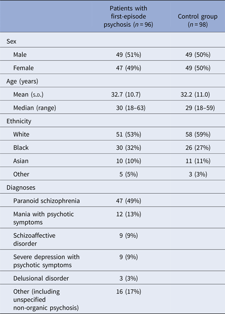
The demographic characteristics of patients with FEP and controls are shown in Table 1.
Characteristics of patients with first-episode psychosis and control participants

Data are n (%) unless otherwise specified.
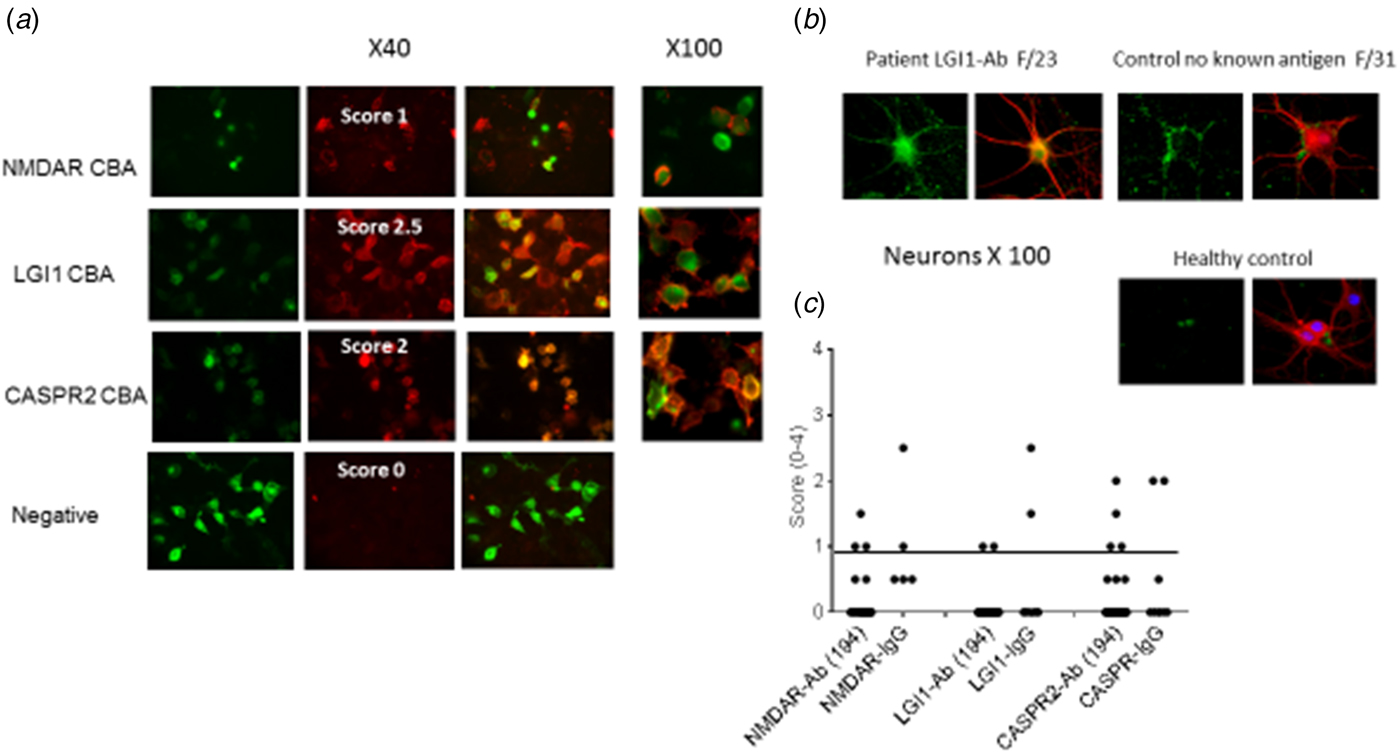
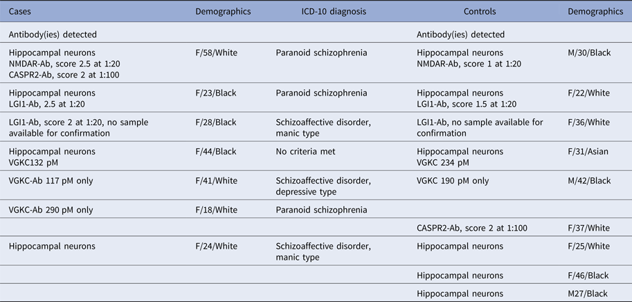
Cases and controls with positive results are summarised in Table 2. Six per cent of cases (n = 6/96) and 6% of controls (n = 6/98) were positive (χ2 = 0.001, p = 0.602) for specific antibodies (NMDAR, LGI1, CASPR2 or VGKC-complex); of these, three cases and three controls had IgG antibodies binding to hippocampal neurons in culture. An additional one case and three controls had unknown neuronal antibodies only. Overall, nine controls and seven patients had antibody positivity or were positive on neuronal staining, but this difference did not meet statistical significance (χ2 = 0.229, p = 0.414). All patients with antibody positivity or positive neuronal staining were female (7/47). Five controls with antibody positivity or with neuronal staining were female and three were male. Examples of the CBAs, neurons and scatter plots of the data are shown in Figure 1.
Cell-based assays and scatter plots of the data. Left. Examples of cell-based assays for antibodies to CASPR2, LGI1 and NMDAR antibodies, and binding to live neuron. In (a). green fluorescent protein is used to indicate the transfection rates (left panels), red indicates the human IgG binding (central panels), and the merged images are shown in the right panels ×40; and at higher magnification on the very right (×100). (b). Examples of sera binding to hippocampal neurons from one case, one control and one healthy individual. Human IgG is detected with green fluorescence, and after washing, fixing and permeabilisation, the neurons are stained red with anti-MAP2. (c). Results of the antibody (Ab) screening assay [with anti-IgG(H&L)] and the IgG-specific assay [with anti-IgG(Fc)]. The latter was only performed on the 14 of 16 sera still available in sufficient volume. Scores are shown for NMDAR and LGI1 antibodies at 1:20, and CASPR2 antibodies at 1:100, as performed for routine diagnostic analysis.

Demographic and clinical characteristics of antibody positive cases and controls

NMDAR, N-methyl-D-aspartate receptor; LGI1, leucine-rich glioma inactiviated-1; CASPR, contactin-associated protein-2; VGKC-Ab, voltage-gated potassium channel-complex antibody (by radioimmunoassay); M, male; F, female.
Of the plasma samples, no antibody-positive cases were detected in cases (n = 30), but one female control sample was positive for LGI1, with no other antibodies identified in control plasma samples (n = 30). When the serum samples alone were considered, therefore, the proportion with antibody positivity or neuronal positivity increased to 8/68 controls (11.8%) and 7/66 patients (10.6%) (χ2 = 0.045, p = 0.525).
NMDAR antibodies
Standardised immunofluorescent CBA for the detection of serum IgG antibodies directed against NMDAR was positive in 1% (n = 1 female) of cases and 1% (n = 1 male) of controls.
VGKC-complex, LGI1 and CASPR2 antibodies
VGKC-complex antibodies were the most prevalent antibody in both cases and controls, found in 3.1% (n = 3) of patients and in 2.0% (n = 2) of controls (χ2 = 0.227, p = 0.490). In cases, VGKC-complex antibody positivity was found in female patients only (n = 3/47) [males (n = 0/49) (χ2 = 3.229, p = 0.113)]. VGKC-complex antibodies were found in one male and one female control.
Two patients (both female) and two controls (both female) had LGI1 antibodies, though none were associated with VGKC-complex antibodies. CASPR2 antibodies were found in one female patient and in one female control. The patient with CASPR2 antibodies was also positive for NMDAR antibodies.
There were no cases or controls with high positive VGKC-complex antibodies (>400 pM). The mean level in cases was 179.7 (s.d. 95.8) pM and 212.0 (s.d. 31.1) pM in controls (t = 0.441, p = 0.689). One case with VGKC-complex antibodies (132 pM) and one control (234 pM) also showed neuronal staining.
Associations with clinical measures
There were no significant differences between cases with any antibody positivity and cases which were antibody negative in mean total PANSS score [64.2 (15.4) v. 59.3 (15.1) (z = 0.460, p = 0.646)]; mean PANSS-positive scores [18.8 (3.9) v. 15.2(6.0) (z = 1.582, p = 0.114)]; mean PANSS-negative score [13.2 (6.6) v. 13.9 (5.3) (z = −0.607, p = 0.544)]; mean GAF symptom scores [29.6 (12.4) v. 28.9 (12.6) (z = 0.335, p = 0.737)] or mean GAF disability scores [30.1 (14.7) v. 31.9 (14.4) (z = −0.502, p = 0.616)].
Discussion
This is the second matched case–control study to assess the prevalence of antibodies in patients with FEP and matched healthy controls. We did not find evidence to support the hypothesis that FEP is associated with an immune-mediated process in a subgroup of patients. Our findings in healthy adults contrast with those of the recent case–control study of 228 FEP patients, in which NMDAR antibodies (n = 7; 3% of total population) were more prevalent in patients with FEP than in the control group (n = 0/105) (Lennox et al. Reference Lennox, Palmer-Cooper, Pollak, Hainsworth, Marks, Jacobson, Lang, Fox, Ferry, Scoriels, Crowley, Jones, Harrison and Vincent2017). Similar to our study, Lennox et al. did not identify a significantly increased prevalence of antibodies against LGI1, or VGKC-complex between the groups. Our rates of patient positivity were similar to, and indeed slightly numerically higher than an earlier case series (Zandi et al. Reference Zandi, Irani, Lang, Waters, Jones, McKenna, Coles, Vincent and Lennox2011), but no higher than in our controls. Our findings therefore fail to support the hypothesis that NMDAR and VGKC-complex antibodies are higher or more prevalent in the sera of patients with FEP than in healthy controls.
This accords with findings in other centres: Steiner et al. (Reference Steiner, Walter, Glanz, Sarnyai, Bernstein, Vielhaber, Kastner, Skalej, Jordan, Schiltz, Klingbeil, Wandinger, Bogerts and Stoecker2013) found no difference in NMDAR antibody rates between groups in a large cohort of people with acute schizophrenia and controls, although the laboratory methods were different to those used here. A large-scale systematic screening did not identify a significant difference in prevalence of NMDAR antibodies between those with schizophrenia (8.6%) and healthy controls (10.8%) (Hammer et al. Reference Hammer, Stepniak, Schneider, Papiol, Tantra, Begemann, Siren, Pardo, Sperling, Mohd Jofrry, Gurvich, Jensen, Ostmeier, Luhder, Probst, Martens, Gillis, Saher, Assogna, Spalletta, Stocker, Schulz, Nave and Ehrenreich2014). In that study, the authors postulated that those with schizophrenia may be rendered vulnerable to the effect of circulating antibodies by previous blood–brain barrier perturbation, perhaps due to previous head injury or infection (Hammer et al. Reference Hammer, Stepniak, Schneider, Papiol, Tantra, Begemann, Siren, Pardo, Sperling, Mohd Jofrry, Gurvich, Jensen, Ostmeier, Luhder, Probst, Martens, Gillis, Saher, Assogna, Spalletta, Stocker, Schulz, Nave and Ehrenreich2014). Likewise, a smaller unmatched case–control study found no NMDAR antibodies in either 80 patients with first-episode schizophrenia or 40 controls (Masdeu et al. Reference Masdeu, Gonzalez-Pinto, Matute, Ruiz De Azua, Palomino, De Leon, Berman and Dalmau2012). Our findings in healthy adults contrast with those of a case–control study of 43 children with FEP (median age 15 years), which identified NMDAR antibodies in six of 43 patients, but in none of the controls (Pathmanandavel et al. Reference Pathmanandavel, Starling, Merheb, Ramanathan, Sinmaz, Dale and Brilot2014).
While we did not find it to be statistical significant, it is intriguing that all seven patients with antibody or neuronal positivity were female. A predominance of females with NMDAR antibodies is evident in patients with NMDAR-antibody encephalitis with 59% of such cases associated with ovarian tumours (Dalmau et al. Reference Dalmau, Gleichman, Hughes, Rossi, Peng, Lai, Dessain, Rosenfeld, Balice-Gordon and Lynch2008). However, subsequent reports tended to find less extreme gender effects (68% female v. 32% males) and lower rates of ovarian tumours (26% of females with NMDAR antibody positivity with ovarian teratoma) (Irani et al. Reference Irani, Bera, Waters, Zuliani, Maxwell, Zandi, Friese, Galea, Kullmann, Beeson, Lang, Bien and Vincent2010b). Our findings are not consistent with prior studies in established psychosis, in which male patients with antibody positivity prevailed (Steiner et al.: 10 male v. 5 female positive; Hammer et al.: 11.5% male positive and 8.7% females) (Steiner et al. Reference Steiner, Walter, Glanz, Sarnyai, Bernstein, Vielhaber, Kastner, Skalej, Jordan, Schiltz, Klingbeil, Wandinger, Bogerts and Stoecker2013; Hammer et al. Reference Hammer, Stepniak, Schneider, Papiol, Tantra, Begemann, Siren, Pardo, Sperling, Mohd Jofrry, Gurvich, Jensen, Ostmeier, Luhder, Probst, Martens, Gillis, Saher, Assogna, Spalletta, Stocker, Schulz, Nave and Ehrenreich2014) and there were equivalent prevalence of IgA and IgM antibodies in cases and controls(Hammer et al. Reference Hammer, Stepniak, Schneider, Papiol, Tantra, Begemann, Siren, Pardo, Sperling, Mohd Jofrry, Gurvich, Jensen, Ostmeier, Luhder, Probst, Martens, Gillis, Saher, Assogna, Spalletta, Stocker, Schulz, Nave and Ehrenreich2014). We identified no difference in IgG antibodies, which are considered highly specific for the full NMDAR-antibody encephalitis, between patients and controls.
Our study excluded people presenting with organic psychosis, and so our sample may have fewer patients with neurological/neuropsychiatric features than a less selected group. Nevertheless, if serum disease-relevant autoantibodies result in uncomplicated psychosis, one would expect to see an excess of these antibodies in the patient group over controls. We did not, however, test for other factors which may render serum antibodies pathological in patients, such as those affecting blood–brain barrier permeability(Hammer et al. Reference Hammer, Stepniak, Schneider, Papiol, Tantra, Begemann, Siren, Pardo, Sperling, Mohd Jofrry, Gurvich, Jensen, Ostmeier, Luhder, Probst, Martens, Gillis, Saher, Assogna, Spalletta, Stocker, Schulz, Nave and Ehrenreich2014), and there is evidence from case series that NMDAR-antibody-positive patients with predominant psychosis at onset can be missed or have delayed diagnosis (Barry et al. Reference Barry, Hardiman, Healy, Keogan, Moroney, Molnar, Cotter and Murphy2011; Tsutsui et al. Reference Tsutsui, Kanbayashi, Tanaka, Boku, Ito, Tokunaga, Mori, Hishikawa, Shimizu and Nishino2012).
We restricted our search to IgG antibodies detected on the CBAs (NMDAR, LGI1, CASPR2) and did not look specifically for other (e.g. IgM, IgA) antibody classes. Although the IgG antibody subtype was the class that was associated with psychosis in a meta-analysis of NMDAR antibody prevalence in FEP (Pollak et al. Reference Pollak, McCormack, Peakman, Nicholson and David2014), it remains possible that subjects with FEP and controls in this study could differ with respect to other antibody classes. Another potential source of underestimate in antibody prevalence in cases was that only serum or plasma was tested. It has previously been shown that in NMDAR-antibody disease, 15% of patients with antibodies in the cerebrospinal fluid (CSF) did not have detectable antibodies in the serum (Gresa-Arribas et al. Reference Gresa-Arribas, Titulaer, Torrents, Aguilar, McCracken, Leypoldt, Gleichman, Balice-Gordon, Rosenfeld, Lynch, Graus and Dalmau2014), although that study used a different method to that used here; nevertheless, it would be important in future studies to obtain CSF.
It has been suggested that differing findings in the field may relate to this variation in methodologies (Rhoads et al. Reference Rhoads, Guirgis, McKnight and Duchemin2011; Masdeu et al. Reference Masdeu, Gonzalez-Pinto, Matute, Ruiz De Azua, Palomino, De Leon, Berman and Dalmau2012). All are based on visualisation of staining, where serum antibody is reacting with a transfected cell expressing the antigen in a natural conformation, but many use fixed permeabilised cells, which may reduce sensitivity and potentially may reduce specificity.
We noted the absence of antibody reactivity in all the plasma samples in cases. This may reflect a true absence of positive samples or could result from an unidentified technical problem. Antibodies can be tested adequately in plasmapheresis samples, but the plasma samples were collected in EDTA tubes; it is theoretically possible that this may have affected the live cell assays. We decided that it was important to report this group as, were we to exclude them, while the proportion of positive cases and controls would increase further, there would still be no difference between groups.
During the course of the study, patients with organic presentations were excluded, which may be one reason for the low proportion of positive results in comparison with Lennox et al. (Reference Lennox, Palmer-Cooper, Pollak, Hainsworth, Marks, Jacobson, Lang, Fox, Ferry, Scoriels, Crowley, Jones, Harrison and Vincent2017). Further, as the proportion of cases and of controls with antibody positivity was small, it remains a possibility that the negative findings may be due to insufficient statistical power. We do not have longitudinal data in relation to outcomes for these patients and none of the patients received immunotherapy concurrent to the identification of antibody positivity. We did not set out to describe clinical impact and course of illness in those with antibody positivity.
Conclusions
We have identified a similar low prevalence of NMDAR and VGKC-complex antibodies in patients with FEP as in controls. Thus, screening of patients with FEP for these antibodies in isolation will generate results that may be difficult to interpret in practice. Further work is needed to understand what other conditions, either clinical or biological, need to exist to render such antibodies clinically relevant in psychosis.
Supplementary material
The supplementary material for this article can be found at https://doi.org/10.1017/S0033291717002689.
Acknowledgements
This paper summarises independent research funded by the National Institute for Health Research (NIHR) under its IMPACT Programme (Grant Reference Number RP-PG-0606-1049) and by the National Institute for Health Research (NIHR) Biomedical Research Centre at South London and Maudsley NHS Foundation Trust and King's College London. F.G. is supported by the National Institute for Health Research (NIHR) Collaboration for Leadership in Applied Health Research and Care South London at King's College Hospital NHS Foundation Trust. Work in the Oxford laboratory is supported partly by the NIHR Oxford Biomedical Centre. The views expressed are those of the authors and not necessarily those of the National Health Service (NHS), the NIHR or the Department of Health. F.G. is also supported by the South London CLAHRC.
Declaration of Interest
F.G. has received honoraria for advisory work and lectures from Roche, BMS, Lundbeck, Otsaka and Sunovion; is a collaborator on a NHS Innovations project co-funded by Janssen and has a family member with professional links to Lilly and GSK, including stock. R.S.-F. is now an employee of Otsuka Pharmaceuticals. E.P. is an employee of Mundipharma Research Limited, Cambridge, UK. R.M.M. has received payment for lectures including service on speakers’ bureaus for BMS, Janssen, AZ. A.V. and the University of Oxford hold patents and receive royalties for antibody tests including LGI1 and CASPR2. The other authors, J.L., K.B., R.M., P.G.-S., E.C., L.J., B.L., M.D.F. and T.N., have no conflict of interest to declare.