INTRODUCTION
Equipped with an on-board megavoltage computed tomography (MVCT) imaging system, helical tomotherapy has become a valuable resource for complex volume irradiation in breast cancers. Images can be acquired using three predefined scan selections: fine, normal and coarse mode with the corresponding slice thicknesses of 2, 4 and 6 mm, respectively.Reference Langen, Papanikolaou and Balog 1 The imaging dose depends on the selected mode as well as the scan length of the imaged anatomy, but it is typically in the range of 1–3 cGy.Reference Shah, Langen, Ruchala, Cox, Kupelian and Meeks 2
Recommended by The American Association of Physicists in Medicine (AAPM) Task Group 75, it was necessary to quantify the additional radiation dose from verification imaging.Reference Murphy, Balter and Balter 3 Accordingly, a small number of studies have accounted for dose values for various organs from MVCT. Shah et al. had computed the imaging dose to patients from thoracic, pelvic and head and neck scans retrospectively. Comparing the dose between neck and thorax regions in lung patients, it revealed that higher imaging dose was deposited in regions of smaller anatomic thickness, whereas lower dose in regions of greater patient separation.Reference Shah, Langen, Ruchala, Cox, Kupelian and Meeks 2 Meanwhile, Quinn et al. investigated the MVCT imaging dose of breast tomotherapy with lithium-fluoride thermoluminescent dosimeters (TLD) by placing them throughout the female anthropomorphic phantom. Both Monte Carlo simulation and phantom studies determined that the imaging dose was strongly correlated to the three predefined scan selections: fine, normal and coarse available on the tomotherapy unit. The findings supported that the MVCT dose delivered via helical tomotherapy was the highest in fine scans and lowest in coarse scans.Reference Shah, Langen, Ruchala, Cox, Kupelian and Meeks 2 , Reference Quinn, Holloway, Hardcastle, Tomé, Rosenfeld and Metcalfe 4 – Reference Shah, Aird and Shekhdar 6 Relative to the normal mode, the organ dose could be scaled down by 0·67 for scans using coarse mode and scaled up by 2·0 for fine scans.Reference Shah, Langen, Ruchala, Cox, Kupelian and Meeks 2
Though the imaging dose contributed from a single MVCT seems minimal, its cumulative absorbed dose to normal tissues associated with daily image guidance may approach or exceed the levels known to increase the probability of secondary cancers,Reference Batumalai, Quinn, Jameson, Delaney and Holloway 7 – Reference Stovall, Smith and Langholz 9 which should not be neglected.
In the midst of reducing patient positioning uncertainties, there are rising concerns on the impact of absorbed dose to critical organs from daily MVCT verification, especially for the large population of long-surviving breast cancer patients. Several published reports had quantified the imaging dose to various critical organs in breast radiotherapy, however, the measurements were either exclusively for patients treated with linear acceleratorsReference Grantzau, Thomsen, Vaeth and Overgaard 10 , Reference Goddu, Yaddanapudi and Pechenaya 11 or limited to a 2-field TomoDirect breast treatment.Reference Quinn, Holloway, Hardcastle, Tomé, Rosenfeld and Metcalfe 4 The scanty information and diverse practice on MVCT verification have restricted the generalisability of the results. Until now, it is still unclear to what extent the daily megavoltage imaging from tomotherapy contribute dose to the normal tissues.
This research project supplements the previous work by addressing the dosimetric impact of daily treatment verification by MVCT in left breast helical tomotherapy. It aims to investigate whether significant difference exists on the radiation dose delivered to organs at risks (OARs) in MVCT verification using three predefined scanning mode, namely fine (2 mm), normal (4 mm) and coarse (6 mm) for left breast cancer tomotherapy; examine the magnitude of cumulative imaging dose in the course of breast tomotherapy.
METHODOLOGY
In the current study, the imaging doses of MVCT were investigated using a humanoid Rando phantom and the highly sensitive thermoluminescent detectors TLD-100.
Study design and instrumentation
Performance test of TLD-100
All lithium-fluoride thermoluminescent dosimeters (TLD-100), size 3·2×3·2×0·4 mm3, were calibrated to ensure that all dosimeters give essentially the same response in a given radiation exposure. The TLDs were irradiated in the Truebeam Linear Accelerator (Varian Medical Systems, Palo Alto, CA, USA) using 6 MV photon beam with SSD of 100 cm and field size of 10×10 cm2.
The TLDs were then read with a Harshaw 5500 TLD reader (Harshaw Thermo Fisher Scientific Inc, USA). The magnitude of the absorbed dose delivered to these TLD chips was calculated relative to that determined by the absolute dose calibration with the 0·6 cm3 ionisation chamber. The calculation accounted for the difference in temperature and pressure, beam quality, chamber polarity, ion recombination and electrometer corrections. After reading, the TLDs were annealed in the oven (Fimel; Paris, France) for an hour at 400 °C and then 100 °C for the next 2 hours.
CT simulation of rando phantom
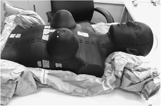
The computed tomography (CT) simulation of the Rando phantom was performed in supine position with slice thickness of 2 mm. A tailor made Vac-Lok was used for immobilisation, as shown in Figure 1. The chest region including the breasts of the female Rando phantom was scanned by a Siemens Somatom Definition Edge CT simulator, from the upper jaw to the umbilicus.
The corresponding slice of female Rando phantom immobilised on Vac-Lok.

Helical tomotherapy planning
The CT images were then imported to the Eclipse treatment planning system (version 10.0). Several region of interests including both lungs, breasts, the couch and body were contoured before planning. A tomotherapy treatment plan for the left breast was created by the treatment planning software provided by accuracy which was based on the convolution/superposition algorithm for dose computation. The plan parameters consisted of a 2·5 cm jaw width in dynamic jaw, 0·287 pitch, and three set modulation factor. The prescription was 2 Gy/fraction to the left breast at D95, 5 fraction/week in 25 fractions to total of 50 Gy. Optimisation parameters were adjusted throughout planning to reduce the dose to the OARs without compromising the target coverage.
Measurement points in Rando phantom
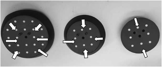
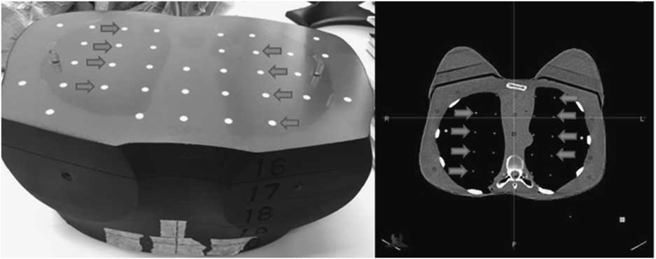
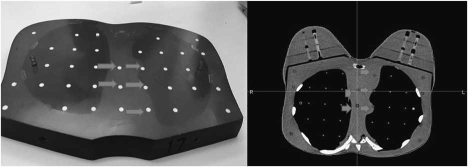
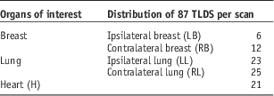
In a total of 98 TLDs, 87 of them were placed into the 11th to 20th slice of the Rando phantom which correspond to the contralateral lung and ipsilateral lung, contralateral breast, left breast and heart to evaluate the organ dose. The TLDs were positioned uniformly along the long axis of the lung region in each slice involved, while the location of heart was estimated with reference to Iaizzo PA,Reference Cook and Weinhaus 12 from T5-T9 at vertebral level. The remaining 11 TLDs chips were used to measure the background radiation. The distribution of 87 TLDs in the organs of interest was presented in Table 1. After the TLDs were annealed and calibrated, they were inserted into the respective slices in the Rando phantom for MVCT scanning. Several examples of TLDs placement were illustrated in Figures 2–4.
A total of 12 thermoluminescent dosimeters were placed in right breast.
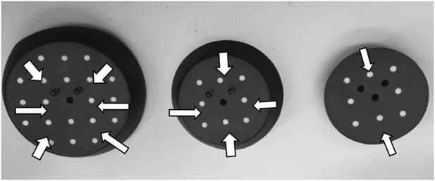
An example of thermoluminescent dosimeters (TLDs) placement for both lungs. Out of 48, eight TLDs were positioned uniformly and symmetrically along the long axis of lungs in the 16th slice of phantom.

An example of TLDs placement for heart. Out of 21, five thermoluminescent dosimeters were positioned in the mediastinum at the 17th slice of phantom.

The distribution of 87 TLD-100 into the organs of interest

MVCT imaging dose measurement
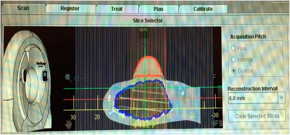
The phantom with pre-loaded TLDs was placed in the Vac-Lok. The treatment plan of the phantom was imported to the TomoHD workstation. Before MVCT scanning, scanning parameters such as the slice thickness classified as fine, normal and coarse values and scan range were selected by researcher. The scan range was selected to cover the entire left breast plus 12 mm margin superiorly and inferiorly. The selection of slice thickness and scan range was illustrated in Figure 5. MVCT was performed separately using (1) fine, (2) normal and (3) coarse mode with absorbed dose measured by the calibrated TLDs placed in the Rando phantom. The phantom was scanned by 3·5 MV photon in the tomotherapy unit. Two measurements were repeated in each scan selections and a total of six MVCT scan were performed.
The selection of slice thickness and scan range on tomotherapy workstation.

Data analysis
According to the readings of the TLDs from performance test, the standard deviation and the mean of TLD readings were calculated. The calibration factor (Gy/nC) was determined as follow:
 $$\eqalignno{ &{\rm Calibration}\,{\rm factor}\,\left( {{\rm Gy}\!/\!{\rm nC}} \right)\cr &{\equals}{{{\displaystyle {\displaystyle \rm 2}\,{\rm Gy{\times}\,\%\,}\,{\rm depth }\,{\rm dose}\left( {{\rm PDD}} \right)\,{\rm at}\atop \,\displaystyle {\rm 5}\,{\rm cm }\,{\rm depth}\left( {{\rm TLDs}\,{\rm location}} \right)}} \over {{\left( {\displaystyle {\rm Mean}\,{\rm of }\,{\rm TLD }\,r{\rm eadings}} \right){\minus}\atop \displaystyle \left( {{\rm Mean }\,{\rm of }\,{\rm control}} \right)}}}$$
With the calibration factor (Gy/nC) obtained from Equation (1), TLDs readings (nC) at each measurement point from MVCT imaging with different scan selections (fine, normal and coarse) were converted to point dose (cGy). The background of the TLDs were subtracted from the readings of TLDs and multiplied by the above calibration factor. While reporting the absorbed dose of various organs, TLDs belonging to the same organ were averaged to obtain a representative response of that organ.
Statistical analysis
Statistical analysis was conducted by using the software Instat (version 3) and 0·05 α levels was chosen in the current study. Assuming the data would not be distributed normally, Kruskal–Wallis test (nonparametric ANOVA) was performed at p<0·05 to examine if there was a significant difference between the three MVCT scanning modes classified as fine, normal and coarse.
RESULTS
MVCT imaging dose
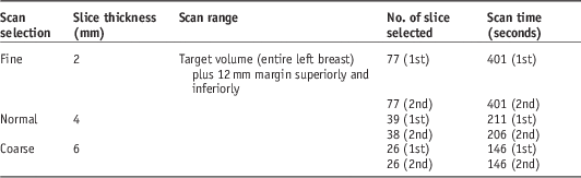
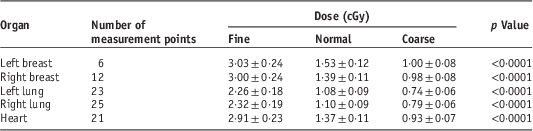
The scanning parameters of the fine, normal and coarse mode are presented in Table 2. The Kruskal–Wallis test indicated that there was a statistically significant difference in absorbed dose between different scan selections (p<0·001). The mean absorbed doses to various organs in the three predefined scan selection are summarised in Table 3.
The scanning parameters of the three scan selection (fine, normal and coarse mode)

The mean absorbed doses to various organs in the three predefined scan selection

The organ dose was inversely proportional to the scanning slice thickness. Utilising fine MVCT scan with 2-mm slice thickness resulted in the highest absorbed dose; whereas the lowest absorbed dose is found in coarse scan with 6-mm slice thickness. Relative to the normal scan selection (4 mm), the absorbed dose to the organs of interests shown in Table 3 can be scaled down, on average, by 0·69 and up by 2·1 for coarse (6 mm) and fine scans (2 mm), respectively. The mean dose to both breasts from a single MVCT is determined to be 3·02, 1·46 and 0·99 cGy for the fine, normal and coarse scanning mode, respectively. The absorbed dose in both lungs is 30% lower than that of the breasts, which is 2·29, 1·09 and 0·77 cGy in fine, normal and coarse scan correspondingly. In addition, the mean absorbed dose to the heart is similar to the right breast for the three scanning mode. The findings revealed that a single MVCT verification before breast tomotherapy contributed approximately ≤3 cGy, ≤1·5 cGy and <1 cGy to organs of interest for fine, normal and coarse mode, respectively.
Cumulative dose of breast tomotherapy with daily verification
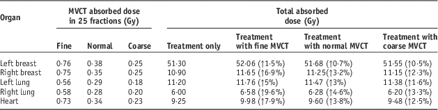
A case of routine left breast tomotherapy had been selected randomly. It was prescribed as 2 Gy per fraction to the left breast in a total of 25 fractions. The mean dose of organs of interest contributed from tomotherapy that collected from dose–volume histogram was summated with the total absorbed dose contributed from daily verification, as shown in Table 4. Though there is a statistically significant difference on MVCT absorbed dose among the three scan selections, the contribution of imaging to total absorbed dose is relatively small among different organs, especially the target volume. In the case of breast tomotherapy, the increase in contralateral breast dose by daily MVCT verification would be 0·75, 0·35 and 0·25 Gy for fine, normal and coarse mode, respectively.
Cumulative megavoltage computed tomography (MVCT) imaging and treatment dose in 25 fractions of left breast tomotherapy

Note: The values in the brackets showed the percentage differences of total absorbed dose from treatment only and treatment plus daily imaging verification using three predefined scan selection.
DISCUSSION
Daily image guidance and consequent patient positioning has been previously shown to be crucial in breast cancer patients. Recognised as an indispensable component throughout the course of tomotherapy, the scan selections for image verification resulted in varied patient absorbed dose, scan time as well as image resolution.
MVCT imaging dose
In concordance with previous work,Reference Shah, Langen, Ruchala, Cox, Kupelian and Meeks 2 this study also showed that the highest absorbed dose to various organs was contributed from fine MVCT scan (2·3–3·0 cGy) followed by normal (1·1–1·5 cGy) and coarse (0·74–1·0 cGy) scan. Utilising coarse mode for verification could halve the imaging dose as compared with the routine normal scan. This is attributed to the wider nominal slice thickness of 6 mm which requires shorter time and hence less radiation dose to scan the same volume than the fine and normal mode.
Treatment verification does not increase the dose to target volume greatly. In agreement with Quinn et al., daily normal scan contributes an increase of <1% to total absorbed dose in the treated breast throughout the course of tomotherapy. For the surrounding OARs, daily imaging increases dose to the contralateral breast, ipsilateral lung, contralateral lung and heart by 3·2, 3·0, 4·6 and 3·8%, respectively, in treatment with normal MVCT scan. Although the imaging dose is relatively small in proportion to the tomotherapy treatment dose, any increment of dose contributes to an increased risk of secondary malignancies, predominantly contralateral breast cancers and heart diseases.Reference Clarke, Collins and Darby 13
Incorporating imaging dose into the treatment planning not only can quantify the cumulative dose received in the course of radiotherapy, and hence better evaluate the magnitude of risk on developing radiation-induced effects. The MVCT imaging dose obtained in the present study may serve as a reference for daily verification in the treatment of breast tomotherapy. Nevertheless, other parameters such as the diversity of patient’s contour and a standardised MVCT scanning protocol need to be further investigated in order to develop a calculation model for computing the imaging dose with treatment planning.
Optimisation on imaging mode
In concordance with published data, the significant difference on organ dose-values among the three predefined scan selections demonstrates that coarse MVCT scan delivered the least additional radiation dose to both target volume and OARs. With the scan time, a 16 cm scan length in a breast cancer patient takes 146 seconds (~2·4 minute) for coarse imaging, which is 1·4 and 2·7 times faster than normal and fine MVCT scan, respectively. For breast cancer patients who have experienced tingling pain, twitching pain and burning sensation caused by damaged nerves from surgery,Reference Monroe and Shea 14 having fine MVCT scan of >6 minutes might be intolerable owing to the lengthy scan time in addition to the treatment time.
However, the optimisation on imaging mode should be subject to tradeoffs between costs and benefits. Researchers argued that normal MVCT scans are superior to coarse scan for the registration of shifts in the superior–inferior directions, with an improvement in residual error of about 0·4 mm evaluated from a head phantom.Reference Woodford, Yartsev and Van Dyk 15 The spatial resolution in the longitudinal direction may be slightly compromised, but the axial resolution of coarse scan does not degrade significantly.Reference Bates, Scaife and Tudor 16
To date, Ho et al. and Woodford et al. agreed that images acquired in coarse scans under its corresponding reconstruction slice thickness showed no significant image quality degradation and low residual error in contrast to normal and fine mode in 4 slices/gantry rotation reconstruction.Reference Woodford, Yartsev and Van Dyk 17 , Reference Ho, Yu and Wong 18 These non-significant differences have pronounced the possibility of selecting the coarse mode for MVCT verification in routine practice so as to keep the radiation dose as minimal as possible.
Recommendation on imaging protocol
In the attempt to keep the radiation exposure from treatment verification ‘as low as reasonably achievable’, the coarse scanning MVCT protocol as standard is recommended. Given that coarse MVCT scan can provide sufficient registration for targets with 3–5 mm margins,Reference Perez, Vijayakumar, Levitt and Purdy 19 having coarse scan not only allows the reduction of radiation dose to normal tissues by 0.7% relative to normal scans, it also shortens the scan-acquisition time. Considering the frequency of MVCT, daily image guidance undoubtedly increases the imaging dose that the patient receives, however the benefits on positioning accuracy would outweigh the risks.
Characterised by the steep dose gradient in tomotherapy plans, accurate patient positioning is essential that small shift in patient’s position may exert influence on the dose coverage of target volume. The breast target volume extends from skin surface to the chest wall and lung interface. Affected by respiratory motion, it accounted to an unavoidable setup error of 3 mm.Reference Sánchez-Rubio, Rodríguez-Romero and Castro-Tejero 20 To reduce the radiation dose, some studies suggested first three fractions or alternate-day imaging protocol.Reference Harris, Donovan and Coles 21 Nevertheless, Goddu et al. observed significant dose differences of 11-mm shifts in the anterolateral and 3-mm shifts in the posteromedial directions if patient setup errors remained uncorrected in helical tomotherapy treatments, leading to dose reduction in target volume.Reference Goddu, Yaddanapudi and Pechenaya 11 Despite the fact that the target position deviates from day to day, daily MVCT imaging allows correction for up to 7 mm of setup error in the anterolateral direction that enables the reduction of the lung volume receiving 20 Gy by 6%.Reference Goddu, Yaddanapudi and Pechenaya 11 Therefore, daily MVCT verification is the absolute way to reduce the random uncertainty during treatment of tomotherapy.
Reducing heart dose has become a concern for left breast irradiation. Darby et al. have stated that the rate of developing major coronary events in 5 years after radiation exposure would be increased by 7·4% for each increment of one Gy from mean dose of 6·6 Gy to the heart.Reference Darby, Ewertz and McGale 22 Using tomotherapy was found to reduce the mean heart dose significantly when compared with tangential IMRT but the megavoltage imaging dose was inevitably against it. For patients with unfavourable cardiac anatomy, who may receive a higher cardiac dose in treatment, having daily coarse MVCT scan at reduced absorbed dose could provide sufficient image resolution with the least dose to the heart. This would reduce the risk of cardiac morbidity after treatment.
However, for patients with seroma after breast conservative surgery, significant volume reduction of 5% or greater in tumour bed were seen during breast irradiation.Reference Chung, Suh and Lee 23 The volumetric change of tumour bed cavity may affect the accuracy of image matching, hence finer settings such as the normal mode should be used if coarse code is not deemed appropriate.
CONCLUSION
Quantifying the additional absorbed dose to various organs in MVCT verification, this study provides the imaging dose to organ in the three predefined scan selections for a breast tomotherapy verification. Relative to the normal scan selection (4 mm), the absorbed dose to the organs can be scaled down by 0·7 and up by 2·1 for coarse (6 mm) and fine scans (2 mm) respectively. Endeavour to keep the radiation exposure ‘as low as reasonably achievable’, optimisation of imaging protocols is of paramount importance when image guidance is considered as an integral component in radiotherapy. The decisions on imaging mode should achieve an optimum balance between imaging dose, image quality and the purpose of verification. The recommendation of undergoing daily coarse mode for MVCT verification in breast tomotherapy not only mitigates the radiation exposure to normal tissues that would subsequently lessen the probability of inducing secondary late complications, but also trims the scan-acquisition time to maximise patient throughput.
Acknowledgement
The authors thank the project advisor, Dr P.M. Wu and medical physicists Ms. Ruby Ho and Dr. Herve Choi at Department of Medical Physics and Research in the Hong Kong Sanatorium & Hospital for their insightful comments and technical advice. Mr. Joseph Lee and Mr. Jacky Wong at Department of Radiotherapy in Hong Kong Sanatorium & Hospital for the continual support throughout the project, scheduling the machine timeslot for TLD irradiation as well as inspiring us with practical advice. The authors also thank the radiotherapists in Hong Kong Sanatorium & Hospital, especially Mr. Jason Lam and Miss Stephanie Chan for assisting with the CT simulation and collection of tomotherapy treatment dose data.
Financial support
This research received no specific grant from any funding agency, commercial or not-for-profit sectors.
Conflicts of Interest
None.