Introduction
Pallasites are stony-iron meteorites essentially composed of 35 to 85 vol.% of olivine crystals embedded in a (Fe,Ni) metallic matrix (Weisberg et al., Reference Weisberg, McCoy, Krot, Lauretta and McSween2006; Boesenberg et al., Reference Boesenberg, Delaney and Hewins2012). Pallasites were originally thought to represent the core–mantle boundary of a differentiated asteroid, subsequently disrupted by impact with a larger body (Yang et al., Reference Yang, Goldstein and Scott2010). As a consequence, the differentiated asteroid was ‘dismembered’ and olivines from its mantle mixed with the molten (Fe,Ni) from the outer core (Yang et al., Reference Yang, Goldstein and Scott2010). Recently, a more plausible theory invokes the impact of a smaller differentiated body with a partially molten core into a larger differentiated body (~200 km radius), leading to the intrusion of the liquid (Fe,Ni) from the former into the dunitic mantle of the latter (Tarduno et al., Reference Tarduno, Cottrell, Nimmo, Hopkins, Voronov, Erickson, Blackman, Scott and McKinley2012). This theory was suggested because of the strong palaeomagnetic field observed in magnetic inclusions in olivines from Main Group pallasites (Tarduno et al., Reference Tarduno, Cottrell, Nimmo, Hopkins, Voronov, Erickson, Blackman, Scott and McKinley2012; Bryson et al., Reference Bryson, Nichols, Herrero-Albillos, Kronast, Kasama, Alimadadi, van der Laan, Nimmo and Harrison2015) that is attributed to the presence of a long-lived core dynamo in the differentiated larger body.
Olivines are silicate minerals with the general formula M'M’’SiO4 where M = Fe2+, Mg, Mn2+, Ca and Ni. Most rock-forming olivines belong to the Fe–Mg solid-solution series.
The main (Fe,Ni) metal phases in pallasite are iron, the α-(Fe,Ni) metal phase (4–7.5 wt.% Ni), and taenite, the γ-(Fe,Ni) phase (27–65 wt.% Ni). In some cases, a mixture of iron and taenite, called ‘plessite’, forms in the retained taenite during slow cooling as a consequence of immiscibility between Fe and Ni (Goldstein and Michael, Reference Goldstein and Michael2006, and references therein). The concentration of minor elements in (Fe,Ni) metal is related to element compatibility and segregation temperature. Thus, (Fe,Ni) metal in pallasites is usually depleted in elements highly compatible with solid metal (Re, Os, Ir, Pt and Ru) and enriched in incompatible elements such as Pd and Au, compared to the early crystallised IIIAB iron meteorites (Mullane et al., Reference Mullane, Alard, Gounelle and Russella2004).
Minor amounts of sulfides [e.g. troilite FeS], phosphides [e.g. schreibersite (Fe,Ni)3P and barringerite (Fe,Ni)2P], phosphates [e.g. merrillite Ca18Na2Mg2(PO4)2, stanfieldite Ca3Mg3(PO4)4 and farringtonite Mg3(PO4)2], chromite FeCr2O4, pyroxene [e.g. orthopyroxenes (Mg,Fe)SiO3] and P-rich olivines (Buseck, Reference Buseck1977) are usually found in pallasites.
Secondary minerals, usually formed in a terrestrial environment either during weathering (e.g. Eggleton, Reference Eggleton1984) or post depositional processes (e.g. Gentili et al., Reference Gentili, Comodi, Nazzareni and Zucchini2014), can be found in altered pallasites. Typical secondary minerals in pallasites are akaganeite β-FeO(OH,Cl), bunsenite NiO, goethite α-FeO(OH), hematite α-Fe2O3, hibbingite γ-Fe2(OH)3Cl, lepidocrocite γ-FeO(OH), maghemite γ-Fe2O3, magnetite Fe3O4 and trevorite NiFe2O4 (Rubin, Reference Rubin1997), which are generally considered to be due to the corrosion of metallic Fe–Ni (Tilley and Bevan, Reference Tilley, Bevan, Taylor and Pain1998). In addition, olivines can alter in aqueous environments to ‘iddingsite’, reddish alteration products made of a cryptocrystalline intergrowth of goethite, that can be transformed by dehydration to hematite, and smectite-type phillosilicates (probably saponite, Eggleton, Reference Eggleton1984) that can be removed by further alteration.
Mineo pallasite
The Mineo pallasite fell on May 3rd 1826 in Mineo, Sicily, Italy (Baldanza, Reference Baldanza1965; Grady, Reference Grady2000). The only sample in a public collection is owned by the Department of Physics and Geology of the University of Perugia, Italy. The Mineo pallasite was studied in detail for the first time by Zucchini et al. (Reference Zucchini, Petrelli, Frondini, Petrone, Sassi, Di Michele, Palmerini, Trippella and Busso2018). Olivine is forsteritic with Fo < 80. It contains 9–11 wt.% FeO and varying quantities of trace elements, especially Ca (ranging from 62 to 473 ppm) that has a positive correlation with the incompatible elements such as Al, Na, K, Ba and Sr, as well as with the compatible Cr (Zucchini et al., Reference Zucchini, Petrelli, Frondini, Petrone, Sassi, Di Michele, Palmerini, Trippella and Busso2018).
The iron–nickel metal is formed primarily as the Fe0.94Ni0.06 metal alloy phase.
Taenite occurs in plessite structures where a Ni-rich phase with average chemical formula of ~Fe0.83Ni0.17 is framed by a continuous taenite rim ~10 μm thick. The Ni-rich phase is also present as a droplet-like structure that hosts: (1) a Ni-poor iron matrix (Fe0.94Ni0.06); (2) taenite (Fe0.81Ni0.20) as submicrometre- to nano-sized ‘droplets’; and (3) a Fe0.60Ni0.40 high-Ni taenite phase as a rim around the taenite regions (Zucchini et al., Reference Zucchini, Petrelli, Frondini, Petrone, Sassi, Di Michele, Palmerini, Trippella and Busso2018).
In addition to the main (Fe,Ni) phases, the metal also contains barringerite and schreibersite. Sulfide phases are rare, represented by only a few grains of troilite (FeS) (Zucchini et al., Reference Zucchini, Petrelli, Frondini, Petrone, Sassi, Di Michele, Palmerini, Trippella and Busso2018). The platinum group elements (PGE) and the highly siderophile elements (HSE) show a depletion in the highly compatible elements and an enrichment in Pd and Au with respect to the early crystallised IIIAB irons (Zucchini et al., Reference Zucchini, Petrelli, Frondini, Petrone, Sassi, Di Michele, Palmerini, Trippella and Busso2018), following the behaviour of the Main Group pallasites (Mullane et al., Reference Mullane, Alard, Gounelle and Russella2004) that traces that of the late-crystallised IIIAB irons (medium octahedrites). However, an enrichment in Re, Os and Ir with respect to typical MG pallasites and late-crystallised IIIAB irons has been observed in the Mineo pallasite (Zucchini et al., Reference Zucchini, Petrelli, Frondini, Petrone, Sassi, Di Michele, Palmerini, Trippella and Busso2018).
In addition to the major and minor mineral phases usually found in pallasites, regions of iron oxides have been observed. Their origin has not been addressed yet. Possible theories include: (1) the consequence of low-temperature alteration during terrestrial chemical weathering of both the (Fe,Ni) metal previously intruded into the olivine cracks and the olivines themselves; or (2) direct precipitation of Fe oxides from an aqueous solution in the cracks of the highly fractured olivines.
This work
This investigation characterised the Fe oxides in the Mineo pallasite using a multi-analytical approach combining Raman spectroscopy, electron microprobe analysis (EMPA), field emission scanning electron microscopy (FE-SEM), transmission electron microscopy (TEM) and 3D Electron Diffraction (3D ED) (Kolb et al., Reference Kolb, Gorelik, Kübel, Otten and Hubert2007).
The latter method was crucial for establishing the nature of the Fe oxides and assigning them to goethite. 3D ED consists of the systematic collection of a 3D electron diffraction data set through either a stepwise or continuous rotation of the sample around an arbitrary, non-crystallographic axis. 3D ED delivers single-crystal-like diffraction data from grains of 50–2000 nm in size and is therefore extremely powerful for the identification of minor components in polyphasic mineralogical aggregates (Gemmi et al., Reference Gemmi, Merlini, Palatinus, Fumagalli and Hanfland2016). This 3D ED method allows separate structural data to be obtained from all classes of nanometric phases. Moreover, compared to the conventional way of acquiring diffraction data through oriented selected-area electron-diffraction patterns (SAED), 3D ED allows a more complete reflection sampling, a reduction of dynamical effects and a decisive acceleration of data acquisition. This results in a reduction of the total electron dose on the sample, which is crucial for beam-sensitive materials. The 3D ED method is currently applied to the structural characterisation of nanocrystalline functional materials (Mugnaioli et al., Reference Mugnaioli, Gemmi, Tu, David, Bertoni, Gaspari, De Trizio and Manna2018; Steciuk et al., Reference Steciuk, Barrier, Pautrat and Boullay2018; Wang et al., Reference Wang, Rhauderwiek, Inge, Xu, Yang, Huang, Stock and Zou2018), pharmaceuticals (Jones et al., Reference Jones, Martynowycz, Hattne, Fulton, Stoltz, Rodriguez, Nelson and Gonen2018; Brázda et al., Reference Brázda, Palatinus and Babor2019) and macromolecules (Nannenga et al., Reference Nannenga, Shi, Leslie and Gonen2014; Xu et al., Reference Xu, Lebrette, Clabbers, Zhao, Griese, Zou and Högbom2019). A comprehensive review of 3D ED was published recently by Gemmi et al. (Reference Gemmi, Mugnaioli, Gorelik, Kolb, Palatinus, Boullay, Hovmöller and Abrahams2019).
In Earth sciences, 3D ED has been used for the structure elucidation of nanoscopic inclusions (Xiong et al., Reference Xiong, Xu, Mugnaioli, Gemmi, Wirth, Grew, Robinson and Yang2020), mineral seeds (Németh et al., Reference Németh, Mugnaioli, Gemmi, Czuppon, Demény and Spötl2018), hydrated phases (Mugnaioli et al., Reference Mugnaioli, Lanza, Bortolozzi, Righi, Merlini, Cappello, Marini, Athanassiou and Gemmi2020a; Krysiak et al., Reference Krysiak, Maslyk, Silva, Plana-Ruiz, Moura, Munsignatti, Vaiss, Kolb, Tremel, Palatinus, Leitão, Marler and Pastore2021) and modulated systems (Lanza et al., Reference Lanza, Gemmi, Bindi, Mugnaioli and Paar2019; Steciuk et al., Reference Steciuk, Škoda, Rohlíček and Plášil2020), even when characterised by very large asymmetric units (Rozhdestvenskaya et al., Reference Rozhdestvenskaya, Mugnaioli, Czank, Depmeier, Kolb, Reinholdt and Weirich2010; Mugnaioli et al., Reference Mugnaioli, Bonaccorsi, Lanza, Elkaim, Diez-Gómez, Sobrados, Gemmi and Gregorkiewitz2020b). 3D ED has been applied to geological samples in different contexts (Mugnaioli and Gemmi, Reference Mugnaioli and Gemmi2018), and in particular for the characterisation of meteorites (Pignatelli et al., Reference Pignatelli, Marrocchi, Mugnaioli, Bourdelle and Gounelle2017, Reference Pignatelli, Mugnaioli and Marrocchi2018; Suttle et al., Reference Suttle, Folco, Genge, Franchi, Campanale, Mugnaioli and Zhao2021), impactites (Campanale et al., Reference Campanale, Mugnaioli, Gemmi and Folco2021), cryptocrystalline oxides (Koch-Müller et al., Reference Koch-Müller, Mugnaioli, Rhede, Speziale, Kolb and Wirth2014) and hydroxides (Viti et al., Reference Viti, Brogi, Liotta, Mugnaioli, Spiess, Dini, Zucchi and Vannuccini2016).
Experimental methods
Meteorite sampling
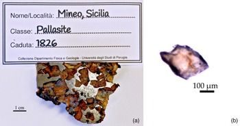
The available portion of the Mineo pallasite is shown in Fig. 1a and the single crystal of olivine with reddish surfaces chosen for the current study in Fig. 1b, that was extracted together with micrometric to nanometric reddish aggregates sampled from the Fe-oxide regions.
(a) Photo of the Mineo pallasite portion owned by the Department of Physics and Geology of the University of Perugia (sample no. Mineo1969.SL3P#002 CNR inventory 213248); (b) isolated olivine single crystal from the Mineo pallasite where a reddish area is observed.

Field emission – scanning electron microscopy (FE–SEM) and electron microprobe analysis (EMPA)
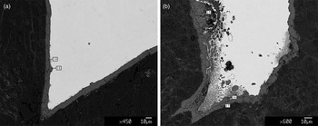
High-resolution back-scattered electron (BSE) FE–SEM images were obtained by a field-emission-gun electron scanning microscope (LEO 1525) and a ZEISS AsB (angle selective back-scattered) Detector, at the Department of Physics and Geology of the University of Perugia, coupled with a Bruker Quantax EDS operating at 15 kV. Chemical analyses of the Fe-oxide areas were performed at the “Ardito Desio” Earth Sciences Department of the University of Milan using a JEOL 8200 Super Probe operating at 15kV acceleration voltage and 5 nA current. The instrument is equipped with five WDS (wavelength dispersive) and one EDS (energy dispersive) spectrometers. In the Fe-oxide areas, five points were analysed (Fig. 2).
BSE SEM images of a portion of the Mineo pallasite. Numbers denote sampling points in the iron oxide rim bordering the Fe,Ni metal (Table 1).

Raman spectroscopic analyses
Raman spectroscopic analyses of the olivine single crystal were performed at the Department of Chemistry, Biology and Biotechnology, University of Perugia. The analytical set up consisted of: back-scattering illumination and collection of the scattered light through an Olympus confocal microscope MOD BX40 (50× objective); 532 nm excitation from the solid-state laser Oxxius LMX (laser power on the sample 45 mW); 1024×256 CCD array Syncerity by Horiba – Jobin Yvon to detect the scattered light. A spectral resolution of ~4 cm–1 was achieved with a 1800 lines mm–1 grating of an iHR320 imaging spectrometer. Spectral measurements were made with continuous scans in the range 200–1800 cm–1, exposure times in the range 30–60 s, and 10 accumulations. The spectrometer was calibrated using the Raman lines of WO3 powder. The collected Raman spectra were baseline-corrected to eliminate the background by using the Peak Analyzer routine of Origin 8.0 software (OriginLab Corporation, Northampton, MA, USA).
Transmission electron microscopy (TEM) and 3D electron diffraction (3D ED)
A few small portions of the Fe-rich areas in the Mineo pallasite, such as the region bordering the olivine to iron interface in Fig. 2, were crushed to reach the optimal grain dimensions needed for the TEM and 3D ED analysis. 3D ED measurements (Mugnaioli and Gemmi, Reference Mugnaioli and Gemmi2018; Gemmi et al., Reference Gemmi, Mugnaioli, Gorelik, Kolb, Palatinus, Boullay, Hovmöller and Abrahams2019) were performed at the Center for Nanotechnology Innovation (NEST, Istituto Italiano di Tecnologia, Pisa, Italy) by a Zeiss Libra TEM operating at 120 kV and equipped with a LaB6 source and a Bruker EDS detector XFlash6T-60. 3D ED acquisitions were done in STEM mode after defocusing the beam in order to have pseudo-parallel illumination on the sample. A beam size of ~150 nm in diameter was obtained by inserting a 5 μm C2 condenser aperture. Conventional ED and 3D ED data, the latter acquired in steady steps of 1°, were recorded by an ASI Timepix detector and analysed by ADT3D software (Kolb et al., Reference Kolb, Mugnaioli and Gorelik2011) and in-house developed MATLAB routines.
Results
Chemical analyses
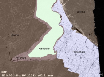
The FE-SEM and EDS analyses show the occurrence of iron oxides bordering the (Fe,Ni) metals, mainly iron (Fig. 3), confirming the results from Zucchini et al. (Reference Zucchini, Petrelli, Frondini, Petrone, Sassi, Di Michele, Palmerini, Trippella and Busso2018).
BSE FE-SEM image of a portion of the Mineo pallasite. The BSE image was coloured according to the mineral assemblage shown by qualitative EDS investigation.

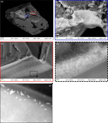
Iron oxide was also observed as reddish areas on the olivine single crystals (Fig. 1b) appearing as light contrast halos and bands, related to the concentration of atoms with a high atomic number such as Fe, as shown by FE-SEM EDS analyses (Fig. 4). The white bands observed on the surface of the olivine crystal were found to be formed by aggregates of Fe-rich nanometric particles (Fig. 4d). In some areas, structures resembling exsolution lamellae were also observed (Fig. 4e).
BSE FE-SEM images of the olivine single crystal isolated from the Mineo pallasite. (a) The whole single crystal. Blue and red rectangles show the location of areas of high Fe concentration enlarged in (b) and (c), respectively. Dashed black and solid boxes in (c) show the areas enlarged in (d) and (e, rotated 180°), respectively.

Electron microprobe analyses within the Fe-oxide areas (Fig. 2) showed the occurrence of high Fe contents, followed by Ni and Mg. Minor elements are also detected, mainly Na, Si and Ca in order of abundance (Table 1).
Compositions (wt.%) from EMPA of Fe oxide at location points 15 to 19 (Fig. 2). Analytical precision is in accordance with the last decimal place.

‘-’ not detected
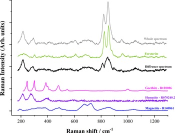
Raman spectroscopic results on the Fe-oxide phases are shown in Fig. 5. Contribution from both Fe-oxide phases and olivine were collected and the contribution of Fe-oxides was isolated by subtracting olivine signals from the whole spectrum. The main forsteritic signals are still quite evident as two main peaks around 800 cm–1; however, the relative intensities of these two are different in the whole spectrum compared to what is observed for the forsterite spectrum. We performed the subtraction in order not to have a negative peak in the 200–1300 cm–1 range and obtained a residual intensity particularly in the 200–300 and 800–900 cm–1 regions. The broad feature at 800–900 cm–1 could be due to the different forsterite structures of the two samples, i.e. a reduced crystallinity of this phase in the Mineo pallasite, compared with the olivine single crystal. The peaks at 212.5 and 282.2 cm–1 might be attributed to hematite; whereas, the features in the range 625–770 cm–1 might be related to magnetite (Hanesch, Reference Hanesch2009). The goethite spectrum was also reported for comparison. However, the features attributed to the Fe oxides (212.5 and 282.2 cm–1) seems to have shorter wavelengths compared to goethite.
Raman analyses on the olivine single crystal from the Mineo pallasite. Collected data on the Fe-oxide-rich phase (‘whole spectrum’ in grey) were processed by subtracting the olivine contribution (collected green spectrum). The difference spectrum is given in black. The compared phases are shown: goethite (pink), hematite (purple) and magnetite (blue). Reference codes of the RRUFF database (Lafuente et al., Reference Lafuente, Downs, Yang, Stone, Armbruster and Danisi2015) for the compared spectra are shown in the figure.

TEM and 3D ED analysis
The mineralogical assignment of alteration phases by conventional analytical techniques was inconclusive due to the sub-micrometric dimension and polycrystallinity of the Fe-rich regions. In fact, preliminary single-crystal X-ray diffraction analysis performed on the isolated single crystal showed no diffraction peak different from those of olivine. The reasons for this behaviour might be either the very low crystallinity or the sub-micrometric dimensions of the high-Fe phases.
Therefore, TEM and 3D ED analyses were performed on these areas to further elucidate their textures and crystal structure. Grains were isolated from the Fe-rich regions within the olivine single crystal and from the Fe-rich areas of the meteorite. The powdered sample was first analysed by TEM-EDS, which allowed identification of particles made only of Fe oxides. Most of the other particles were olivine fragments and, unexpectedly, CaCO3 crystals.
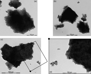
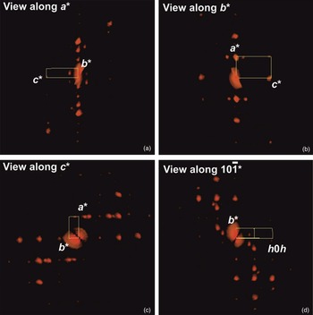
The isolated Fe-oxide grains in Fig. 6 are nano-polycrystalline aggregates. Most crystals are nanometres in size and too small and aggregated to perform 3D ED analysis, but occasionally it was possible to find coherent crystalline areas of some hundreds of nanometres in width. Only when a fragment hosted a dominant coherent crystalline area of at least 100 nanometers in size, could a unit cell be defined (Fig. 7). Spurious extra reflections, with an intensity sometimes comparable with the dominant crystalline area, were always present and originated from surrounding Fe-oxide crystals. Automatic routines for cell-parameter determination systematically failed and a cell consistent with most of the collected reflections could be obtained only after a careful inspection of the 3D reconstructed data sets. In all cases, we observed a unit cell with parameters a = 4.8(1) Å, b = 10.0(2) Å, c = 2.8(1) Å and α ≈ β ≈ γ ≈ 90°. In addition, extinction rules consistent with space group Pbnm were observed. This evidence points to the orthorhombic structure of goethite FeO(OH).
TEM images of different nano-sized Fe-oxide grains (a, b, c). In (d) a zoomed portion of (c) is shown.

Reconstructed 3D ED data for a mostly coherent Fe-oxide grain, consistent with goethite lattice. (a) View along a*; (b) view along b*; (c) view along c*, showing missing reflections 0kl : k = 2n + 1, consistent with the (100) b-glide plane of goethite; and (d) view along 10$\bar{1}$ *, showing missing reflections h0l: h + l = 2n + 1, consistent with the (010) n-glide plane of goethite. An important fraction of reflections does not belong to the main goethite domain, whose unit cell is sketched in yellow. Cell vector projections are labelled in white. The number of spurious reflections is emphasised by the fact that reflections belonging to the main goethite domain overlay in projection. One should consider that these images are not conventional 2D electron diffraction patterns, but projections of the whole 3D diffraction reconstruction.

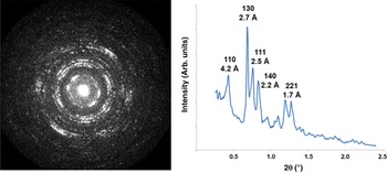
In order to verify that this phase identification could be extended to the more cryptocrystalline portion of Fe-oxide aggregates, several ring diffraction patterns (conceptually comparable with powder diffraction) were collected from polycrystalline Fe-oxide areas. For these measurements, the beam was enlarged to ~500 nm to sample more crystallites and obtain a better intensity distribution (Fig. 8). The diffraction rings were consistently indexed with typical goethite interplanar spaces (see for example RRUFF ID R050142; Lafuente et al., Reference Lafuente, Downs, Yang, Stone, Armbruster and Danisi2015).
Electron diffraction rings of a polycrystalline Fe-oxide aggregate, showing interplanar distances consistent with goethite. The plot on the right was obtained by an intensity integration of the rings. Measured interplanar distances and related indexes are indicated.

Finally, 3D ED was performed on CaCO3 crystals with sizes <100 nm, allowing identification of the cell parameters for calcite. This mineral generally appears in close association with goethite aggregates.
Discussion
The results from EMPA gave an average composition of the Fe- and Ni-oxides of 67±4 and 5±1 wt.%, respectively, and detected other minor elements as Mg, Si, Ca and Na. The low totals might be due to the presence of undetectable chemical species, such as OH and water. As regards the Raman analyses, the Fe-oxide grains were attributed to hematite, with a slight magnetite contribution. In contrast, coupling 3D ED and TEM-EDS analyses, we were able to unequivocally assign the observed Fe-oxide grains to goethite, confirming the supposed presence of hydrated Fe oxides suggested by the EMPA. The reason for the discrepant results from the Raman analysis may be the alteration of goethite to hematite by laser-induced effects, as shown by Seifert et al. (Reference Seifert, Thomas, Rhede and Forster2010), giving rise to aggregates of hematite with possible magnetite contributions, as observed in Fig. 5.
The occurrence of goethite in the mineral assemblage of the Mineo pallasite is reasonably assigned to terrestrial alteration, also confirmed by the presence of calcite, which was probably precipitated from circulating water.
The EMPA on the goethite areas showed the presence of minor elements, such as Ni, Mg, Si, Ca and Na, that might be due either to their incorporation into the Fe-oxide crystal structure, or to the presence of accessory minerals. The former hypothesis suits Ni very well, as it is a major element of the (Fe,Ni) metal, however it does not explain the occurrence of the other chemical species that could be related to the presence of other mineralogical components, probably coming from interactions with the terrestrial environment. The likely occurrence of cryptocrystalline mineral associations, such as iddingsite, resulting from olivine alteration, cannot be excluded. However, on the one hand, the observed minor elements occurrence that cannot be related to smectite-type phyllosilicates, and, on the other hand, the textural occurrence of the goethite areas mainly bordering on the (Fe,Ni) metal (Fig. 3), are good reasons to attribute goethite to oxidation of the metallic component.
Goethite was also observed on the grain boundaries between the fragmented olivines where the (Fe,Ni) metal probably intruded during pallasite formation, as shown by the reddish areas on the isolated single crystal in Fig. 1b. This observation is not common for pallasite, where olivines usually occur as macrocrystals with size ranges from a few hundreds of micrometres to a few centimetres (Scott, Reference Scott1977). Thus, further analyses of major-, minor- and trace-elements distribution in olivines, as well as on the fragmentation of olivines and other constituent phases in the Mineo pallasite, are needed.
This study shows how for polyphasic and cryptocrystalline samples, even simple mineralogical identification can benefit strongly from TEM methods. In particular, the combination of TEM-EDS and 3D ED can deliver chemical and crystallographic information on the same nanometric portion of the sample, allowing recognition of any mineral species without ambiguities even for low-symmetry systems. This protocol may become a routine path for discriminating and validating new nanometric mineral species, and possibly will expand our understanding of geological and planetary phenomena.
Conclusions
The detailed characterisation of Fe-oxide alteration in Mineo pallasite shows once more how the combination of TEM-EDS and 3D ED allows unambiguous mineral phase identification at the nanoscale. Chemical and structural information can be retrieved from single nano-grains, even when these are minor constituents of a polycrystalline aggregate. This is an obvious advantage when compared with spectroscopic and X-ray diffraction methods that deliver a global signal from the whole ensemble of material. Future applications of such an approach look especially promising for rocks forming in a short time and in unequilibrated conditions, such as alteration bands, fault mirrors, impact rocks and meteorites.
Results of the present work show that the Fe-oxide regions are goethite, derived from chemical weathering in a terrestrial environment of the (Fe,Ni) metal that possibly also intruded on the grain boundaries between the fragmented olivines during pallasite formation.
Acknowledgements
This work was supported by the Department of Physics and Geology of the University of Perugia which allowed the sampling and studying of the meteorite collection. The Smithsonian Natural Museum of Natural History is acknowledged for kindly providing the San Carlos reference standard for olivine chemical analyses. E.M. and M.G. acknowledge the Regione Toscana for funding the purchase of the Timepix detector through the FELIX project (Por CREO FESR 2014-2020 action). Prof. Hilary Downes, anonymous referees and the Principal Editor Dr. Stuart Mills are acknowledged for their suggestions, which helped us to improve the quality of this manuscript.