Atopic dermatitis is a pruritic chronically relapsing skin disease that develops on the background of a dominance of Th2 cells (Ref. Reference Leung and Bieber1). Life-time prevalence of atopic dermatitis is about 20% in children and 1–3% in adults (Ref. Reference Maintz and Novak2). A subgroup of atopic dermatitis patients may develop one or more episodes of a severe viral super-infection of the skin, caused by herpes simplex virus (HSV). This condition is named atopic dermatitis complicated by eczema herpeticum (Refs Reference Mackley3, Reference Wollenberg4). Eczema herpeticum was reported for the first time in 1887 by Moritz Kaposi and was termed ‘eczema infantum’ (Ref. Reference Nagy5). Kaposi described disseminated, partially grouped, vesicopustular eruptions with a predominance in the facial area. He recognised similarities of this infection with a varicella virus infection of the skin (Ref. Reference Wollenberg6). Hence, the term ‘Kaposi's varicelliforme eruption’ is still used in clinical practice, and applies to the viral complications of different skin diseases (Ref. Reference Wollenberg6), such as T-cell lymphoma (Ref. Reference Brion7), allergic contact dermatitis (Ref. Reference Paradisi8), psoriasis (Ref. Reference Santmyire-Rosenberger and Nigra9), rosacea (Ref. Reference Kucukyilmaz10), autografted skin (Ref. Reference Manders and Chetty11), skin injury after burning (Ref. Reference Nishimura12) or as a complication after laser treatment in immunosuppressed patients (Ref. Reference Rodriguez-Serna13). Juliusberg recognised similarities of the clinical features of eczema herpeticum and varicella virus infection several years later (Ref. Reference Thulliez14) and therefore coined the term ‘Pustulosis acuta varioliformis’ for a vesiculo-papulous eczema of the face in a child. The term eczema herpeticum, which followed after identification of HSV in the vesicles, is now used for disseminated HSV infection of the skin as a complication of atopic dermatitis or other types of eczematous skin disease, whereas Kaposi's varicelliform eruption is primarily used for complications of other skin diseases caused by a disseminated HSV infection (Ref. Reference Wollenberg6).
Herpes simplex virus
HSV belongs to the alpha herpesviridae and is a member of the double-stranded DNA virus family (Ref. Reference Novak and Peng15). It consists of an envelope with different glycoproteins (gG) and spikes (gD, gB) and has a diameter of 100 nm. HSV type 1 (HSV1) is an oro-facial virus whereas HSV type 2 (HSV2) primarily infects the genital region. However, infections of the genital region by HSV1 and oro-facial region by HSV2 do occur. The genomes of HSV1 and HSV2 have nearly 50% nucletotide identity. Seroprevalence of HSV is high and is estimated to be about 80–90% for HSV1 and 12–15% for HSV2 in some studies (Ref. Reference Wutzler16). An outstanding feature of HSV is its capability to persist after primary infection of the skin or mucosa for a lifelong period in the ganglia of the nervous system causing secondary HSV infections after a phase of latency. Both the extraordinary high seroprevalence of HSV as well as its capacity to be reactivated by exogenous and endogenous factors explain why HSV in particular, in contrast to other virus types, might be responsible for severe complications of chronic inflammatory skin diseases with high prevalence, such as atopic dermatitis. In most cases, primary infection with HSV1 occurs at mucocutaneous locations as a mild and subclinical infection during infancy. Primary target cells of HSV are epithelial cells. Three different types of surface molecules that allow viral entry of HSV into cells have been identified so far: (1) members of the Ig superfamily nectin-1 and nectin-2 (Ref. Reference Sakisaka17); (2) herpes virus entry mediator (HVEM), which belongs to the TNF family; and (3) specific sites in heparan sulphates (Ref. Reference Campadelli-Fiume18). The typical clinical picture of HSV infection is caused by intracellular oedema of infected cells, which then degenerate. The nuclei of infected cells show eosinophilic nuclear inclusion and marginal nuclear chromatin. HSV-infected cells then form multinucleated clusters of giant cells, which serve as diagnostic tool. Reactivation of HSV is facilitated by stress, mechanical damage, UV radiation or immunosuppression. Although viral proof shows that eczema herpeticum is most often associated with HSV1, HSV2 is detectable in some rare cases of eczema herpeticum. A specific genotype of HSV1, the F35 strain, was associated significantly more often with eczema herpeticum in a Japanese study (Ref. Reference Yoshida and Umene19). This implies that specific HSV virus strains might play a role in eczema herpeticum, but this aspect needs further evaluation in large cohorts of patients, including eczema herpeticum patients of Caucasian origin.
Clinical features of eczema herpeticum
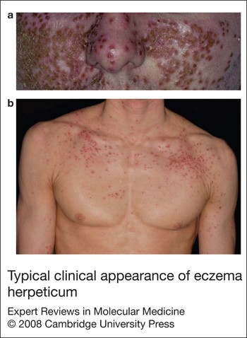
Factors predisposing the emergence of eczema herpeticum in a subgroup of patients with atopic dermatitis, which occurs most frequently within the second and third decade of life, are still unclear (Refs Reference Wollenberg4, Reference Bork and Brauninger20). No seasonal onset of eczema herpeticum has been detected in a retrospective study that included 100 patients (Ref. Reference Wollenberg4). Although eczema herpeticum could be identified as a secondary HSV infection in a majority of patients, a higher fever and more pronounced lymphopaenia combined with the respective serological markers indicated eczema herpeticum as the primary HSV infection in some atopic dermatitis patients (Ref. Reference Wollenberg4). Viral spreading in eczema herpeticum takes place from cell to cell by viraemia or via the lymphatic system. In most cases, eczema herpeticum starts with small monomorphic dome-shaped papulovesicles developing into pustules and punched-out crusts, which might in severe cases spread from the facial area over the entire body (Fig. 1). The vesicles develop into erosions and frequently become secondarily infected with bacteria, primarily Staphylococcus aureus. Eczema herpeticum is accompanied by fever, malaise, lymphadenopathy and rapid impairment of general health (Ref. Reference Wollenberg4). Predilection is the head and neck area as well as the thorax. Over 2–6 weeks, the lesions dry out, become crusted and heal as sharply bordered pits or erosions. A subgroup of about 10–20% of atopic dermatitis patients with eczema herpeticum has two or more recurrent eczema herpeticum episodes, which might have a milder course (Ref. Reference Wollenberg4). Interestingly, DNA of HSV has been detected in the oral cavity of patients with eczema herpeticum, implying that subclinical or asymptomatic HSV lesions might exist in the oral mucosa in these patients (Ref. Reference Yoshida and Amatsu21). However, this finding might not be specific for eczema herpeticum, because HSV has also been detected for example, in the oral cavity of pemphigus vulgaris patients (Refs Reference Takahashi22, Reference Schlupen23).
Typical clinical appearance of eczema herpeticum. Typical clinical features of eczema herpeticum are monomorphic dome-shaped vesicles (a), as well as yellowish crusts and sharply bordered erosions, which spread over the body (b).

A subgroup of atopic dermatitis patients identified to be at risk of eczema herpeticum displayed early onset of atopic dermatitis, a high number of sensitisations and increased total serum IgE levels (Refs Reference Wollenberg4, Reference Peng24). In addition, some studies showed that this group had an early onset of atopic dermatitis with a persistent, more severe, course into adulthood, mirrored by a higher scoring of atopic dermatitis (SCORAD)(Refs Reference Wollenberg4, Reference Peng24).
Many of these patients displayed a predilection of atopic dermatitis for the head and neck region as well as particular sensitisation to the lipophilic yeast Malassezia sympodialis, implying that concomitant interactions of different microbial antigens or a specific micromilieu in the region of primary virus infection might contribute to the onset of the disease (Ref. Reference Peng24). Whether these predisposing factors differ between children and adult patients with eczema herpeticum, has not yet been addressed, and epidemiological data from larger cohorts of patients are needed to confirm these findings.
Diagnosis of eczema herpeticum
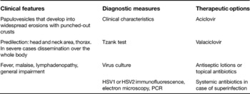
Diagnosis of eczema herpeticum is based on clinical features, and can be confirmed by viral identification (Table 1). Differential diagnoses include impetigo, varicella infection, chicken pox or eczema vaccinatum. Multinucleated giant cells serve as a diagnostic tool in the Tzank test. They can be visualised using light microscopy by staining cells taken from the base of skin lesions with Giemsa or Wright stain. Virus identification by culture of skin lesions represents another diagnostic option that can be combined with quantitative measures, such as plaque titration or evaluation of the sensitivity of HSV to antiviral agents. Therapy-resistant strains of HSV in eczema herpeticum have not yet been described. Monoclonal antibodies directed against HSV1 or HSV2 can be used for immunofluorescence staining of the virus. Electron microscopy is widely established for the detection of the virus in blister fluid, and represents a rather unspecific but rapid diagnostic tool. The presence of HSV1 or HSV2 can also be shown by detection of viral mRNA by PCR. Viral IgM antibody titres in the sera of the patients further confirm the diagnosis in the case of a primary HSV infection. However, IgM titres may be within normal ranges during reinfection with HSV, which is more frequently the cause of eczema herpeticum. No matter which method is chosen for viral identification, samples for all diagnostic procedures should be taken from relatively fresh lesions and before the commencement of any antiviral therapy to avoid any false-negative results.
Since the introduction of systemic antiviral treatments, fatalities and severe systemic complications, such as systemic viraemia and multiorgan involvement (Ref. Reference Sanderson25), have been observed less often with eczema herpeticum. However, during the 1970s and 1980s, the number of eczema herpeticum patients hospitalised for treatment increased (Ref. Reference Bork and Brauninger20). By contrast, a study that included a retrospective analysis from 1980 to 1996 could not confirm any increasing incidence of eczema herpeticum in relation to the rising incidence of atopic dermatitis in general (Ref. Reference Wollenberg4).
Diagnosis and treatment of eczema herpeticum

Risk factors and molecular pathogenesis
Impaired mechanical skin barrier function
A constitutionally reduced epidermal skin barrier, which is impaired by noncompliance with regard to topical treatment with emollients, might contribute to a higher susceptibility to virus infections in atopic dermatitis patients. However, currently there is no direct experimental evidence to support this hypothesis. Local immunosuppression induced by the use of topical glucocorticosteroids or topical immunomodulators has been controversially proposed to promote virus infection of the skin in atopic dermatitis (Refs Reference Wollenberg4, Reference Bork and Brauninger20, Reference Wahn26).
The level of innate immunity
Antimicrobial peptides, such as defensins and cathelicidins, are important for both innate and adaptive immunity. Antimicrobial peptides can be released by keratinocytes, mast cells, eccrine gland epithelia, neutrophil and other cell types (Ref. Reference Bardan27).
Moreover, significantly lower expression of the cathelicidin LL-37 has been revealed in skin biopsies of atopic dermatitis patients complicated with eczema herpeticum compared with patients presenting with simple atopic dermatitis (Ref. Reference Howell28) (Fig. 2). This suggests that an LL-37 deficiency in the skin predisposes a subgroup of atopic dermatitis patients to eczema herpeticum. Another important cofactor with an impact on defensin and LL-37 expression is the Th2-prone micromilieu in the skin in the acute phase of atopic dermatitis. In vitro experiments have shown a downregulation of the antimicrobial peptide human β-defensin (HBD)-3 by IL-4, IL-13 and IL-10, all of which are present in the skin of patients with atopic dermatitis (Ref. Reference Howell29).
Increased susceptibility to herpes simplex virus in atopic dermatitis. Predisposing factors that might contribute to increased susceptibility to HSV infection in atopic dermatitis are an impaired skin barrier, which allows viruses to invade into the skin, a reduced release of antimicrobial peptides, such as defensins by keratinocytes and a Th2-overbalanced cytokine milieu in the blood and the skin. All of these result in a reduced number of plasmacytoid dendritic cells (PDCs) within the epidermis and impaired viral defence. Reduction of the capacity to produce IFN-α/β after IgE-receptor (IgE-R) crosslinking of PDCs might later induce apoptosis of PDCs after allergen challenge. Abbreviation: IFN, interferon.

Experiments with skin explants from different eczematous skin diseases have shown a more rapid viral replication and spread of HSV in atopic dermatitis skin compared with healthy skin (Ref. Reference Goodyear30). These findings imply that some unidentified factors in an inflammatory skin micromilieu, such as increased expression of viral entry structures or factors of cellular adhesion, might contribute to the rapid viral spreading observed in atopic dermatitis.
Predisposing factors in adaptive immunity
Other factors that are associated with an increased risk for HSV infections of the skin are higher levels of total IgE and Th2 cytokines in the blood and periphery such as the skin, mirrored partially by higher allergen-specific IgE serum levels (Refs Reference Wollenberg4, Reference Peng24). Experiments with peripheral blood mononuclear cells (PBMCs) revealed their reduced capacity to produce interferon (IFN)-α in response to stimulation with inactivated HSV in the subgroup of atopic dermatitis patients at risk for eczema herpeticum (Ref. Reference Peng24). These data provide evidence for a missing induction of an antiviral immune response at the cellular and humoral level. Locally in the skin, the reduced number of plasmacytoid dendritic cells (PDCs) in the epidermis in atopic dermatitis compared with that in other inflammatory skin diseases, such as contact dermatitis, psoriasis or lupus erythematodes might contribute to a reduced cellular defence to HSV (Ref. Reference Wollenberg31). The Th2-prone micromilieu in atopic dermatitis skin might be one reason for this phenomenon, because it has been shown that Th2 cytokines such as IL-4 and IL-10 induce rapid cell death of PDCs in vitro (Ref. Reference Rissoan32), whereas IFN-γ prevents it. PDCs are important for viral defence because they are equipped with pattern-recognition receptors to sense viral antigens and thus link innate and adaptive immunity. Recognition of HSV is mediated in part by toll-like receptor (TLR) 9 on PDCs (Ref. Reference Hochrein33) and in response to viral activation, the cells release type I interferons. Furthermore, PDCs express the high-affinity receptor for IgE (FcɛRI), and the expression of this receptor correlates with IgE serum levels (Ref. Reference Novak34). A counter-regulation of FcɛRI and TLR9 (Ref. Reference Schroeder35) might explain why preactivation of PDCs via FcɛRI lowers their capacity to produce IFN-α or IFN-β, which is crucial for defence against virus infection (Fig. 2) (Ref. Reference Novak34). A reduced number of circulating CD56+ natural killer cells and lower IL-2R expression on peripheral blood cells has been observed in children with eczema herpeticum (Ref. Reference Goodyear36) and might also contribute to their higher susceptibility to HSV infections. However, studies confirming these results in larger cohorts and focusing on the functional consequences of these findings are required.
Higher risk for other virus infections in patients with atopic dermatitis
Patients with atopic dermatitis also tend to have other disseminated viral infections, such as eczema molluscatum, caused by the molluscipox virus or eczema vaccinatum, caused by the vaccinia virus (Refs Reference Wollenberg6, Reference Engler37, Reference Copeman and Wallace38). Since atopic dermatitis patients are at high risk of developing eczema vaccinatum, atopic dermatitis represents a contraindication to smallpox vaccination. Both vaccination or accidental vaccinia inoculation lead to the eruption of disseminated blisters, pustules accompanied by fever and severe systemic reactions, no matter whether atopic dermatitis was active or inactive in the patient at the time of inoculation (Refs Reference Wollenberg6, Reference Engler37, Reference Nell39). Therefore, the normal host immune response towards virus infections in comparison with the immune response in atopic dermatitis patients has to be studied to understand which immunological changes in atopic dermatitis patients might lead to higher risk for virus infections in general. For this purpose, models for vaccinia virus infections of the skin have been established. Deficiency of macrophage inflammatory protein (MIP)-3α in the skin of atopic dermatitis patients, the Th2-dominated micromilieu and chronic inflammation in the skin, as well as defective T-cell responses, have all been found to cause rapid viral spreading and replication in atopic dermatitis (Refs Reference Kim40, Reference Scott41, Reference Freyschmidt42, Reference Howell43).
Clinical implications
The most important aspect in the management of eczema herpeticum is the administration of systemic antiviral therapy as early as possible during the initial phase. Clinical efficacy of aciclovir has been proved in a double-blind placebo-controlled study, with significant reductions in terms of the duration of eczema herpeticum, and healing results (Ref. Reference Sanderson25). Bioavailability of oral acyclovir is relatively low, hence acyclovir is recommended for intravenous treatment over a time period of 7 days, whereas valaciclovir has a better absorption rate and should be given orally (Ref. Reference Wollenberg6). The efficacy of prophylactic treatment with oral aciclovir or valaciclovir in patients with recurrent episodes of eczema herpeticum has not been studied systematically, therefore no general conclusion can be drawn at present. In cases in which bacterial superinfection impairs eczema herpeticum, systemic antibiotic treatment should also be used. Local therapy includes antiseptic lotions or topical antibiotics (Ref. Reference Wollenberg6). Topical and systemic glucocorticoids, as well as calcineurin inhibitors, UV treatment and immunosuppressants, such as cyclosporin A, should all be avoided in the initial phase of eczema herpeticum owing to unwanted immunosuppression, which might allow rapid viral spreading (Ref. Reference Wollenberg6).
Research in progress and outstanding research questions
Knowledge of the ability of the innate and adaptive immune system to limit or counteract viral infection has grown during recent years. These insights have enabled us to focus on putative mechanisms that might be deficient or modified in patients prone to severe viral infection, such as the subgroup of patients with atopic dermatitis at risk for eczema herpeticum. Early identification of atopic dermatitis patients at risk for viral complications is important in order to develop effective prevention strategies and to evolve novel therapeutic options targeting components of the immune systems that sustain the effective viral defence. Detailed characterisation of the factors promoting viral spreading in atopic dermatitis patients would provide mechanisms of relevance for other viral infections of the skin in these patients, which should be considered in a risk–benefit analysis before conducting trials of preventive viral vaccines in large cohorts. This would avoid severe complications, such as eczema vaccinatum, a complication in patients with a history of atopic dermatitis or manifest atopic dermatitis (Ref. Reference Engler37).
Acknowledgements
This study was supported in part by grants from the DFG NO 454/1-4, NO454/2-4, BONFOR and federal funds from the National Institute of Allergy and Infectious Diseases, National Institutes of Health, Department of Health and Human Services to T.B. and N.N. under contract number HHSN266200400029C. N.N. is supported by a Heisenberg-Professorship of the DFG NO454/3-1 and NO454/5-1. We thank Andreas Neubauer, Laura Maintz and Christina Kissenbeck for critically reading this manuscript.