Toxoplasma gondii is capable of infecting many warm-blooded mammals, including humans. Acquired infection occurs through ingestion of food and water contaminated with the cyst or oocyst stages of the parasite. Acute infection is characterised by proliferation of tachyzoites and is known to cause various diseases, including lymphadenitis and congenital infection of fetuses (Ref. Reference McCabe, Remington, Mandell, Douglas and Bennett1). Interferon-gamma (IFN-γ)-mediated immune responses limit the proliferation of tachyzoites (Ref. Reference Suzuki2), but the parasite establishes chronic infection by forming cysts, which can contain hundreds to thousands of bradyzoites, primarily in the brain. Chronic infection with T. gondii is one of the most common parasitic infections in humans, and it is estimated that 500 million to 2 billion people worldwide are chronically infected (Refs Reference Denkers and Gazzinelli3, Reference Boyer, Marcinak, McLeod, Long, Pickering and Prober4). The tissue cysts remain largely quiescent for the life of the host, but can reactivate and cause life-threatening toxoplasmic encephalitis (TE) in immunocompromised patients, such as those with AIDS, neoplastic diseases and organ transplants (Refs Reference Israelski, Remington, Remington and Swrltz5, Reference Wong, Remington, Broder, Mergan and Bolognesi6). In immunocompetent individuals, recent studies suggest that T. gondii is an important cause of cryptogenic epilepsy (Refs Reference Yazar7, Reference Stommel8) and is involved in the aetiology of schizophrenia (Refs Reference Torrey9, Reference Wang10, Reference Singh11). Although the cyst stage of the parasite is not affected by any of the current drug treatments and has been generally regarded as untouchable, we recently showed that perforin-mediated activity of CD8+ T cells can induce removal of this stage of the parasite from the brain (Ref. Reference Suzuki12). In this review, we discuss the mechanisms of IFN-γ- and perforin-mediated immunity against T. gondii in the brain.
Entry of T. gondii into the intestine and dissemination into the brain
Infection with T. gondii occurs through oral uptake of cysts or oocysts. In the small intestine, bradyzoites or sporozoites released from the cysts or oocysts invade the epithelium, convert into tachyzoites and multiply. Using murine models, it was recently demonstrated that tachyzoites are preferentially detected in CD11c+ cells in the lamina propria on day 2 and in the mesenteric lymph nodes from day 3 to day 7 of infection (Ref. Reference Courret13). Therefore, infected CD11c+CD11b+/− dendritic cells (DCs) appear to be a carrier of the parasite, disseminating infection from the lamina propria to mesenteric lymph nodes. In contrast to the gut-associated lymphoid tissues, CD11c−CD11b+ monocytes are the major cell population that contains tachyzoites in the blood (Ref. Reference Courret13). Therefore, this cell population an important role in disseminating the infection to various organs, including the brain. In support of this possibility, 1 day after an intravenous injection of CD11b+ blood cells from infected mice into uninfected animals, the parasite is detectable in mononuclear cells obtained from the brains of recipient animals (Ref. Reference Courret13). This is in contrast to an intravenous injection of a small number of free tachyzoites, in which the parasite was not detected 1 day later. Treatment of infected mice with anti-CD11b monoclonal antibody at 6 and 7 days after infection resulted in a 50% reduction in the number of parasites detected in their brains 8 days after infection, suggesting the involvement of CD11b integrin in parasite dissemination to the brain. Because macrophages also express CD11b on their surfaces and contribute to dissemination of the parasite to the lymph nodes (Ref. Reference Da Gama14), this cell population might also be involved in carrying tachyzoites to the brain after infection.
DCs also play a role in disseminating the parasite into the brain. DCs are the main cell population in gut-associated lymphoid tissues during the early stage of infection, as mentioned above. Tachyzoite-infected DCs show hypermotility in vitro (Ref. Reference Lambert15). When infected DCs are injected intravenously into uninfected mice, the parasite can be detected in the brains of the recipients (Refs Reference Courret13, Reference Lambert15).
Transepithelial migration of tachyzoites might also be involved in passage of the parasite through the blood–brain barrier. Using in vitro models with monolayers of several different cell lines (Madin Darby Canine Kidney cells, colorectal adenocarcinoma-derived Caco2 cells and placenta-derived BeWo cells), interaction of intercellular adhesion molecule 1 with the parasite adhesin MIC2 was shown to be important for transmigration of tachyzoites through a monolayer of host cells (Ref. Reference Barragan, Brossier and Sibley16). The contribution of this interesting mechanism in dissemination of tachyzoites into the brain remains to be elucidated.
IFN-γ from T cells is required to control T. gondii tachyzoites in the brain
IFN-γ is the absolute requirement for controlling tachyzoite proliferation during the acute stage of T. gondii infection. T cells are also essential for resistance against acute infection because athymic nude and SCID mice, which lack T cells, succumb to acute infection, and their mortality is associated with the proliferation of large numbers of tachyzoites in various organs, including the brain. In resistance to acute infection in general, CD8+ T cells are the major efferent limb of protective cellular immunity, although CD4+ T cells are also involved. The protective activity of T cells is predominantly mediated by IFN-γ. Both CD4+ and CD8+ T cells infiltrate the brain of mice after infection, and these T cells are the main source of IFN-γ (Refs Reference Schluter17, Reference Suzuki18, Reference Hunter19).
T cells and IFN-γ are essential for maintaining the latency of chronic infection with T. gondii in the brain and for preventing reactivation of infection (TE). CD8+ T cells are known to be the main mediator of resistance, and this is consistent with evidence that the H-2L d, an MHC class I gene, confers resistance to TE in mice (Refs Reference Suzuki20, Reference Brown21). The protective activity of the T cells is through their production of IFN-γ (Ref. Reference Wang22). T cells bearing the T-cell receptor Vβ8 are the most abundant population that produces IFN-γ in the brains of infected BALB/c mice (Ref. Reference Wang23). Furthermore, adoptive transfer of Vβ8+ T cells alone into infected nude mice prevents the development of TE (Refs Reference Wang23, Reference Kang24). When immune Vβ8+ T cells were divided into CD4+ and CD8+ T-cell populations, the CD8+ population conferred much greater resistance to development of TE than the CD4+ population (Ref. Reference Wang23). The protective activity of total Vβ8+ T cells was greater than that of CD8+Vβ8+ T cells (Ref. Reference Wang23). Therefore, the CD8+ population has a major role in the activity of Vβ8+ immune T cells against reactivation of infection in the brain, although the CD4+ population works additively or synergistically with the CD8+ population. The importance of CD4+ T cells for optimum IFN-γ production by cerebral CD8+ T cells was also shown in CB6 (BALB/c × C57BL/6) mice infected with T. gondii (Ref. Reference Lutjen25). The mechanisms by which CD4+ T cells enhance IFN-γ production and the protective activity of CD8+ T cells are unclear at present. One possible mechanism is that interleukin (IL)-4 produced by CD4+ T cells upregulates IL-12 production of DCs. IL-4, alone or with IFN-γ, efficiently enhances the production of bioactive IL-12 in mouse and human DCs (Ref. Reference Hochrein26) (see the ‘IL-4’ section below). Because IL-12 is important for the maintenance of IFN-γ production in T cells that mediate resistance to chronic infection (Ref. Reference Yap, Pesin and Sher27), it is possible that IL-4 produced by CD4+ T cells is involved in the activity of this T-cell population to enhance IFN-γ production by CD8+ T cells during the chronic stage of T. gondii infection. This possibility is supported by evidence that STAT6, a molecule involved in IL-4 signalling, is important for activation of CD8+ T cells and their IFN-γ production (Ref. Reference Jin28).
It was previously shown that adoptive transfer of CD4+ immune T cells conferred partial protection against reactivation of infection in recipient athymic nude mice, whereas the same number of a total population of immune T cells completely prevented the reactivation of infection (Ref. Reference Kang and Suzuki29). Therefore, a large number of CD4+ immune T cells alone could confer a protection against reactivation of T. gondii infection. However, it is unclear how long the protective effect lasts in the absence of CD8+ T cells. The presence of both T-cell subsets is crucial for long-term maintenance of the latency of chronic infection in the brain.
Several signalling molecules have been shown to be important for induction and maintenance of protective T-cell responses during infection with T. gondii. In T-cell-receptor signalling in response to the parasite, mice deficient in two Tec kinases, resting lymphocyte kinase and inducible T-cell kinase, had increased mortality associated with increased brain cyst numbers and decreased IFN-γ production by splenocytes after in vitro stimulation with a low dose of T. gondii antigens (Ref. Reference Schaeffer30). Protein kinase C-theta is another enzyme involved in signalling in the T-cell response. Infection of mice deficient in this enzyme (Prkcq −/−) resulted in impaired production of IFN-γ in both CD4+ and CD8+ T cells, and the animals succumbed to necrotising TE (Ref. Reference Nishanth31). Impaired IFN-γ production by T cells in infected Prkcq −/− mice is associated with decreased activation of transcription factors, including nuclear factor (NF)-κB, activator protein 1 and mitogen-activated protein kinase pathways. T-cell expression of myeloid differentiation factor 88 (MyD88) is also important for antigen-specific IFN-γ production by T cells after infection (Ref. Reference LaRosa32), although the mechanisms by which MyD88 mediates T-cell response need to be determined.
The NF-κB family transcription factors RelB, c-Rel, NF-κB1 and NF-κB2 are all involved in regulating T-cell responses to T. gondii infection (Refs Reference Caamano33, Reference Mason, Liou and Hunter34, Reference Caamano35, Reference Harris36). For example, infected NF-kB1-deficient mice developed TE associated with a local decrease in the number of CD8+ T cells and IFN-γ production (Ref. Reference Harris36). Transfer of naive T cells from deficient animals to SCID mice conferred less protection against infection than the T cells from wild-type animals. c-Rel-deficient mice survived the acute phase of infection, but developed severe TE associated with decreased numbers of CD4+ T cells and reduced production of IFN-γ in their brains during the later stage of infection (Ref. Reference Mason, Liou and Hunter34).
Epitopes recognised by CD8+ T cells, including H-2d-restricted epitopes of GRA6, GRA4, ROP7, SAG1 and SAG3 of T. gondii, have been identified in mice (Refs Reference Blanchard37, Reference Caetano38, Reference Frickel39, Reference Kirak40). The reactivity of CD8+ T cells to GRA4, GRA6 and ROP7 peaked at 2, 4 and 6–8 weeks after infection (Refs Reference Blanchard37, Reference Frickel39), and these changes would probably reflect changes in antigens available in association with conversion of T. gondii from the tachyzoite to the cyst stage during the course of infection. The GRA6 epitope HF10 has potent activity to stimulate IFN-γ production by CD8+ T cells obtained from infected mice with an H-2d haplotype, and immunisation with this epitope peptide prevented the mortality of B10.D2 (H-2d) but not C57BL/6 (H-2b) mice after challenge infection (Ref. Reference Blanchard37). Recently, the H-2b-restricted epitope of tgd057 was also identified (Refs Reference Kirak40, Reference Wilson41). Tgd057-specific CD8+ T cells obtained from ES-cloned mice after somatic cell nuclear transfer of individual nuclei from tgd057-tetramer+ CD8+ T cells into ES cells also mediated significant protection to lethal challenge infection when transferred into recipient C57BL/6 mice (Ref. Reference Kirak40). In humans, HLA-A02 supertype-restricted epitopes of SAG2C, SAG2D, SAG2X, SAG3, GRA6, GRA7, MIC1, MICA2P and SPA, and HLA-A03 supertype-restricted epitopes of SAG1, SAG2C, GRA6, GRA7 and SPA were recently identified (Refs Reference Cong42, Reference Cong43, Reference Tan44). In addition, immunisation of transgenic mice expressing these human HLA class I molecules [HLA-A0201 or HLA-A1101 (an HLA-A03 supertype allele)] with those identified epitope peptides conferred a protection associated with reduced parasite burden against challenge infection (Refs Reference Cong42, Reference Cong45). Therefore, these epitopes appear to be promising candidates for a human vaccine to induce protective immune responses against T. gondii infection.
With the use of T. gondii transfected to express ovalbumin (OVA) and OT-1 CD8+ T cells specific to OVA peptide, in conjunction with two-photon microscopy of living brain tissue, antigen-specific cerebral CD8+ T cells are shown to make transient contact with granuloma-like structures containing parasites and with individual CD11b+ antigen-presenting cells (Ref. Reference Schaeffer46). Another study showed that the movement of brain-infiltrating OT-1T cells is closely associated with an infection-induced reticular system of fibres (Ref. Reference Wilson47). In the study, 7–14 days after the transfer of OT-1 cells into infected mice, a reduction in parasite burden in the brains of the recipients occurred (Ref. Reference Wilson47). However, the parasite burden gradually increased thereafter in association with an increase in PD-1 expression in transferred OT-1 cells, suggesting that T cells recruited to the brain during T. gondii infection downregulate their ability to act as effector cells over time.
IFN-γ production by innate immune cells for control of tachyzoites in the brain
IFN-γ production by cells other than T cells was suggested to be important for the prevention of reactivation of T. gondii infection in the brain (TE) in chronically infected mice (Ref. Reference Kang and Suzuki29). In the study, athymic nude, SCID and IFN-γ-deficient (Ifng −/−) mice were infected with T. gondii and treated with sulfadiazine to establish chronic infection, and received adoptive transfer of immune spleen or T cells from infected wild-type mice. After discontinuation of sulfadiazine to initiate reactivation of infection, infected athymic nude and SCID mice did not develop TE, whereas infected IFN-γ-deficient mice developed the disease (Ref. Reference Kang and Suzuki29). Before cell transfer, IFN-γ mRNA was detected in the brains of nude and SCID mice but not in the brains of Ifng −/− mice. IFN-γ mRNA was also detected in the brains of infected SCID mice depleted of NK cells, and such animals did not develop TE after receiving the immune cells (Ref. Reference Kang and Suzuki29). These results suggest that IFN-γ production by non-T, non-NK cells, in addition to T cells, is important for the prevention of reactivation of T. gondii infection in the brain. CD11b+ CD45low microglia and CD11b+ CD45high blood-derived macrophages are identified as the main non-T, non-NK cells that express IFN-γ in the brains of mice infected with T. gondii (Refs Reference Suzuki48, Reference Wang and Suzuki49). Therefore, it is possible that IFN-γ production by microglia or macrophages has an important role in prevention of TE in collaboration with T cells. Because microglia reside within the brain parenchyma, IFN-γ production by these brain-resident cells might have an important role as an innate defence system that senses the early stage of tachyzoite proliferation and upregulates chemokine production to facilitate T-cell accumulation into the site of parasite growth. Information regarding the production of IFN-γ by microglia in response to T. gondii is still limited. However, microglial expression or production of this cytokine has been observed in response to various stimuli, including lipopolysaccharide (LPS), β-amyloid protein structure and IL-12 (Refs Reference De Simone, Levi and Aloisi50, Reference Lindberg51, Reference Kawanokuchi52). Microglial expression of IFN-γ was also observed in brain samples from multiple sclerosis patients (Ref. Reference Cannella and Raine53). Therefore, it appears that microglia could produce this cytokine in various disease settings. Further studies are needed to define how IFN-γ production by microglia contributes to prevention of TE in collaboration with T cells.
IFN-γ-mediated effector mechanisms to control tachyzoites in the brain
Microglia
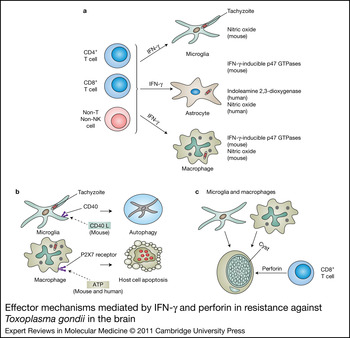
Microglia appear to be major effector cells in the prevention of proliferation of T. gondii tachyzoites in the brain. Both human (Ref. Reference Chao54) and murine (Ref. Reference Chao55) microglia become activated in vitro to inhibit intracellular proliferation of tachyzoites after treatment with IFN-γ and LPS. TNF-α and IL-6 are involved in activation of human microglia (Ref. Reference Chao54). Nitric oxide (NO) mediates the inhibitory effect of activated murine microglia on intracellular replication of tachyzoites because treatment of these cells with NG-monomethyl-l-arginine (which blocks the generation of NO) ablates their inhibitory activity (Ref. Reference Chao56) (Fig. 1a). In murine microglia activated by a combination of IFN-γ and TNF-α, both NO-dependent and NO-independent mechanisms are involved in their resistance (Ref. Reference Freund, Zaveri and Javitz57). In contrast to these observations in murine microglia, it was reported that NO is not involved in the inhibitory effect of activated human microglia against T. gondii (Ref. Reference Chao54). In vivo, after T. gondii infection, IFN-γ mediates the activation of microglia to produce TNF-α (Ref. Reference Deckert-Schluter58). This, in combination with autocrine TNF-α, is probably one of the resistance mechanisms in the brain against T. gondii.
Effector mechanisms mediated by IFN-γ and perforin in resistance against Toxoplasma gondii in the brain. (a) IFN-γ-mediated effector mechanisms against tachyzoites in the brain. Microglia activated by IFN-γ and lipopolysaccharide (LPS) inhibit proliferation of tachyzoites through nitric oxide (NO) production by inducible nitric oxide synthase (NOS2). The involvement of both NO-dependent and NO-independent mechanisms is suggested in the resistance of murine microglia activated by a combination of IFN-γ and TNF-α. The mechanism of the NO-independent inhibitory effect are unknown. Murine astrocytes activated by IFN-γ inhibit proliferation of tachyzoites by Irgm3 (IGTP). TNF-α and IFN-γ synergistically activate indoleamine 2,3-dioxygenase (IDO) in human glioblastoma cell lines and native astrocytes, and this IDO activity results in a strong toxoplasmostatic effect in the activated cells. NO production by NOS2 was first shown to be a mechanism of macrophages activated by IFN-γ to restrict tachyzoite growth in vitro. However, recent studies indicated that p47 GTPases such as IGTP and IIGP1 are critical for the activity of activated macrophages to restrict tachyzoite growth. Studies using peritoneal macrophages from mice primed with live attenuated tachyzoites show that the IFN-γ-induced, IGTP-mediated killing is preceded by parasitophorous vacuole (the vacuole containing tachyzoites) indentation, vesiculation, disruption and stripping of the parasite plasma membrane. Denuded parasites are then enveloped in autophagosome-like vacuoles, which ultimately fuse with lysosomes. In peritoneal macrophages activated in vitro by IFN-γ and LPS, autophagy protein ATG5 is required for recruitment of IIGP1 to the parasitophorous vacuole membrane and parasite clearance. (b) IFN-γ-independent effector mechanisms against tachyzoites in the brain. CD40–CD40L interaction induces autophagy-mediated control of tachyzoites within microglia. Microglia from mice lacking the autophagy molecules, Beclin-1 or ATG7, fail to kill intracellular tachyzoites in vitro after stimulation through CD40. P2X7 receptor activation by extracellular ATP kills tachyzoites in infected macrophages, which occurs in parallel with host cell apoptosis. Although this killing mechanism itself does not require IFN-γ, P2X7 receptor expression can be upregulated by this cytokine. (c) Perforin-mediated effector mechanisms against tachyzoites in the brain. Tissue cysts of T. gondii are formed mostly within neurons and astrocytes in the brain. Immune T cells can eliminate the cysts from the brain when the T cells are transferred into infected immunodeficient animals that have already developed large numbers of cysts. The T-cell-mediated immune process is associated with accumulation of microglia and macrophages around tissue cysts. Because the accumulated phagocytes penetrate the cyst, these cells appear to be the main effector cells that destroy the cysts and eliminate them from the brain after initiation of this process by immune T cells. CD8+ immune T cells have potent activity to remove the cysts, and their anticyst activity requires perforin. Perforin is the main molecule that mediates cytolysis of target cells by CD8+ cytotoxic T cells. Therefore, CD8+ T cells appear to induce elimination of T. gondii cysts through their perforin-mediated cytotoxic activity.

Granulocyte-macrophage colony stimulating factor (GM-CSF) and transforming growth factor-β (TGF-β) are other cytokines that appear to be involved in the induction of the effector functions of microglia. Expression of mRNA for these cytokines increases in the brain after infection (Refs Reference Deckert-Schluter59, Reference Hunter, Roberts and Alexander60). Murine microglia can be activated by treatment with GM-CSF (Ref. Reference Fischer61) or a combination of IFN-γ and TGF-β (Ref. Reference Chao55) in vitro, and they then inhibit intracellular multiplication of tachyzoites. The inhibitory effect of microglia activated by GM-CSF is due to their synthesis of NO (Ref. Reference Fischer61).
Interaction of CD200 with its receptor (CD200R) negatively regulates microglial function during T. gondii infection (Ref. Reference Deckert62). Infection induces an upregulation of CD200R on microglia in the brain. Infected CD200-deficient mice have significantly increased numbers of microglia, as well as increased expression of TNF and inducible NO synthase (NOS2), when compared with infected wild-type animals. The increased microglial activation is associated with moderately decreased intracerebral parasite burden and increased survival rate.
The presence of an IFN-γ-independent CD40-autophagy pathway to control intracellular replication of tachyzoites in microglia was recently reported (Ref. Reference Portillo63) (Fig. 1b). Microglia from mice lacking the autophagy molecules Beclin-1 or ATG7 failed to kill intracellular tachyzoites in vitro after stimulation through CD40, whereas these microglia were able to kill the parasite when stimulated with IFN-γ and TNF-α. In addition, these mutant mice are more susceptible to cerebral and ocular toxoplasmosis than wild-type animals, despite upregulation of IFN-γ, TNF-α and NOS2.
Astrocytes
Astrocytes are another effector cell population in the prevention of T. gondii growth in the brain. Human astrocytes become activated after treatment with IFN-γ and IL-1β to inhibit the intracellular proliferation of tachyzoites (Ref. Reference Peterson64). This inhibitory effect is mediated by NO. TNF-α induced a significant reduction in intracellular multiplication of the parasite in human astrocytoma-derived cells, whereas IL-1α induced an increase in parasite multiplication (Ref. Reference Pelloux65). TNF-α and IFN-γ were synergistic in the activation of indoleamine 2,3-dioxygenase (IDO) in human glioblastoma cell lines and native astrocytes (Ref. Reference Daubener66). This IDO activity results in a strong toxoplasmostatic effect in glioblastoma cells activated by treatment with a combination of these cytokines (Ref. Reference Daubener66) (Fig. 1a).
Murine astrocytes are also able to inhibit the proliferation of tachyzoites in vitro. Pretreatment of murine astrocytes with IFN-γ resulted in 65% inhibition of tachyzoite replication (Ref. Reference Halonen, Chiu and Weiss67). In contrast to human astrocytes, the inhibitory effect of activated murine astrocytes is not mediated by NO or by IDO (Ref. Reference Halonen, Chiu and Weiss67), but by Irgm3 (IGTP), which is a p47 GTPase (Ref. Reference Halonen, Taylor and Weiss68) (Fig. 1a). After infection of IFN-γ-activated astrocytes with tachyzoites, multiple p47 GTPases, including Irga6 (IIGP1) and IGTP, accumulate at vacuoles containing the parasite (Refs Reference Martens69, Reference Melzer70). Vacuolar accumulations of these enzymes culminate in disruption of the parasitophorous vacuoles (the vacuole containing tachyzoites) and finally of the parasite itself. IGTP and in part IIGP1 are required for the IFN-γ-mediated restriction of T. gondii growth in murine astrocytes (Refs Reference Martens69, Reference Melzer70). The importance of astrocytes in controlling T. gondii in the brain was demonstrated in GFAP-Cre gp130fl/fl mice lacking expression of gp130, the signal-transducing receptor for IL-6 family cytokines, in their astrocytes (Ref. Reference Drogemuller71). These infected mice developed a lethal necrotising TE associated with loss of GFAP+ astrocytes in inflammatory lesions and impaired parasite control. Furthermore, in vitro studies with astrocytes isolated from the same mice showed that they inhibited the growth of T. gondii efficiently after stimulation with IFN-γ, but neighbouring uninfected cells became apoptotic. Therefore, astrocytic gp130 expression is crucial for the survival of GFAP+ astrocytes in the brain of infected mice, and this protective effect of gp130 in astrocytes appears to contribute to controlling the cerebral proliferation of tachyzoites.
Macrophages
Activation of macrophages by IFN-γ is crucial for controlling tachyzoite proliferation during the acute stage of T. gondii infection (Ref. Reference Suzuki2). Because macrophages, in addition to T cells, migrate into the brain after infection (Refs Reference Suzuki48, Reference Schluter72, Reference Wilson73), this cell population is probably involved in resistance to cerebral toxoplasmosis. NO production by NOS2 was first shown to be a mechanism of macrophages activated by IFN-γ to restrict tachyzoite growth in vitro (Ref. Reference Adams74). However, recently, the importance of IGTP for their protective activity became clear (Fig. 1a). IGTP is required for the killing of tachyzoites by macrophages activated by IFN-γ (Refs Reference Butcher75, Reference Ling76). Studies using peritoneal macrophages from mice primed with live attenuated tachyzoites showed that the IFN-γ-induced, IGTP-mediated killing was preceded by parasitophorous vacuole indentation, vesiculation, disruption and stripping of the parasite plasma membrane (Ref. Reference Ling76). Denuded parasites are then enveloped in autophagosome-like vacuoles, which ultimately fuse with lysosomes (Ref. Reference Ling76). In peritoneal macrophages activated in vitro by IFN-γ and LPS, autophagy protein ATG5 is required for recruitment of IIGP1 to parasitophorous vacuole membrane and parasite clearance (Ref. Reference Zhao77). However, autophagosomes enveloping tachyzoites were not detected in these cells. Differences in how macrophages become activated might be related to the different observations in these studies.
P2X7-mediated killing of tachyzoites by human and murine macrophages was recently reported (Ref. Reference Lees78). P2X7 receptor activation by extracellular ATP kills tachyzoites in infected cells, and this killing occurs in parallel with host cell apoptosis (Fig. 1b). Although this killing mechanism itself does not require IFN-γ, expression of the P2X7 receptor can be upregulated by this cytokine. Therefore, this mechanism might also have a role as part of the IFN-γ-mediated resistance to T. gondii. The importance of P2X7 in resistance to the parasite is supported by evidence for association of polymorphisms in the gene encoding the receptor P2RX7 with clinical signs of congenital toxoplasmosis (Ref. Reference Jamieson79).
Regulatory role of IFN-γ in recruitment of T cells into the brain
T cells need to enter the brain to perform their protective activity against TE, although the blood–brain barrier limits access of lymphoid cells from the periphery. IFN-γ has an important role in induction of vascular cell adhesion molecule-1 (VCAM-1) on cerebral vessels and recruitment of immune T cells into the brain in TE-resistant BALB/c mice during the chronic stage of infection (Ref. Reference Wang80). The numbers of VCAM-1-expressing vessels were significantly greater in the brains of infected wild-type mice than in those of Ifng −/− mice, and significantly fewer CD8+ T cells were recruited into the brains of the latter than into those of the former. Treatment of infected Ifng −/− mice with recombinant IFN-γ restored the expression of VCAM-1 on their cerebral vessels and recruitment of CD8+ T cells into their brains, confirming the importance of this cytokine for upregulation of VCAM-1 expression and CD8+ T-cell trafficking. In adoptive transfer of immune spleen cells, pretreatment of the cells with antibodies against α4 integrin markedly inhibited the recruitment of CD8+ T cells into the brain of chronically infected wild-type mice. Therefore, IFN-γ-induced expression of endothelial VCAM-1 and its binding to α4β1 integrin on CD8+ T cells are important for the recruitment of T cells into the brain during the chronic stage of T. gondii infection. By using a similar cell transfer method, the importance of α4β1 integrin for T-cell recruitment into the brain was also observed in TE-susceptible C57BL/6 mice (Ref. Reference Wilson47). However, a TE-susceptible strain of mouse with neonatal inactivation of VCAM-1 gene did not show any impaired leukocyte recruitment into the brain after infection (Ref. Reference Deckert81). These results might suggest that VCAM-1-mediated T-cell recruitment into the brain during T. gondii infection is compensated by other cell adhesion molecules over time, or neonatal inactivation of VCAM-1 might result in expression of a molecule, which is not usually expressed in wild-type mice, to compensate for the absence of VCAM-1.
In addition to the regulatory activity of IFN-γ on expression of VCAM-1, this cytokine has an important role in induction of expression of various chemokines in the brains of mice during acute systemic (Refs Reference Amichay82, Reference Strack83) and chronic infection (Ref. Reference Wen84) with T. gondii. During the chronic stage of infection, mRNA and protein levels of CXCL9, CXCL10 and CCL5 are abundant in the brains of infected BALB/c mice (Ref. Reference Wen84). Combined in situ hybridisation and immunohistochemistry demonstrated the expression of CXCL10 and CCL2 in astrocytes and of CXCL9 and CCL5 in microglia (Ref. Reference Strack85). CXCR3, the receptor for CXCL9 and CXCL10, is expressed predominantly on activated T cells, and this chemokine receptor is considered to have the primary role in recruitment of effector T cells into the site of Th1-type immune response (Refs Reference Park86, Reference Sallusto87). CCL5 is one of three ligands that bind to CCR5, and in addition to CXCR3, CCR5 is preferentially expressed on Th1 cells and has an important role in infiltration of these T cells in Th1-type reactions (Ref. Reference Moser and Loetscher88). CCR5 is also expressed on macrophages, which are important effector cells activated by IFN-γ to prevent the proliferation of T. gondii tachyzoites (Ref. Reference Suzuki2). Therefore, expression of CCL5 in combination with CXCL9 and CXCL10 could have an important role in recruiting T cells to maintain a type-1 immune response and in facilitating migration of effector macrophages into the brain to control the parasite. During the acute stage of T. gondii infection, CXCL10 has been shown to induce a massive influx of T cells into the lungs and liver, and thus controls the parasite in these organs (Ref. Reference Khan89). Another chemokine receptor, CCR2, the only known functional receptor for CCL2, is also involved in regulating cell migration into the brain after T. gondii infection (Ref. Reference Benevides90). CCR2-deficient (Ccr2 −/−) mice presented greater parasitic burden associated with reduced CD4+ and Mac1+ and increased CD8+ cell migration in the peripheral organs during the acute phase and in the brain at the initial phase of chronic infection. Infected Ccr2 −/− mice also had reduced NOS2+ cell numbers in these organs.
In addition to the adhesion molecules and chemokines described above, metalloproteinases (MMPs) and their inhibitor (tissue inhibitor of metalloproteinase-1: TIMP-1) appear to be involved in the regulation of T-cell infiltration into the brain during T. gondii infection. Expression of MMP-8, MMP-10 and TIMP-1 is upregulated in the brains of mice after T. gondii infection, and infected TIMP-deficient mice have decreased perivascular accumulation of lymphocyte populations, increased CD4+/CD8+ ratio and decreased parasitic burden in the brain (Ref. Reference Clark91). The mechanisms by which MMPs and TIMP-1 regulate T-cell trafficking into the brain during infection remain to be elucidated.
IL-6 also appears to be involved in the cerebral migration of T cells during T. gondii infection. Lymphocyte preparations isolated from the brains of infected Il6 −/− mice had significantly lower ratios of γδ T cells and CD4+ αβ T cells but higher ratios of CD8+ αβ T cells than those of infected control mice (Ref. Reference Suzuki18). Of interest, no differences were detected in the ratios of these T-cell subsets in the spleens of these animals (Ref. Reference Suzuki18). These results suggest that IL-6 is another player that regulates T-cell infiltration into the brain during T. gondii infection, although the mechanisms of this regulatory function are unclear.
Participation of other cytokines in controlling tachyzoites in the brain
IL-12
IL-12 is critical for induction of IFN-γ production after infection with T. gondii. Neutralisation of IL-12 with antibodies against this cytokine results in 100% mortality in mice infected with an avirulent strain of T. gondii, and the mortality is associated with decreased IFN-γ production (Ref. Reference Gazzinelli92). DCs, macrophages and neutrophils produce IL-12 in response to the parasite. In DCs, the binding of a profilin-like protein of the parasite to Toll-like receptor (TLR) 11 stimulates IL-12 production (Ref. Reference Yarovinsky93). Mice deficient in the gene encoding MyD88, an adaptor molecule essential for most TLR as well as IL-1 and IL-18 signalling, are susceptible to acute infection with an avirulent strain, and their susceptibility is associated with impaired IL-12 responses to the parasite (Ref. Reference Scanga94). The binding of IL-12 to its receptor leads to the activation of signal transducer and activator of transcription (STAT) 4, and this signalling cascade is crucial for IFN-γ-producing cells. In agreement with this, STAT4-deficient mice are susceptible to acute infection with T. gondii, and their mortality is associated with a defect in the ability to produce IFN-γ in response to infection (Ref. Reference Cai95). Tpl2 (tumour progression locus 2) belonging to p38 mitogen-activated protein kinase is also involved in this IL-12-induced IFN-γ production (Ref. Reference Watford96). In addition to the importance of IL-12 for induction of IFN-γ production by T cells, IL-12 is also important for the maintenance of IFN-γ production in T cells mediating resistance to chronic infection (Ref. Reference Yap, Pesin and Sher27).
Lipoxin A4 (LXA4), an eicosanoid product generated from arachidonic acid, is an important downregulator of IL-12 production to prevent pathogenic inflammatory responses in the brain during the chronic stage of T. gondii infection. Wild-type mice produce high levels of serum LXA4 beginning at the onset of chronic infection. 5-Lipoxygenase (5-LO) is an enzyme that is critical in the generation of LXA4, and mice deficient in 5-LO (Alox5 −/−) succumbed to infection during the chronic stage, displaying marked inflammation in their brains (Ref. Reference Aliberti, Serhan and Sher97). The increased mortality of Alox5 −/− animals is not due to defective control of the parasite but due to enhanced inflammatory responses associated with increases in IL-12 and IFN-γ, and their death is completely prevented by the administration of a stable LXA4 analogue. Therefore, LXA4 is important for the downregulation of proinflammatory responses during the chronic stage of T. gondii infection. Recent studies showed that lipoxins activate two receptors (AhR and LXAR) in DCs, and this activation triggers expression of suppressor of cytokine signalling (SOCS)-2. SOCS-2-deficient mice succumb to chronic infection with T. gondii in association with increased IL-12 and IFN-γ responses and reduced brain cysts (Ref. Reference Machado98), as observed in Alox5 −/− animals (Ref. Reference Machado98).
IL-4
Although IFN-γ-mediated immune responses have an essential role in resistance to T. gondii infection, Th2-type immune responses are also involved in protective immunity. IL-4-deficient (Il4 −/−) mice all died during the late stage of infection, whereas control mice survived (Ref. Reference Suzuki99). Histological study showed significantly greater numbers of cysts and areas of acute focal inflammation associated with tachyzoites in the brains of Il4 −/− mice than in control mice during the later stage of infection (Ref. Reference Suzuki99). These results indicate that IL-4 is protective against development of TE by preventing the formation of cysts and the proliferation of tachyzoites in the brain. Spleen cells of control mice at 8 weeks after infection were found to produce significantly greater amounts of IFN-γ after stimulation in vitro with T. gondii antigens than measured in Il4 −/− mice (Ref. Reference Suzuki99). Therefore, IL-4 has a role in enhancing IFN-γ production during the late stage of infection. The impaired ability of Il4 −/− mice to produce IFN-γ probably contributes to their susceptibility for development of severe TE.
STAT6 is a molecule involved in IL-4 and IL-13 signalling. In agreement with the observations in Il4 −/− mice, significantly greater numbers of T. gondii cysts were recovered from the brains of STAT6-deficient (Stat6 −/−) than from wild-type mice. CD8+ T cells obtained from the cerebrospinal fluids and spleens of infected wild-type mice produced greater amounts of IFN-γ than T cells from infected Stat6 −/− animals (Ref. Reference Jin28). In addition, transfer of CD8+ T cells from wild type to Stat6 −/− mice reduced cyst numbers in the brains of recipients. Transfer of splenic adherent cells from wild type to Stat6 −/− mice induced activation of CD8+ T cells and decreased brain cyst numbers. Therefore, STAT6 signalling is important in CD8+ T-cell activation after T. gondii infection, possibly through regulation of the activity of antigen-presenting cells. In this regard, it has been shown that IL-4, alone or together with IFN-γ, efficiently enhances the production of bioactive IL-12 in mouse and human DCs (Ref. Reference Hochrein26). The lack of this positive regulatory effect of IL-4 on IL-12 production by DCs most probably contributes to the reduced resistance of Il4 −/− and Stat6 −/− mice to T. gondii infection described above.
Another study reported that greater numbers of cysts and more severe histological changes were observed in the brains of control mice than in Il4 − /− mice in the late stage of infection, although the former were more resistant to death during the acute stage (Ref. Reference Roberts100). Because the strains of T. gondii differ between the studies mentioned, the strains and virulence of the parasite might have contributed to the different outcomes in the studies.
IL-10
IL-10 is an important negative regulator of inflammatory responses (Ref. Reference Kuhn101). IL-10-deficient (Il10 −/−) mice all die during the acute stage of infection with T. gondii and their mortality is associated with development of severe immunopathology mediated by Th1 immune responses in the liver (Ref. Reference Gazzinelli102) and intestine (Ref. Reference Suzuki103). Therefore, IL-10 is crucial for downregulating IFN-γ-mediated immune responses and preventing the development of pathology caused by the immune responses. When Il10 −/− mice were treated with sulfadiazine to control the proliferation of T. gondii in the early stage of infection, the animals survived the acute stage but developed lethal inflammatory responses in their brains in the later stage of infection (Ref. Reference Wilson73). The importance of IL-10-dependent signalling for survival of mice during the acute and chronic stages of infection is confirmed by treating infected wild-type animals with antibodies against anti-IL-10 receptor (Ref. Reference Jankovic104). Therefore, IL-10 has an important downregulatory role in preventing immunopathology and mortality during the course of infection with T. gondii.
Various populations of T cells, such as Th2 and Tregs, are able to produce IL-10. However, conventional IFN-γ-secreting T-bet+Foxp3− Th1 cells were shown to be the main producers of IL-10 in T. gondii-infected mice. These IL-10+IFN-γ + CD4+ T cells possess potent activity to prevent intracellular multiplication of tachyzoites within macrophages, while profoundly suppressing IL-12 production by antigen-presenting cells (Ref. Reference Jankovic104). In addition, these IL-10-producing T cells are generated from IL-10−IFN-γ + CD4+ T cells after restimulation with tachyzoite antigens. These results indicate that IL-10 production by CD4+ T cells after infection with T. gondii does not require a distinct regulatory Th subset, but can be generated in Th1 cells as part of the effector response to the parasite, which provides a crucial negative-feedback loop for prevention of pathogenic overstimulation of the Th1 response.
There is further interesting evidence on the existence of a negative-feedback loop of Th1 response in T. gondii infection. Tyk2, a member of the Jak family of nonreceptor tyrosine kinases, mediates the biological effects of IL-12 and IFN-αβ and promotes IFN-γ production by Th1 cells, and Tyk2-null mice are susceptible to infection with the parasite. However, Tyk2 is also required for the production of IL-10 by immune CD4+ T cells after challenge infection of vaccinated mice (Ref. Reference Shaw105). The Tyk2-dependent production of IL-10 is mediated by IFN-γ, indicating negative autoregulation of the Th1 effector response in infection. Therefore, Tyk2 has a dual function mediating induction and suppression of the Th1 effector response, most likely for maximising pathogen clearance while minimising immunopathology.
IL-17 and IL-27
A unique subset of CD4+ T cells, Th-17, produces IL-17, and this T-cell population has been suggested to mediate inflammation in models of autoimmune diseases such as multiple sclerosis and rheumatoid arthritis. In infection with T. gondii, IL-17 has been shown to have both protective and pathogenic roles. IL-17-receptor-deficient mice are more susceptible to acute infection with the parasite (Ref. Reference Kelly106). Their mortality was associated with increased parasite burden in the organs, including the brain, and with a defect in the migration of neutrophils to infected sites during the early stage of infection. Therefore, IL-17-mediated induction of neutrophil migration to infected sites during the initial stage of infection appears to have an important role in resistance against acute infection with T. gondii. However, the activity of Th-17 cells needs to be appropriately downregulated by IL-27 to prevent the development of severe inflammatory changes in the brain during the later stage of infection. Chronically infected IL-27-receptor-deficient mice developed severe inflammation in their brains mediated by CD4+ T cells, and the pathology was associated with a prominent IL-17 response (Ref. Reference Stumhofer107). In addition, treatment of naive primary T cells with IL-27 in vitro suppressed the development of Th-17 cells induced by IL-6 and TGF-β, and the suppressive effect was dependent on the intracellular signalling molecule STAT1 (Ref. Reference Stumhofer107). Therefore, IL-27 has a crucial role in the prevention of Th-17-mediated inflammatory responses in the brain during the chronic stage of T. gondii infection.
IL-33
IL-33 is a member of the IL-1 family with the ability to downregulate IFN-γ production by Th1 cells and upregulate Th2 response. Mice deficient in T1/ST2, a component of the IL-33 receptor, demonstrated increased susceptibility to T. gondii infection that correlated with increased pathology and greater parasite burden in the brain (Ref. Reference Jones108). Real-time PCR analysis of cerebral cytokine levels revealed increased mRNA levels of IFN-γ, TNF-α and NOS2 in infected T1/ST2-deficient animals. The mechanisms mediated by IL-33 receptor to control T. gondii in the brain remain to be analysed.
Perforin-mediated Immune responses to the cyst stage of T. gondii in the brain
The basis of persistence of chronic infection with T. gondii is the tissue cyst, which remains largely quiescent for the life of the host, but can reactivate and cause disease. This stage of the parasite life cycle is not affected by any of the current drug treatments and has generally been regarded as untouchable. However, we recently showed that the immune system can eliminate T. gondii cysts from the brains of infected hosts when immune T cells are transferred into infected immunodeficient animals that have already developed large numbers of cysts (Ref. Reference Suzuki12). This T-cell-mediated immune process was associated with accumulation of microglia and macrophages around tissue cysts. Because the accumulated phagocytes penetrate within the cyst, these cells appear to be the main effector cells that destroy the cysts and eliminate them from the brain after initiation of this process by immune T cells. CD8+ immune T cells possess potent activity to remove the cysts. Of interest, the initiation of this process by CD8+ T cells does not require the production of IFN-γ, the main mediator that prevents proliferation of tachyzoites during acute infection, but does require perforin. Perforin is the major molecule that mediates cytolysis of target cells by CD8+ cytotoxic T cells. Therefore, our results suggest that CD8+ T cells induce elimination of T. gondii cysts through their perforin-mediated cytotoxic activity. A study using two-photon microscopy in C57BL/6 mice with OVA-expressing T. gondii showed that OVA-specific CD8+ T cells accumulated in regions containing isolated parasites (tachyzoites), but not around cysts in the brain. C57BL/6 mice are genetically susceptible to chronic infection with the parasite, and continuous tachyzoite proliferation occurs in the brain. Therefore, it is possible that the activity of cerebral CD8+ T cells of these animals is downregulated in this environment and does not have potent anticyst activity.
Perforin-mediated cytolytic activity by T cells in resistance against T. gondii infection was less appreciated previously when compared with the absolute requirement of IFN-γ to control tachyzoite proliferation during the acute stage of infection. Perforin-knockout mice survive acute infection (Refs Reference Wang22, Reference Denkers109). In vitro studies demonstrated that the lysis of tachyzoite-infected cells by cytotoxic CD8+ T cells results in release of viable parasites (Ref. Reference Yamashita110). However, the situation of the parasite in the cyst stage seems different. Whereas the cyst resides within an infected cell (Refs Reference Ferguson and Hutchison111, Reference Ghatak and Zimmerman112), as do tachyzoites, bradyzoites within the cyst are surrounded by a thick cyst wall, which is unique to the cyst. Therefore, cytolysis of cyst-containing cells by perforin-mediated activity of CD8+ T cells followed by quick accumulation of large numbers of microglia and macrophages would probably provide the parasite little time to escape from a coordinated attack by T cells and phagocytes. A three- to fourfold increase in brain cyst numbers is reported in perforin-knockout mice in the later stage of infection (Ref. Reference Denkers109). The absence of perforin-dependent anticyst activity of CD8+ T cells in these animals might have contributed to their observation.
Outstanding research questions
IFN-γ-mediated immune responses are essential for controlling the proliferation of tachyzoites during the acute stage of infection and for prevention of reactivation of infection with T. gondii (TE). To maintain the latency of chronic infection in the brain and prevention of TE, IFN-γ production by T cells is essential. CD8+ T cells are the main mediator of protective immunity, and CD4+ T cells are important for optimum IFN-γ production by cerebral CD8+ T cells. However, the detailed role of each of CD4+ and CD8+ T-cell populations and their interactions in protective immunity in the brain during the course of infection still remain to be elucidated. The recent discovery of H-2d- and H-2b-restricted CD8+ T-cell epitopes provides a powerful tool for this analysis. However, additional information regarding T. gondii epitopes for both CD4+ and CD8+ will facilitate a better understanding of the development and maintenance of protective T-cell responses in the brain during the course of infection, in association with conversion of T. gondii from the tachyzoite to the cyst stage. It is important to depict how T-cell responses differ between TE-resistant and TE-susceptible hosts. We also need a better understanding of the mechanisms of recruitment of T cells into the brain through the blood–brain barrier and into the site of parasite growth within the brain.
With regard to T. gondii epitopes recognised by human CD8+ T cells, HLA-A0201- and HLA-A03-restricted epitopes were recently identified. In addition, immunisation of transgenic mice expressing these human HLA class I molecules [HLA-A0201 or HLA-A1101 (an HLA-A03 supertype allele)] with those identified epitope peptides conferred a protection associated with reduced parasite burden against challenge infection. These epitopes appear to be promising candidates for a human vaccine to induce protective immune responses. Using the information from murine models and searching for additional or more dominant epitopes could improve the efficacy of the vaccination to confer protection against T. gondii infection.
With regard to effector mechanisms to control T. gondii in the brain, several cell types, such as microglia, astrocytes and macrophages, become activated by IFN-γ to prevent intracellular proliferation of tachyzoites through NO, IDO, p47 GTPases and/or unidentified molecules. IFN-γ-independent mechanisms that require CD40 or P2X7 receptor were recently shown to participate in controlling tachyzoites in microglia and macrophages, respectively. However, the information currently available is mostly derived from in vitro studies using these cell types. The role of these effector mechanisms to control tachyzoite growth in each of microglia, astrocyte, neuron and macrophage in vivo is needed to understand the mechanisms to control T. gondii in the brain.
In addition to IFN-γ-mediated protective immunity against tachyzoites, it was recently shown that the immune system uses perforin-mediated activity of CD8+ T cells to remove tissue cysts from the brain. Therefore, resistance to T. gondii in the brain requires a coordinated collaboration of IFN-γ- and perforin-mediated immune responses. It is crucial to elucidate how perforin-mediated activity of CD8+ T cells eliminates T. gondii cysts from the brain, and how these protective mechanisms are coordinated to compose effective resistance to the parasite in the brain. Elucidating these mechanisms will be crucial to developing a new method to prevent and eradicate T. gondii infection.
Acknowledgements
We thank the peer reviewers for their critical reading of this manuscript and helpful comments. This work was supported by grants from the National Institutes of Health (AI073576, AI078756 and AI077887) and from The Stanley Medical Research Institute (08R-2047). We thank Sara Perkins and James Hester for their assistance with preparing the manuscript.