Chest pain is a common presenting complaint among adolescents and rarely signifies cardiac diseaseReference Khalilian, Emami Moghadam, Torabizadeh, Khalilinejad and Moftakhar1. However, cardiac aetiologies for adolescent chest pain should always be considered as they may be life-threatening. Myocarditis often presents with chest pain and marked non-ischemic inflammation.Reference Kindermann, Barth and Ukena2 Separately, isolated coronary artery anomalies can also present with chest pain but can range from being asymptomatic to sudden cardiac death.Reference Frescura, Basso and Thiene3,Reference Buccheri, Cortese and Piraino4 Notably, a coronary artery anomaly is one of the leading causes of cardiac arrest in young athletes.Reference Landry, Allan and Connelly5 We present a case of a young man presenting with non-exertional chest pain found to have myocarditis as well as an anomalous origin of the right coronary artery with an interarterial course. We suspect his acute presentation was due to myocarditis, which subsequently led to the discovery of his anomalous right coronary artery, allowing for potentially life-saving intervention.
An 18-year-old active young man presented after two episodes of chest pain occurring over a 48-hour period. The pain was substernal, non-radiating, “stabbing” in nature, and occurred at rest. These episodes self-resolved after 30 minutes and were accompanied by lightheadedness and headache. There was no history of chest trauma or recent illness. He denies using any medications, supplements, or recreational drugs. There was no family history of myocardial infarction, stroke, or sudden cardiac death.
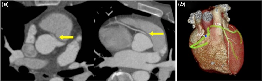
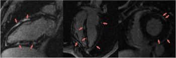
On presentation, there was no chest tenderness on exam. Electrocardiogram showed normal sinus rhythm without ST segment changes, and the chest radiograph was unremarkable. Laboratory studies were significant for initial troponin-I of 61 ng/mL and subsequent troponin-T of 5.86 ng/ml, pro-BNP of 437 pg/ml, creatine kinase of 1623 U/L, and high-sensitivity c-reactive protein of 13.9 mg/L. Transthoracic echocardiogram revealed a left ventricular ejection fraction of 50% and did not comment on the coronary arteries. CT angiography demonstrated an anomalous origin of the right coronary artery arising from the left coronary sinus and coursing between the aorta and right ventricular outflow tract (Fig 1). Subsequent cardiac MRI demonstrated extensive epicardial and myocardial enhancement with oedema consistent with myocarditis (Fig 2). The patient underwent an unroofing procedure of the right coronary artery, during which he was found to have a small calibre right coronary artery coursing intramurally through the aortic wall, with a “slit-like” ostium positioned superior to the left and right commissure.
CT angiogram of coronary arteries with contrast showing right coronary artery originating from left sinus ( a ), also highlighted in 3D imaging generated by CT angiogram ( b ).

Cardiac MRI showing extensive epicardial and myocardial enhancement in delay post-contrast images consistent with myocarditis.

Discussion
The discovery of elevated troponins in the setting of substernal chest pain led to an initial concern for acute coronary syndrome, an extremely rare diagnosis in an otherwise healthy 18-year-old man. CT angiogram was obtained to evaluate the coronary arteries, which were not visualised on echocardiography. The CT revealed that the right coronary artery was noted to arise from the left coronary sinus and course between the aorta and right ventricular outflow tract, which could lead to compression of the right coronary and impaired perfusion during exertion resulting in myocardial infarction.Reference Lorenz, Mookadam and Mookadam6 Additionally, the artery was noted to be small in calibre with an intramural course and to have a slit-like ostium, characteristics which increase the risk of impaired perfusion due to decreased luminal diameter in the proximal artery in combination with external compression.Reference Opolski7
Although the patient was found to have a high-risk lesion, compression of the right coronary artery leading to myocardial ischemia would be expected to occur during or shortly after exertion and thus it was unclear whether this was an adequate explanation for the patient’s non-exertional chest pain. Cardiac MRI was obtained to further evaluate for cardiac muscle scarring secondary to a prior ischemic event or for evidence of myocarditis, either of which could explain the patient’s non-exertional chest pain. MRI revealed extensive epicardial and myocardial inflammation more consistent with myocarditis (Fig 2), rather than an endocardial or transmural pattern following the arterial distribution, as expected an arterial supply-dependent infarction.Reference Laissy, Messin and Varenne8,Reference Rajiah, Desai and Kwon9 These MRI findings, in addition to the elevated c-reactive protein, support a diagnosis of myocarditis as the aetiology for the patient’s chest pain. In infectious myocarditis, the damage and dysfunction of the cardiac vasculature is associated with aberrant regulation of vascular tone including coronary vasospasm.Reference Woudstra, Juffermans and van Rossum10 In this case, the fortuitous discovery of the anomalous origin of the right coronary artery with interarterial and intramural course, as well as notably the slit-like ectopic ostium course, warranted immediate surgical intervention.
Conclusion
This case demonstrates myocarditis presenting as a mimicker of an acute myocardial infarction, leading to the diagnosis of an anomalous origin of the right anomalous coronary artery with interarterial course. It highlights the value of cardiac MRI in diagnosing an unclear aetiology of chest pain and the importance of surgical intervention of anomalous right coronary artery to mitigate the risk of sudden cardiac death.
Acknowledgement
The authors would like to thank Dr. Merjuan Uzunyan, pediatric cardiologist, for his great assistance in caring for this child.
Financial support
The authors have no financial relationships relevant to this article to disclose.
Conflict of interest
The authors have no conflicts of interest to disclose.
Ethical standards
Not applicable.