Coronary CT angiography allows easy evaluation of coronary artery morphology. The heart rate should be controlled by beta-blocker to obtain clear images and reduce radiation exposure.Reference Mark, Berman and Budoff 1 Children undergoing coronary CT angiography especially need a beta-blocker because their basal heart rate is higher than that in adults, they are easily excited, and they have small coronary vessels. However, no standard dose and no protocol for administering the beta-blocker have been established for children, as they are primarily used in adults.Reference Sabarudin and Sun 2 The doses and protocols vary among facilities. The beta-blocker should be provided at an appropriate dose and usage because it could engender severe side effects. The aim of this study was to clarify the appropriate dose and provide a standard protocol for beta-blocker administration in children.
Materials and methods
Study population
This retrospective study was performed at Nihon University Itabashi Hospital in Japan from January, 2009 to August, 2016. A total of 63 children, comprising 50 boys and 13 girls, aged 4–15 years who underwent coronary CT angiography for evaluation of coronary artery lesions due to presumed Kawasaki disease comprised the study group. We excluded patients younger than 3 years because of their inability to hold their breath. The children with contraindications to beta-blocker administration – such as severe asthma, past history of some allergic reaction to beta-blockers, severe aortic stenosis, ventricular systolic dysfunction, atrioventricular block, and calcium channel blocker ingestion – were excluded from the study. The children who had already been prescribed a daily beta-blocker were also excluded, as it would interfere with assessing the effect of the beta-blocker at the time of scanning. Finally, children whose vital signs around the time of scanning were missing from their medical records were also excluded.
The institutional review board of Nihon University Itabashi Hospital approved the study protocol (RK-160913-20). The first and second authors take complete responsibility for the integrity of the date and accuracy of the data analysis.
Study protocol
The coronary CT angiography scan images were obtained using a 320 detector row Aquilion ONE® system (Toshiba, Otawara, Japan) with the following scanning parameters: slice configuration 320×0.5 mm, gantry rotation time 350 ms, retrospective or prospective electrocardiography-triggered scan, tube voltage 80–120 kVp (100 kVp for those <10 years old, adjusting for each patient’s body frame), and tube current 180–580 mA. The intravenous bolus injection technique was used at acquisition.
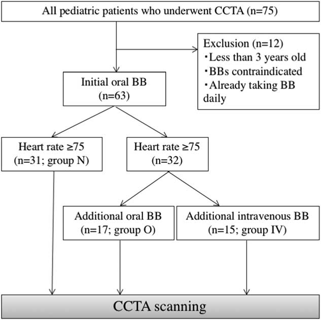
After acquisition, off-line reconstruction was performed with the appropriate workstation. The contrast medium ioversol (320 mg I/ml) was injected during scanning. The patients were assigned to three groups according to the beta-blocker administration protocol in Figure 1. All study patients were given an initial dose of oral beta-blocker 1 hour before coronary CT angiography. An additional beta-blocker was given if the heart rate exceeded 75 beats/minute just before scanning.
Study protocol. A total of 12 patients were excluded for the reasons of age, contraindications, and/or daily use of a beta-blocker (BB). Finally, 63 patients were enrolled. All study patients were given an initial dose of oral BB (metoprolol or atenolol) 1 hour before cardiac computed tomography angiography (CCTA). Additional BB was given orally or intravenously if the heart rate exceeded 75 beats/minute just before scanning. The patients were grouped as: without additional BB (group N); with additional oral BB (metoprolol or atenolol) (group O); with additional intravenous BB (landiolol) (group IV).

The patients were grouped on the basis of their beta-blocker protocol: scanning without additional beta-blocker (group N), with additional oral beta-blocker (metoprolol or atenolol) (group O), and with additional intravenous beta-blocker (landiolol) (group IV). As an additional administration, only an oral beta-blocker was selected from 2009 to 2011, and an intravenous beta-blocker was selected only after official approval for the use of landiolol during CT scanning was granted in 2011. An additional dose of oral beta-blocker in the range of 0.4–0.8 mg/kg was prescribed in clinical settings, whereas for intravenous beta-blocker the additional dose was 0.125 mg/kg. To clarify the appropriate beta-blocker dosage for heart rate control in children, we compared the image quality and the amount of radiation exposure for each group. To predict the occurrence of adverse events, all patients were carefully observed and their condition, including vital signs and clinical symptoms, was monitored.
Image quality
We compared the image quality in the three groups. Image quality was classified into five grades that depended on the degree of visualisation of each coronary branch (Table 1). Two paediatric cardiologists with advanced imaging training assessed the image quality. We used a correlation analysis to identify the factor that contributed to image quality and then a multiple regression analysis to confirm the precise parameters to use to estimate image quality.
Evaluation of image quality.

The image quality was classified into five grades according to the visualisation of each coronary branch: 5, whole branches are well visualised; 4, both proximal and middle branches are well visualised, but only parts of the distal branches are visualised; 3, almost all proximal and middle branches are visualised; 2, only proximal arteries are visualised; 1, all branches are difficult to evaluate
Dose of radiation exposure
We compared radiation exposures in the three groups. The dose–length product represented the dose of radiation exposure. We used a correlation analysis to identify the factor that contributed to the radiation exposure dose and then a multiple regression analysis to confirm the precise parameters to use for estimating the radiation exposure dose.
Adverse events
We compared the occurrence of adverse events owing to administration of the beta-blocker in the three groups. We categorised adverse events as follows.
∙ Clinical symptoms: fatigue, dyspnoea, chest discomfort, somnolence, headache, vertigo, nausea, vomiting, any symptoms related to an allergic reaction, and the effect of the beta-blocker.
∙ Hemodynamics: bradycardia, hypotension with symptoms, variation of the heart rate, and mean blood pressure on admission with no administration of beta-blocker compared with that just before coronary CT angiography scanning.
Statistical analysis
The three groups were compared using a one-way factorial analysis of variance. Pairwise comparisons were made using the Tukey–Kramer adjustment and Student’s t-test, as appropriate. Correlations between the parameters and image quality and radiation exposure were analysed using Pearson’s correlation coefficient. Stepwise multiple regression analysis was performed using a stepwise forward selection method, excluding covariates with univariate values of p>0.2. Results were considered statistically significant at the level of 0.05. Statistical analysis was performed using JMP® 13 software (SAS Institute, Cary, North Carolina, United States of America)
Results
Patients’ characteristics
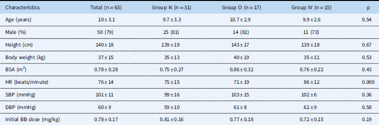
Patients’ characteristics of the groups are shown in Table 2. Altogether, 63 patients were enrolled in this study and were classified into three groups according to their participation in the three beta-blocker protocols, as explained in the legend to Figure 1: without additional beta-blocker (group N, n=31), with additional oral beta-blocker (group O, n=17), and with additional intravenous beta-blocker (group IV, n=15). Overall, the average patient age was 10.0±3.1 years, and 50 of 63 patients (79%) were male. The average dose of the initial beta-blocker was 0.78±0.17 mg/kg. No significant difference was found among the three groups with respect to age, height, body weight, systolic blood pressure, diastolic blood pressure at the time of admission, or the initial beta-blocker dose. There was a significant difference in the baseline heart rate between groups N and IV (p=0.032) and groups O and IV (p=0.009).
Patients’ characteristics.

BB=beta-blocker; BSA=body surface area; DBP=diastolic blood pressure; Group N=no extra BB needed; Group O=extra oral BB needed; Group IV=extra intravenous BB needed; HR=heart rate; SBP=systolic blood pressure
Results are given as numbers (%) or means±SD
The baseline vital signs were obtained on admission to hospital with no administration of BB
Image quality
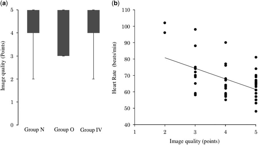
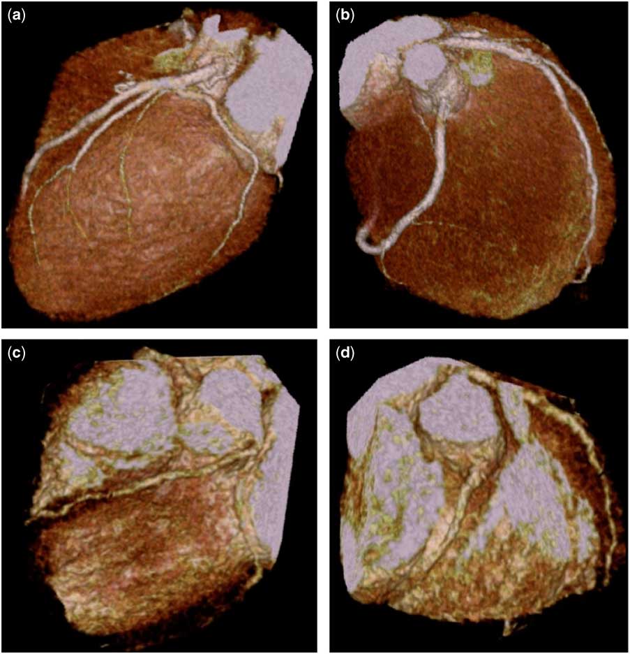
Figure 2a shows the image quality for the groups: 4.32±0.82 for group N, 4.18±0.92 for group O, and 4.33±0.94 for group IV. There were no significant differences among the groups. Significant correlations with image quality were seen for the patient’s height (p=0.035, r=0.266), heart rate on admission (p=0.049, r=−0.249), and heart rate just before acquisition (p<0.001, r=−0.533) (Fig 2b). The multiple regression analysis of the overall patients indicated that the heart rate just before acquisition (p<0.001) and height (p=0.036) were significantly associated with image quality. Stepwise multiple regression analysis for variable selection revealed that only the heart rate just before acquisition was needed for the most appropriate regression formula (R2=0.284; root mean squared error=0.757). The images evaluated as fully adequate (5 points) and barely adequate (2 points) are presented in Figure 3 as examples.
Image quality. ( a ) Points assigned for image quality were 4.3 ± 0.8 for group N (no extra beta-blocker (BB) needed), 4.1 ± 0.9 for group O (extra oral BB needed), and 4.3 ± 0.9 for group IV (extra intravenous BB needed). There were no significant differences within each group. ( b ) There was a significant inverse correlation between image quality and the post-imaging heart rate (r = −0.533).

Examples of image quality. ( a and b ) Imaging conditions of a fully adequate (5 points) example from a 7-year-old boy weighing 23.8 kg and having a heart rate of body 55 beats/minute at acquisition (scan conditions: 80 kVp tube voltage, 150 mA tube current). Dose–length product decreased to 25.7 mGy*cm as a result of these conditions. (c and d): imaging conditions of a barely adequate (2 points) example from a 7-year-old boy weighing 16.8 kg and having a high heart rate (96 beats/minute) at acquisition (scanning conditions: 80 kVp tube voltage, 100 mA tube current). Dose–length product was 89.4 mGy*cm, which was relatively higher than that of the first patient because of segment reconstruction.

Radiation exposure
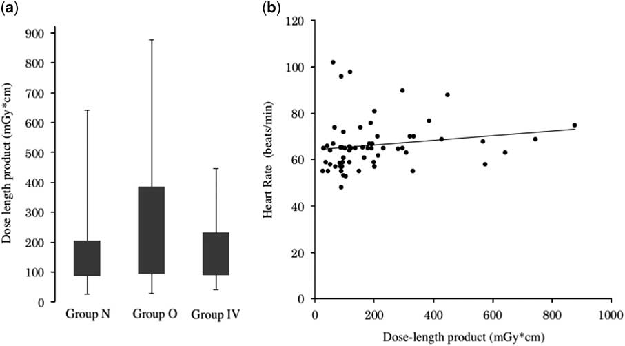
Figure 4 shows the dose–length product for the groups. There were no significant differences in the dose–length product in each group. There was no significant correlation with the heart rate just before acquisition (p=0.45, r=0.096) (Fig 4b).
Radiation exposure. ( a ): mean dose–length products were 167 ± 142 mGy*cm in group N (no extra beta-blocker (BB) needed), 270 ± 245 mGy*cm in group O (extra oral beta-blocker (BB) needed), and 282 ± 437 mGy*cm in group IV (extra intravenous BB needed). There were no significant differences within each group. ( b ) there were no significant correlations with any parameters including the post-imaging heart rate (r=0.096).

Adverse events
Regarding clinical symptoms, there were no adverse events related to allergic reactions in any of the 63 patients, reflecting the effects of the beta-blocker.
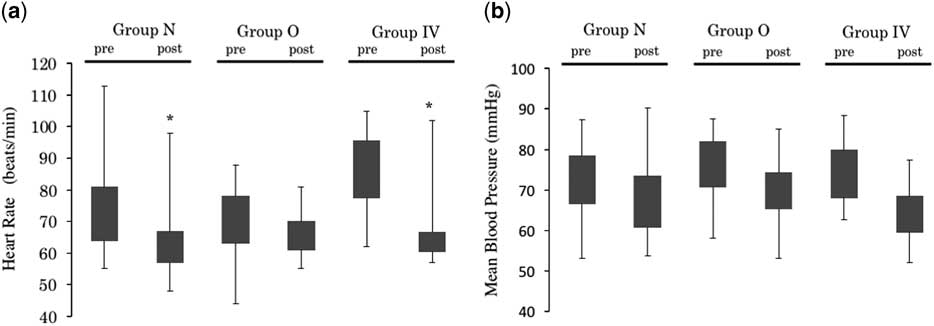
The hemodynamics values are shown in Figure 5. There were variations in the heart rate and mean blood pressure between the time of admission and just before scanning in each group. The heart rate just before scanning was significantly lower than that on admission in the N and IV groups, whereas the mean blood pressure just before scanning was not significantly different from that on admission in these two groups. There were no adverse events based on low blood pressure, such as bradycardia with symptoms.
Variations in the heart rate and mean blood pressure. Heart rate and mean blood pressure variations from the time of admission (pre) to just before scanning (post) in each group. ( a ) the mean heart rate just before scanning (post) was significantly decreased compared with that on admission (pre) in groups N (no extra BB needed) and IV (extra intravenous BB needed). There was no significant difference in the heart rate just before scanning (post) in each group. ( b ) there was no significant difference in the mean blood pressure just before scanning (post) compared with that on admission (pre) in any of the groups (*p < 0.05 versus the value at admission). BB: beta-blocker; group O: extra oral BB needed.

Discussion
We believe that administering additional beta-blocker is useful for stabilising a patient’s heart rate and improving the quality of images without increasing radiation exposure. This is true even in patients with poor heart rate control – based on our results that no decline of image quality and no increase in radiation exposure were revealed in the groups administered additional beta-blocker, compared with the group given only initial beta-blocker.
The dose–length product in this study was not correlated with the heart rate just before acquisition, although there is a relatively strong inverse correlation between the heart rate just before acquisition and image quality. The latter may be explained by segment reconstruction owing to incomplete breath-holding and body movement of patients with a lower heart rate, as well as low voltage/low current scanning of patients with a higher heart rate. Appropriate heart rate control is important, especially in children, when applying imaging methods with low radiation exposure, including prospective electrocardiography-gated scanning or half-reconstruction.
One of the difficulties of performing coronary CT angiography is the variation in peak contrast time in the presence of a giant coronary aneurysm. Indeed, the acquisition technique must be optimised individually for patients with a giant coronary aneurysm in whom a contrast delay might be anticipated, even though the intravenous bolus injection technique was used at acquisition. In addition, the difficulties seen more often in children than adults during acquisition include the children’s higher base heart rates as this age group is easily excited, has smaller coronary vessels, and sometimes has little control of their body movement and breath-holding. High-end modalities allow easy attainment of visual images even in patients with a high heart rate.Reference Machida, Tanaka and Fukui 3 Using image reconstruction methods, we can apply segment reconstruction in patients with a high heart rate. However, with multi-segment reconstruction increased radiation exposure because of multiple scans and incomplete breath-holding are problematic. In addition, end-systolic phase scanning is known to improve the image quality in patients with a high heart rate, but it tends to be more problematic than that of images obtained during the end-diastolic phase in patients with a low heart rate.Reference Araoz, Kirsch and Primak 4 In contrast, prospective electrocardiography-gated scanning is expected to provide easily visualised images with low-dose radiation exposure.Reference Gutstein, Wolak and Lee 5 Prospective electrocardiography-gated scanning should be applied in the patients with appropriately controlled heart rate, such as the patients in this study. It is necessary to control the heart rate using beta-blockers based on adequate background knowledge.
There are few reports of using beta-blockers for paediatric coronary CT angiography. Although some regimens using a beta-blocker and general anaesthesia have been reported,Reference Rigsby, deFreitas and Nicholas 6 the techniques vary among facilities, and specific procedures have not been established. Experience using beta-blocker safely in children has been frequently reported, however, including those with congestive heart failureReference Alabed, Sabouni, Dakhoul Al, Bdaiwi and Frobel-Mercier 7 and perioperatively for those with CHD.Reference Tokunaga, Hiramatsu and Kanemoto 8
For appropriate heart rate control, we suggest that all patients be given metoprolol or atenolol orally 1 hour before coronary CT angiography at a dose of around 0.8 mg/kg. In addition, if the heart rate exceeds 75 beats/minute just before scanning, landiolol should be given intravenously. We chose landiolol as the additional beta-blocker for safety and utility because of its short half-life period.Reference Khan, Cummings, Gutierrez, Bhalla, Woodard and Saeed 9 , Reference Fujito, Takayanagi, Kimura, Yokoyama and Yamada 10 In terms of dose optimisation related to body weight, the use of landiolol avoids excessive administration in comparison with oral beta-blocker, which is administered according to the dose carried by each tablet. Indeed, landiolol significantly reduced the heart rate but did not lower the blood pressure in our experience. Moreover, we were unable to compare the present protocol with the protocol whereby landiolol was used as the initial beta-blocker. Nevertheless, we believe that the two phases of beta-blocker administration might be safe for the patients with difficult heart rate control. It is also important to help the child relax with a reasonable approach such as musicReference Ng, Karimzad and Menezes 11 and/or proper preparation.Reference Gulec and Ozcengiz 12
Patients younger than 3 years were excluded from this study because of their high base heart rate and inability to hold their breath. However, coronary CT angiography for patients younger than 3 years is still necessary to evaluate coronary artery lesions in patients with Kawasaki disease and CHD including transposition of the great arteries. Hence, additional investigation regarding the appropriate use of beta-blockers for children younger than 3 years is desirable.
We fixed the cutoff heart rate for applying additional beta-blocker as 75 beats/minute, considering it the lower limit of the heart rate in young children. We thought it was reasonable for the targeted children who are unable to control their heart rate at <65 beats/minute. Our reasoning was based on the fact that the half-scan method with our modality could control the children’s heart rates at <75 beats/minute, thereby providing a control technique suitable for capturing a minimum two-segment reconstruction. The efficacy of the beta-blocker is occasionally restrictive because the base heart rate is higher in young children. Thus, it is important to focus on the points that should be observed before CT scanning – for example, collateral circulation, pattern of calcification, intimal thickening, and the possibility of thrombus – which should be fully discussed among the paediatric cardiologists and radiology technicians before attempting advanced acquisition in patients with coronary artery lesions due to Kawasaki disease. Such measures could avoid the use of unnecessary excessive sedation and radiation exposure for children during coronary CT angiography.
Acknowledgements
The authors thank Nancy Schatken, BS, MT(ASCP), from Edanz Group (www.edanzediting.com/ac) for editing a draft of this manuscript
Financial Support
This research received no specific grant from any funding agency or from commercial or not-for-profit sectors.
Conflicts of Interest
None.
Ethical Standards
The authors assert that all procedures contributing to this work comply with the ethical standards of the Helsinki Declaration of 1975, as revised in 2008.