Introduction
The carotid blow-out syndrome can be defined as rupture of the extra-cranial carotid artery or one of its main branches. It is one of the most life-threatening complications associated with head and neck carcinoma.Reference Roh, Suh, Kim, Lee, Choi and Choi 1
Carotid blow-out syndrome has been classified into three levels of severity.Reference Chaloupka, Putman, Citardi, Ross and Sasaki 2 , Reference Citardi, Chaloupka, Son, Ariyan and Sasaki 3 The least severe level – threatened bleeding – involves exposure of the carotid artery due to wound dehiscence. Rupture is inevitable unless the vessel is covered by healthy, vascularised tissue. The second degree – imminent rupture or sentinel haemorrhage – is characterised by an episode of transcervical or transoral haemorrhage, typically from a pseudo-aneurysm, which usually resolves spontaneously or with surgical packing. The third degree – acute rupture – is characterised by profuse haemorrhage that is extremely difficult or impossible to control by pressure or packing. Acute rupture usually presents as an unexpected, massive haemorrhage that can only be controlled by surgical ligature or artery repair.
The incidences of mortality and neurovascular morbidity associated with this complication are as high as 40 and 60 per cent, respectively.Reference Chaloupka, Putman, Citardi, Ross and Sasaki 2 – Reference Koutsimpelas, Pitton, Külkens, Lippert and Mann 4
Factors associated with carotid blow-out syndrome in head and neck carcinoma include post-operative complications such as infection and neck wound dehiscence following neck dissection. It is most common in patients who have received previous radiotherapy.Reference Roh, Suh, Kim, Lee, Choi and Choi 1 , Reference Koutsimpelas, Pitton, Külkens, Lippert and Mann 4 – Reference Temam, Pape, Janot, Wibault, Julieron and Lusinchi 8 Carotid blow-out syndrome may also occur when local or regional disease compromises the vascular axis.Reference Roh, Suh, Kim, Lee, Choi and Choi 1 , Reference Koutsimpelas, Pitton, Külkens, Lippert and Mann 4
We present two patients with pharyngolaryngeal carcinoma treated with concomitant chemoradiotherapy, who both suffered carotid blow-out syndrome despite having none of the usual predisposing factors. Events related to the onset of carotid blow-out syndrome and its fatal consequences are analysed.
Case reports
Case one
A 64-year-old man with a history of smoking and alcoholism presented with a tumour involving the right side of the epiglottis and aryteno-epiglottic fold and the inner wall of the pyriform sinus. Mobility of the glottis was preserved, and he had one unilateral neck node. The tumour was staged as supraglottic tumour T2 N1. The patient was treated with chemoradiotherapy (70 Gy of external radiotherapy plus three cycles of cisplatinum 100 mg/m2).
A computed tomography (CT) scan three months after treatment showed post-radiotherapy changes and no evidence of local or regional tumour. The patient had moderate odynophagia and reflex otalgia. Endoscopic examination revealed a fibrinous ulcer on the right lateral wall of the pharynx.
At four-month follow up, a direct laryngoscopy was performed due to persistence of ulceration and pain, and biopsies were taken from the ulcer margins. Necrotic tissue and chronic inflammation were observed, with no evidence of tumour recurrence. Endoscopy showed a non-tumoural, fibrotic ulcer extending from the inferior pole of the tonsil along the lateral wall of the pharynx to the outer wall of the pyriform sinus.
The patient was monitored periodically. At eight months post-treatment, the ulcer showed no changes.
However, two weeks later, and in the absence of any precipitating factors, the patient presented following an episode of non-quantified, self-limiting, oral bleeding. On arrival at the emergency department, he suffered a massive arterial haemorrhage from the mouth and died.
A post-mortem examination was not carried out.
Case two
A 52-year-old man with a history of smoking and moderate drinking but no other relevant medical history was diagnosed with a right pyriform sinus tumour, staged as T3 N1, with an infiltrating lesion affecting both the internal and external walls of the pyriform sinus and hemilaryngeal fixation. He underwent chemoradiotherapy treatment (70 Gy of external radiotherapy plus three cycles of cisplatinum 100 mg/m2).
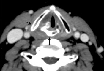
Three months later, a CT scan showed sclerotic changes involving the right side of the arytenoid, thyroid and cricoid cartilages, with mucosal oedema but no evidence of residual tumour (Figure 1). The patient complained of severe odynophagia and reflex otalgia, requiring analgesia. Endoscopic examination showed mobility of the right hemilarynx and hypopharyngeal mucosal oedema.
Axial computed tomography scan of case two, three months after chemoradiotherapy, showing sclerotic changes involving the right side of the arytenoid, thyroid and cricoid cartilages (arrows). Mucosal oedema is present but there is no evidence of residual tumour.

As the symptoms persisted, an examination was carried out under general anaesthesia. Direct laryngoscopy showed ulceration with fibrinous deposits on the right wall of the pyriform sinus, and exposure of the thyroid and cricoid cartilages. Multiple biopsies were obtained from the ulcer margins, showing necrosis and inflammatory changes but no evidence of neoplasia.
A provisional diagnosis of chronic ulceration induced by radiotherapy was made, and the patient was given antibiotics (amoxicillin and clavulanic acid) and anti-inflammatory treatment for two months. His right pyriform sinus oedema and pain gradually improved.
Further assessments up until 10 months post-treatment continued to show a symptomatic improvement, with no evidence of tumour recurrence.
However, 2 weeks after his 10-month follow-up appointment, and without any warning signs or symptoms, the patient suffered a sudden, massive oral haemorrhage at home and died.
Post-mortem examination showed ulceration on the lateral pharyngeal wall with carotid axis rupture. No tumoural infiltration was detected.
Discussion
We describe two patients with pharyngolaryngeal carcinoma who were treated with chemoradiotherapy and who later presented with a residual ulcer on the lateral pharyngeal wall. Surgical examination and biopsies of these ulcers excluded neoplastic infiltration, and conservative treatment was pursued. However, both patients died due to carotid blow-out syndrome.
Radiotherapy is an effective treatment for head and neck carcinoma. However, its side effects include erythema, erosion and ulceration of skin and mucosa, and bone and cartilage necrosis in the most severe cases.Reference Maruyama, Arai, Hoshida, Yoneda, Furukawa and Yoshizaki 9 , Reference Okamura, Kamiyama, Takiguchi, Kimizuka, Ishikawa and Kishimoto 10 Radiotherapy can cause vascular damage via obliteration of the vasa vasorum, premature atherosclerosis, adventitial fibrosis and arterial wall thinning. It also causes fragmentation of elastic fibres in the tunica media, as well as sub-endothelial oedema and vacuolisation.Reference Roh, Suh, Kim, Lee, Choi and Choi 1 , Reference Maruyama, Arai, Hoshida, Yoneda, Furukawa and Yoshizaki 9 – Reference Luo, Teng and Chang 11
Carotid blow-out syndrome is a major complication of head and neck surgery. Risk factors include healing problems with wound dehiscence, cutaneous flap necrosis or pharyngocutaneous fistula, as well as the presence of foreign bodies such as tracheostomy cannulas or nasogastric tubes.Reference Roh, Suh, Kim, Lee, Choi and Choi 1 , Reference Koutsimpelas, Pitton, Külkens, Lippert and Mann 4 – Reference Lesley, Chaloupka, Weigele, Mangla and Dogar 7 , Reference Okamura, Kamiyama, Takiguchi, Kimizuka, Ishikawa and Kishimoto 10 , Reference Luo, Teng and Chang 11
Previous radiotherapy is a predisposing factor for carotid blow-out syndrome. It favours neck wound complications associated with vascular rupture and reduces arterial wall resistance.Reference Roh, Suh, Kim, Lee, Choi and Choi 1 , Reference Upile, Triaridis, Kirkland, Archer, Searle and Irving 5 , Reference Lesley, Chaloupka, Weigele, Mangla and Dogar 7 , Reference Temam, Pape, Janot, Wibault, Julieron and Lusinchi 8 According to Maran et al., radiotherapy increases the risk of carotid blow-out syndrome in head and neck carcinoma patients sevenfold.Reference Maran, Amin and Wilson 12 Another risk factor related to carotid blow-out syndrome is tumour infiltration of the vascular axis.Reference Roh, Suh, Kim, Lee, Choi and Choi 1 , Reference Koutsimpelas, Pitton, Külkens, Lippert and Mann 4
The singularity of our clinical cases resides in the absence of previous surgery or tumour recurrence as predisposing factors for vascular rupture. The only risk factor in our two patients was chronic ulceration on the lateral pharyngeal wall. As their symptoms improved and recurrence was ruled out, we decided to keep these patients under observation only. However, the ulceration progressed in both cases, reaching the vascular axis and precipitating carotid rupture.
Cases of internal carotid rupture secondary to osteoradionecrosis have been described in the absence of surgical treatment or tumour recurrence in patients with nasopharyngeal carcinoma treated with radiotherapy.Reference Lam, Abdullah, Wormald and Van Hasselt 13 , Reference Cheng, Lee, Chiang, Ho and Kuo 14 However, to our knowledge there have been no previously reported cases of delayed carotid blow-out syndrome at cervical level following chemoradiotherapy in patients with laryngeal or hypopharyngeal carcinoma in the absence of the usual risk factors.
Angiographic studies would probably have led us to approach these cases in a more active manner. Such studies may have revealed the threat of rupture by showing local swelling and/or irregular, focal narrowing of the affected artery.Reference Luo, Teng and Chang 11 However, these angiographic changes can be subtle or absent.
In the light of our experience, we believe that, in the presence of radiogenic ulceration in areas at risk of this type of vascular accident, an active approach is advisable to prevent complications. After ruling out tumour recurrence by endoscopy and biopsy, we believe that a tissue implant between the vascular axis and the pharynx could be justified. Pathak et al. described the interposition of a sternocleidomastoid muscle flap between the pharynx and the vascular axis in an attempt to prevent possible carotid blow-out syndrome in patients treated with total laryngectomy and neck dissection.Reference Pathak, Viallet and Nason 15
In cases of lateral pharyngeal wall ulceration as described above, we propose necrotic tissue debridement and reconstruction with a vascularised flap. Reconstruction options include local flaps (as described by Pathak et al.), regional flaps (e.g. a pectoralis muscle flap) and microvascularised free flaps.Reference Pathak, Viallet and Nason 15 The aims of surgical intervention would be to reconstruct the pharyngeal defect and protect the vascular axis.
-
• Carotid blow-out syndrome usually occurs post-operatively or due to tumoural vascular axis compromise
-
• Post-chemoradiotherapy pharyngolaryngeal cancer patients with non-tumoural, lateral, hypopharyngeal ulceration are at risk
-
• In such patients, the vascular axis should be protected
Several treatment approaches have been recommended for carotid blow-out syndrome, depending on its stage. In cases of threatened rupture, various authors have recommended: direct reconstruction with grafts; resection and anastomosis; aneurysmography; and carotid stents.Reference Koutsimpelas, Pitton, Külkens, Lippert and Mann 4 , Reference Chang, Lirng, Luo, Wang, Wu and Guo 6 , Reference Lesley, Chaloupka, Weigele, Mangla and Dogar 7
Management is more complex in acute cases due to haemodynamic instability, hypotension, cerebral ischaemia, and disseminated intravascular coagulopathy secondary to massive blood loss. In these cases, surgical revascularisation is not usually possible due to the emergency setting, poor tissue quality and haemodynamic instability. Treatment is usually limited to arterial ligature, which may result in neurological complications and spread of necrosis.Reference Roh, Suh, Kim, Lee, Choi and Choi 1 , Reference Koutsimpelas, Pitton, Külkens, Lippert and Mann 4 , Reference Chang, Lirng, Luo, Wang, Wu and Guo 6
Unfortunately, such treatment was not possible in our two cases. One patient died suddenly at home, while the other had a self-limited bleeding episode but suffered a massive haemorrhage on arrival at our centre, leaving no chance for treatment.