Introduction
The overall burden of stroke is well known. It is the third commonest cause of death in the United Kingdom,1 and the commonest cause of severe acquired physical disability in old age. The incidence of stroke is approximately 1–2 per 1000 population per year in the United KingdomReference Rothwell, Coull and Giles2 and that approximates to a ‘stroke a day’ in most typical district general hospitals. As such it is something all who take part in emergency medicine should know about, particularly geriatricians, as stroke becomes increasingly common with old age. In the last decade, developments on many fronts have markedly changed the way in which we approach the diagnosis and subsequent management of stroke.
The aim of this review is to give an overview of these developments, from the most recent evidence available. It is important to bear in mind that as the population ages, stroke will be seen more frequently in frailer older people. Treatments that appear beneficial for a younger, more robust patient may have a different balance of risk and benefit in the frailer older person, and this must always be borne in mind when applying the available evidence.
Making the diagnosis
It has become increasingly important to make the diagnosis of stroke as quickly, but also as accurately, as possible. If a person presents within the first few hours of their symptoms, it opens up the possibility of using reperfusion strategies that could minimize or prevent permanent damage. Even if those symptoms resolve, it enables secondary prevention strategies to be commenced quickly and effectively. Making the diagnosis must involve taking an accurate history, especially around timing of onset of symptoms, performing a rapid but thorough clinical examination and performing neuroimaging. As well as making a positive diagnosis of stroke, this enables the identification of stroke mimics such as tumour or infection, which can represent up to 20% of patients presenting with stroke symptoms.Reference Libman, Wirkowski, Alvir and Rao3 If symptoms have completely resolved at 24 hours, the patient is classified as having a transient ischaemic attack (TIA). If symptoms persist, the diagnosis is one of stroke. TIA and ischaemic stroke are essentially the same diseaseReference Koudstaal, van Gijn, Frenken, Hijdra, Lodder and Vermeulen4 and as such, treatment such as secondary prevention will apply equally to TIA and ischaemic stroke.
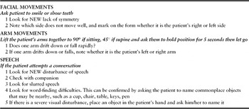
Simple screening procedures have been developed to raise public awareness of stroke symptoms. One of the easiest and most effective is the FAST test. Adapted from the Cinncinati pre-hospital stroke scale,Reference Kothari, Pancioli, Liu, Brott and Broderick5 it consists of three simple steps (Table 1). Paramedics using the scale achieved an accuracy of stroke diagnosis of 79%.Reference Harbison, Hossain and Jenkinson6 As well as improving speed of delivery to hospital by paramedics, it has been the basis of public health campaigns to help patients recognize the symptoms of stroke and contact emergency services earlier. Because of such strategies, patients with ‘acute neurovascular syndrome’ are being seen within the first few hours of symptoms, which creates potential for hyperacute interventions that can radically alter the natural history of the event.
Instructions for use of Face Arm Speech Test (adapted from Harbison et al.Reference Harbison, Hossain and Jenkinson6)

Brain imaging
Scanning patients with stroke quickly (immediately or within 24 hours), compared with any later time, confers the highest number of quality-adjusted life years (QALYs).Reference Wardlaw, Seymour and Cairns7 This is because early accurate diagnosis avoids needless investigations and allows appropriate management to be started immediately, which includes the imparting of information to patients and their relatives. For the majority of patients, the main question still to be determined by imaging is whether the stroke is due to primary intracerebral haemorrhage (PICH) or ischaemic infarction, as it is not possible to distinguish reliably between them on clinical grounds alone. Computerized tomography (CT) fulfils this role quickly and effectively. Acute haemorrhage on CT shows up as hyperdense (white) almost immediately from onset of stroke symptoms but, as haemorrhage is broken down, it gradually becomes isodense, then hypodense and indistinguishable from ischaemic infarction. This can occur within a matter of days for minor haemorrhage.Reference Wardlaw, Keir and Dennis8 CT will identify those with even minor PICH and occasionally identify patients with subdural and subarachnoid haemorrhage, as well as identify early signs of cerebral ischaemia or infarction.
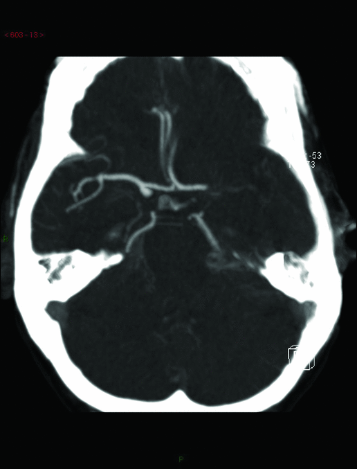
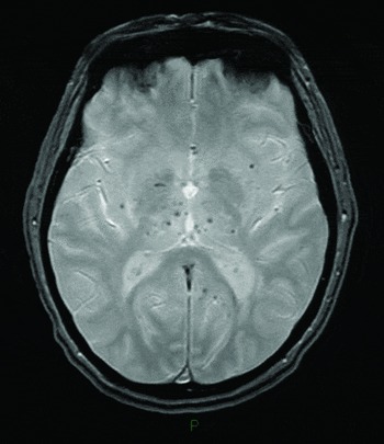
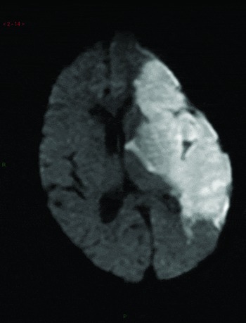
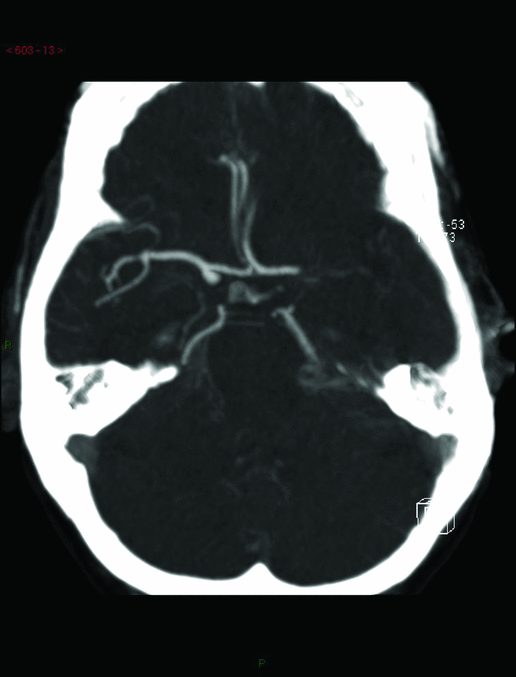
There are other imaging modalities that can provide information on the nature of a stroke. The type of imaging used to characterize a stroke may be determined simply by local availability, but if there is a choice available, to make best use of the imaging, it is important to think of the nature of the information needed and the safety of the patient. Magnetic resonance imaging (MRI) takes longer than CT, so for a patient with a large stroke who may be at risk of hypoxia, especially when lying flat on their back, the most rapid imaging procedure may be favoured. Angiography using either CT or MRI is less invasive than traditional intra-arterial angiography and can provide valuable anatomical information (Figure 1). Acute haemorrhage on MRI is not as easy to identify, but certain MR sequences, particularly gradient-echo (also known as T2*, Figure 2) are extremely sensitive to the breakdown products of haemoglobin and thus important for identifying previous haemorrhage.Reference Dimigen, Keir, Dennis and Wardlaw9 Identifying ischaemic stroke on imaging is easier with diffusion-weighted MRI (DWI, Figure 3), a technique that detects very small movements of water in and out of ischaemic brain cells.Reference Keir, Wardlaw, Bastin and Dennis10 There is also much interest in perfusion imaging, both with CT and MRI, used in combination with DWI. The aim is to identify potentially salvageable brain tissue (the ‘penumbra’) that may be rescued using reperfusion strategies.Reference Hacke, Albers and Al-Rawi11–Reference Davis, Donnan and Parsons13 However, there are still a number of unanswered questions that have to be addressed before diffusion-perfusion studies become the imaging modality of choice for management of acute reperfusion.Reference Rivers, Wardlaw and Armitage14
CT angiogram demonstrating occluded left middle cerebral artery (MCA) (image courtesy of Dr AJ Farrell, SINAPSE Collaboration, Edinburgh University)
MRI gradient-echo sequence demonstrating scattered microbleeds (image courtesy of Dr AJ Farrell, SINAPSE Collaboration, Edinburgh University)

MRI diffusion-weighted imaging sequence demonstrating ischaemia in the left MCA territory (image courtesy of Dr AJ Farrell, SINAPSE Collaboration, Edinburgh University)

Management of acute stroke
Once diagnosis of acute neurovascular syndrome has been established, treatment will depend upon the underlying pathology and will differ depending upon whether the stroke was ischaemic or haemorrhagic (primary intracerebral haemorrhage, PICH). The underlying mechanism of ischaemic stroke is presumed to be thrombosis arising from: small perforating vessels (microatheroma); larger intracranial vessels (e.g. a ruptured plaque in the basilar artery); or an embolic event from thrombosis elsewhere (e.g. from the carotid, vertebral arteries and arch of the aorta, or embolism from the heart or beyond). Rarer causes of ischaemia include: thrombi arising from the venous system passing into the cerebral circulation via a patent foramen ovale in the heart, and ischaemia due to cerebral venous thrombosis.
Acute reperfusion treatment
Thrombolysis with intravenous recombinant tissue plasminogen activator (rt-PA) is now recommended for use in selected patients with acute ischaemic stroke.15 Evidence from randomized controlled trials comparing the outcome of patients with acute ischaemic stroke treated with rt-PA (mostly administered up to 6 h) versus placebo have shown a significant reduction in the odds of being dead or dependent (modified Rankin 3 to 6) at 3–6 months. This is despite the increased risk of intracranial haemorrhage. Treatment within 3 h of stroke appeared most effective.Reference Wardlaw, Murray, Berge, del Zoppo, Yamaguchi and Berge16 This benefit was confirmed in the SITS-MOST study: a pan-European post-marketing surveillance study required by the European Medicines Agency following the licensing of rt-PA, confirming it as an effective therapy for selected patients with acute ischaemic stroke.Reference Wahlgren, Ahmed and Davolos17 In specialized centres, endovascular clot removal, either by lysis or mechanical removal, is favoured for selected patients. Although favourable results have been seen, evidence in the form of randomized trials is lacking. Questions remain of its benefit over intravenous rt-PA, which will usually be able to be delivered more quickly, and the safety of the various devices that are being used for clot retrieval.
With regard to intravenous rt-PA, important issues still need to be addressed: whether thrombolysis is as safe in the older patient (only 0.5% of patients that have been randomized into trials were over 80), whether any stroke subtype benefits more than another, and what the latest time window is in which treatment is still beneficial. ECASS-III explored the safety of intravenous rt-PA between 3 and 4.5 h of stroke onset. It found that patients who were given rt-PA had a more favourable outcome (Rankin 0 to 1) without a significant difference in mortality, although the benefit was less than that seen if treatment was within 3 h. Age exclusions were still applied, however.Reference Hacke, Kaste and Bluhmki18 IST-3 is an ongoing international trial, without age exclusions, seeking to determine whether a wider range of patients may benefit from thrombolysis up to 6 h post-stroke onset. To date, it has randomized over 800 patients over the age of 80 and will make a major contribution to the evidence base for this group.Reference Sandercock, Lindley and Wardlaw19
Currently, licensed indications limit treatment to those who can be assessed, scanned and start treatment under the supervision of a specialized team, within 3 h of stroke onset. To deliver thrombolysis at all, it is clear that a well-organized process has to be in place. Central to this is a dedicated team with enough expertise available 24 h per day. In smaller hospitals, this is not always achievable and time pressures governing treatment delivery preclude transfer to a specialist centre. One solution to this is telemedicine: patients with a stroke and their imaging results can be linked to a specialist using video or teleconferencing. This enables the expert to assess the situation and give advice remotely.Reference Tatlisumak, Soinila and Kaste20
Anticoagulation
Meta-analysis of over 23,000 patients randomized into 24 trials of early anticoagulation in ischaemic stroke has concluded that there is no evidence of benefit in any subgroup of ischaemic stroke.Reference Counsell and Kamal21 Haemorrhagic transformation is a natural phenomenon after stroke. Haematoma formation occurs in about 1% of untreated ischaemic stroke patients and lesser bleeding seen on imaging occurs in about 5–8% of patients.Reference Lindley, Wardlaw, Sandercock, Rimdusid, Lewis and Signorini22 The use of anticoagulants will increase this baseline rate.
Anti-platelet drugs
Routine aspirin, started with an initial dose of at least 300 mg within 48 h of stroke onset reduces the rate of early recurrent stroke, by around 1%. Modest though this benefit may be, the public health impact is considerable. Routine aspirin given to 100,000 patients will prevent around 1000 patients dying or becoming disabled from stroke.Reference Chen, Sandercock, Pan, Counsell, Collins and Liu23
Neurosurgery in the treatment of acute stroke
Perhaps the most well-established role for surgery is the use of an intraventricular shunt to prevent or alleviate acute hydrocephalus, caused by an intraventricular bleed, or a posterior fossa infarct or haematoma. Surgery to manage the haematoma of a PICH directly has proven more challenging. The STICH trial of early surgical intervention in PICH was the largest of its kind and successfully recruited 1033 patients who were randomized to early surgery or initial conservative treatment. Of 468 patients randomized to early surgery, 26% had a favourable outcome compared with 24% of the 496 randomized to initial conservative treatment.Reference Mendelow, Gregsonow and Fernandes24 This overall result was not significant, but subgroup analysis demonstrated that patients without intraventricular haemorrhage or hydrocephalus were more likely to achieve a favourable outcome. This has provided the impetus for STICH II, which is recruiting patients within 48 h of onset of symptoms with superficial haematomas within a specific volume range.25
Another surgical intervention whose role has become more established recently is that of hemicraniectomy. Removal of a sizeable part of the skull can provide lifesaving cerebral decompression in patients with massive cerebral oedema, but can leave them with severe disability, making many stroke physicians wary of undertaking such a major procedure. Three European trials (DECIMAL, DESTINY and HAMLETReference Vahedi, Vicaut and Mateo26–Reference Hofmeijer, Kappelle and Algra28) have randomized patients under 55 or 60 years of age with life-threatening cerebral oedema to hemicraniectomy within 48 h of symptoms onset or best medical management. These trials, although not identical in design, had enough similarities to allow them to pool their results. They found that hemicraniectomy reduced the absolute risk of mortality by 51%, although survivors were left with disability that could be severe.Reference Vahedi, Hofmeijer and Vicaut29 Hemicraniectomy does have its place in the management of the younger patient with severe stroke, but should not be undertaken lightly; consent must be informed and there is not the evidence available to suggest that it is an effective treatment for the older patient. Indeed, the problem of cerebral compression may be attenuated in the more elderly population as the age-associated cerebral atrophy provides more space for the hemisphere to expand in the closed box of the skull.
Stroke units
Patients with mild, moderate or severe stroke should be managed on a stroke unit. The Cochrane systematic review of 31 trials involving over 6936 patients demonstrated that, at final (median 1 year) follow-up, stroke unit care reduced odds of death, odds of death and disability, and odds of death or institutionalized care. Outcomes were independent of age, sex and stroke severity and appeared better in stroke units based on a discrete ward.30
Physiological parameters and neuroprotection
Laboratory and clinical studies have demonstrated that the brain damage that happens after stroke occurs over a period of time rather than instantly. This raises the issue of whether any interventions within this time period can protect any vulnerable, but potentially salvageable, brain. Trials of drugs for ‘neuroprotection’ have been uniformly disappointing, although poor trial design and lack of power mean that a small but real absolute benefit may have been missed. A number of the physiological changes seen in acute stroke are known to have potentially deleterious effects on vulnerable brain, so close attention should be paid to these parameters in the hope that a degree of neuroprotection can be provided.
Temperature
Fever or higher body temperature is associated with worse outcome after stroke or neurological injury.Reference Greer, Funk, Reaven, Ouzounelli and Uman31 A 1 degree increase from admission body temperature has been shown to independently predict a 30% relative increase in long-term mortality.Reference Kammersgaard, Jorgensen, Rungby, Reith, Nakayama and Weber32 However, the Cochrane systematic review of trials of therapeutic cooling in stroke (423 participants) found no effect on death or dependency and a non-significant increase in infections.Reference Den Hertog, van der Worp, Tseng and Dippel33 Further trials are ongoing.
Glycaemic control
Hyperglycaemia is present in around 40% of patients presenting with acute stroke, most of whom remain hyperglycaemic during their in-patient stay. Hyperglycaemia is independently associated with an increased risk of death at 30 days, 1 year and 6 years after stroke. Hyperglycaemic patients have a longer in-patient stay.Reference Williams, Rotich, Qi, Fineberg, Espay and Bruno34 GIST-UK was a randomized controlled trial exploring whether close glycaemic control using variable-dose insulin (GKI) reduced death or disability at 90 days. Aiming for a sample size of 2355 patients, it was powered to detect a difference of 6% in the primary outcome of death at 90 days. The trial was stopped after 933 patients had been recruited due to slow enrolment and, on intention-to-treat analysis, a lack of difference between treatment and control.Reference Gray, Hildreth and Sandercock35
Oxygenation status
Hypoxia is a threat for patients with acute stroke for a number of reasons, including central disturbance of respiration, sleep apnoea, hypoventilation, left ventricular failure and pulmonary embolus.Reference Roffe, Sills, Wilde and Crome36 Although a trial of supplemental oxygen cast doubt on its merits (albeit without reaching levels of significance),Reference Ronning and Guldvog37 it would seem logical to provide supplemental oxygen in patients with hypoxia. Hyperbaric oxygen has also been used with acute stroke: meta-analysis of six trials of only 283 patients found no evidence of benefit, although the limited amount of data means that this could not be discounted.Reference Bennett, Wasiak, Schnabel, Kranke and French38
Hydration
Raised plasma osmolality on admission is associated with increased stroke mortality.Reference Bhalla, Sankaralingam, Dundas, Swaminathan, Wolfe and Rudd39 In spite of lack of evidence of the benefit of aggressive volume expansion or haemodilution,Reference Asplund40 it is appropriate that close attention is paid to hydration status and dehydration avoided.
Blood pressure
The issue of blood pressure control in acute stroke is a topic of much debate. Up to 40% of acute stroke patients will be taking anti-hypertensives at presentation, the majority of patients with stroke will be hypertensive during their acute presentation, and in many blood pressure will return to normal within a week.Reference Robinson and Potter41 Analysis of over 17,000 patients in IST1 found that both high and low blood pressures were independent prognostic factors for poor outcome.Reference Leonardi-Bee, Bath, Phillips and Sandercock42 One observational study found that a drop in blood pressure in acute stroke was also a poor prognostic factor.Reference Castillo, Leira, Garcia, Serena, Blanco and Davalos43 More recently, however, CHHIPS, a pilot study of 180 patients exploring the safety and feasibility of blood pressure lowering in acute stroke, suggests that active lowering of blood pressure in hypertensive patients in acute stroke may be beneficial. Using labetalol (oral or intravenous) or lisinopril (oral or sublingual), it found no increase in death or dependency in the treatment groups at 2 weeks, and a possible mortality benefit at 3 months.Reference Potter, Robinson and Ford44 ENOS is an ongoing randomized, controlled, parallel-group, single-blind study exploring blood pressure lowering with a GTN patch and whether to withhold or continue existing anti-hypertensive medication within the first 7 days of stroke. It has randomized over 1300 patients to date.45 COSSACS is another ongoing trial exploring stopping or continuing existing anti-hypertensive medication for the first 2 weeks following stroke.46
Prevention of venous thromboembolism
Deep vein thrombosis (DVT) and pulmonary embolism (PE) are common complications of stroke: those that are clinically recognized being the tip of the iceberg. Subclinical veno-thromboembolism has been found in around 40% of stroke patients, with an even higher prevalence seen in those with a lower Barthel index.Reference Kelly, Rudd, Lewis, Coshall, Moody and Hunt47 Heparin given subcutaneously reduces the risk of DVT in acute ischaemic stroke, but the excess of intracranial and extracranial haemorrhage seen largely offsets any benefit.Reference Counsell and Kamal21 In the UK, heparin is not currently recommended routinely for all patients as DVT prophylaxis, but in some countries it is used. A randomized trial exploring the merit of low molecular weight (LMW) heparin compared with unfractionated heparin found a reduction in DVT with LMW (10% versus 18%) and an increase in major extracranial bleeding.Reference Sherman, Albers and Bladin48 Aspirin has been shown to give a very small reduction in the risk of PE in the first 2 weeks after stroke.49 Graduated compression stockings have been shown to reduce the risk of PE in peri-operative patients,Reference Wells, Lensing and Hirsh50 but that population is obviously very different from those having had a stroke. The CLOTS 1 trial randomized 2518 patients with acute stroke to wearing of thigh-length graduated compression stockings or to no stockings. It found no significant reduction of risk of symptomatic or asymptomatic DVT and an increase in skin damage in patients wearing stockings.51 Further trials by the same collaborators are underway, looking at whether there is any benefit of intermittent pneumatic compression.52
Nutrition
The swallow is commonly affected in acute stroke, and stroke patients, even if they are able to swallow, commonly have mobility issues that prevent independence in eating or drinking. There has been debate about the best and safest way to maintain nutrition in stroke patients. For instance, is it better to maintain nutrition from the outset in someone with an unsafe swallow, or does that encourage aspiration? Should enteral feeding be via a nasogastric tube or a percutaneous feeding tube (PEG)? Should those patients able to swallow receive extra supplements to boost their nutritional status? The FOOD trial was designed to address these questions. It found that: for patients who were able to swallow safely, no difference was found between those taking sip feed supplements (120 ml three times a day) and those who did not,Reference Dennis, Lewis and Warlow53 there was no statistical difference between the policy of early or delayed enteral feeding for dysphagic patients in the first 7 days after stroke, although early nasogastric feeding was associated with a (non-significant) 6% lower death rate, and there was no statistical difference in outcome at 6 months between patients randomized to feeding via a nasogastric tube compared with those with a PEG.Reference Dennis, Lewis and Warlow54 The implications of these results are that routine food supplementation is not indicated. In the first week of stroke it may be reasonable to use either nasogastric feeding or to avoid tube feeding, depending upon the clinical status of the patient. However, if dysphagia is prolonged, a nasogastric feeding tube is preferred unless there are compelling practical reasons to use a percutaneous feeding tube (PEG). If oral feeding cannot be recommenced due to poor recovery, patients and their carers need to be consulted regarding the long-term pros and cons of PEG tube feeding.
Secondary prevention of stroke
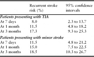
Patients presenting with TIA or minor stroke have a relatively high risk of early stroke (Table 2).Reference Coull, Lovett and Rothwell55 The benefits of early targeted secondary prevention were demonstrated in EXPRESS, a prospective two-phase trial exploring the effect of speeding up access to, and subsequent interventions from, a neurovascular clinic. This showed a reduction in risk of recurrent stroke at 90 days from 10.3% at baseline to 2.1% after urgent assessment, and immediate treatment strategies were implemented.Reference Rothwell, Giles and Chandratheva56 Therefore it is vital that patients with TIA or minor stroke are assessed as soon as possible. The major aspects of secondary prevention are discussed below.
Risk of recurrent stroke after TIA/minor stroke (adapted from Coull et al.Reference Coull, Lovett and Rothwell55)

Smoking
There should be a strong effort to stop smoking. Although there will never be an appropriate randomized controlled trial, the evidence from a large body of epidemiology suggests that half of all smokers will die prematurely, but giving up smoking will halve future risks of vascular events. These benefits probably apply to all ages and to all pathological subtypes of stroke.Reference Peto, Lopez, Boreham, Thun and Heath57
Blood pressure
Following both cerebral ischaemia and PICH, long-term blood pressure lowering is crucial. Over a wide range of blood pressures, a lower blood pressure is correspondingly associated with a lower risk of stroke for patients with or without a neurovascular history.Reference MacMahon, Peto and Cutler58, Reference Rodgers, MacMahon and Gamble59 Data from two trials in patients with stroke or TIA have also shown that blood pressure lowering is beneficial even in those with blood pressure in the classically ‘normal’ range. PROGRESS randomized 6105 patients to perindopril with or without the addition of the diuretic indapamide or placebo. It found a 28% relative risk reduction in stroke in the active treatment group (10% strokes versus 14%, with greater benefit seen in those taking combination therapy).60 HOPE randomized 9297 patients to ramipril or placebo and found that stroke risk was reduced by 32% with active treatment (3.4% strokes versus 4.9%).61 These risk reductions were found in both hypertensive and normotensive individuals. The strength and consistency of the evidence for blood pressure lowering suggests that the choice of agent is not important provided that safe and effective agents are used. However, in the older patient, as falls and syncope become more common, it may not always be possible to apply ideal blood pressure lowering strategies. It remains important, therefore, to consider all other aspects of secondary prevention.
Cholesterol
As with blood pressure, lowering blood cholesterol over a wide range of levels is associated with significant vascular risk reduction.Reference Manktelow and Potter62 The Heart Protection Study (20,536 patients) randomized patients between the ages of 40 and 80 to simvastatin at 40 mg or placebo, and found significant reductions in ischaemic stroke and all major vascular events (28 and 24%, respectively)63 in the treatment group. The effect of statins in the older person was specifically addressed by PROSPER, which randomized patients over the age of 75 years and found a significant reduction in cardiovascular risk, but no reduction in stroke.Reference Shepherd, Blauw and Murphy64
Carotid stenosis
Severe symptomatic carotid stenosis carries a high risk of stroke. In the ECST (European Carotid Surgery Trial), 30% of patients with recent stroke symptoms and a 90–99% stenosis on the ipsilateral side to the affected hemisphere had had a stroke within 3 years.65 Evidence from ECST and the North American Symptomatic Carotid Endarterectomy Trial,66 demonstrated that endarterectomy of recently symptomatic severe carotid stenosis almost completely abolishes the risk of ischaemic stroke ipsilateral to the operated artery over the next 2–3 years. Consideration for endarterectomy is not straightforward, however, as the operation itself carries a number of risks including stroke and as ECST demonstrated, many high-risk patients do not have a stroke. Indeed, the data showed that the unoperated risk rapidly attenuated within 2 years, and by 3 years it was the same as in patients with mild risk. It is therefore crucial for the Stroke Physician, when advising an individual considering endarterectomy, to know as much about what that individual's risk is both with and without an operation to enable a sensible estimate to be made about the immediate surgical risk compared with the longer term, unoperated risk. Previously, the risk figures calculated from the trials were that if the combined risk of surgery and angiography were 10%, and the unoperated risk at 2 years was 20%, and if successful surgery reduced that risk to near zero, then doing ten operations would cause one stroke and avoid two; net gain one stroke avoided. Although this gave some idea of the merit of endarterectomy, this crude calculation was less than adequate for the individual under consideration. Further meta-analysis using the ECST and NASCET pooled data (5893 patients), which used the same method of measurement of stenosis and definitions of outcome events, has refined these estimates. It found that age, sex and timing of surgery modified the efficacy of surgery. For patients with greater than 50% stenosis, benefits were greatest in men (number needed to treat to prevent one ipsilateral stroke in 5 years was 9, versus 36 for women), in the older patient (number needed to treat was 5 over the age of 75 years versus 18 for younger than 65 years) and for those undergoing surgery soon after the event (number needed to treat was 5 if operated on within 2 weeks of their last ischaemic event, versus 125 after more than 12 weeks).Reference Rothwell, Eliasziw, Gutnikov, Warlow and Barnett67 The importance of timing of surgery confirms that if one is going to operate, one should do it quickly, or not at all.
Anti-platelet drugs
Anti-platelet therapy is now firmly established for patients with ischaemic stroke.Reference Collaboration68 Over the last few years, the question of whether combinations of anti-platelet drugs have additional benefit has been explored in detail. ESPRIT randomized 2739 patients to aspirin (30–325 mg) with or without modified-release dipyridamole at 200 mg. It found a reduction in composite vascular end-points of 13% to 10% in the group of patients receiving both, equivalent to an absolute risk reduction of 1% per year. More people in the combined treatment arm discontinued trial medication, mainly due to headache (34.1 versus 13.5%).69 Clopidogrel, a thienopyridine derivative, has been shown to be as effective as aspirin in reducing further vascular events in people with vascular risk factors.70 A more recent study (PRoFESS, a randomized non-inferiority trial of over 20,000 patients) compared the combination of aspirin and modified-release dipyridamole with clopidogrel. It found no difference in recurrent stroke between the two treatment groups.Reference Diener, Sacco and Yusuf71 The combination of aspirin and clopidogrel has not been found to be generally helpful in the secondary prevention of stroke. MATCH (7599 patients) demonstrated that the combination of aspirin (75 mg) and clopidogrel (75 mg) compared with clopidogrel alone did not have any additional benefit in terms of reduction of combined vascular end-point. There was also a significant increase in life-threatening haemorrhage.Reference Diener, Bogousslavsky and Brass72 CHARISMA randomized over 15,000 patients with cardiovascular risk factors to aspirin and clopidogrel, or aspirin and placebo, and found no additional benefit with combination treatment.Reference Bhatt, Fox and Hacke73
Atrial fibrillation
Atrial fibrillation (AF) is common and carries an increased risk of stroke.Reference Wolf, Abbott and Kannel74 Warfarin is superior to aspirin in the prevention of stroke in patients with non-valvular AF, reducing the risk of stroke, myocardial infarction or vascular death by over one-third.Reference Aguilar and Hart75 This benefit comes with risk, however; treating 1000 patients for 1 year with warfarin rather than aspirin would prevent 23 strokes but cause an additional nine major haemorrhages.Reference Van Walraven, Hart and Singer76 Also, warfarin is associated with the inconvenience and expense of regular monitoring. The risk and inconvenience associated with warfarin has led to a perceived under-utilization of it in the management of AF, particularly in the older population. Studies looking more specifically at risk as well as benefit have helped inform decision-making strategies. One prospective study of the risk associated with warfarin in its first year of use in older people (average age 77 years) found higher rates of haemorrhage than had previously been found in younger cohorts.Reference Hylek, Evans-Molina, Shea, Henault and Regan77 Of note was that a third of patients over 80 were also on an anti-platelet drug. In BAFTA, 973 patients over the age of 75 (mean age 81) were randomized to dose-adjusted warfarin (INR 2.0 to 3.0) or aspirin, and it was found that in patients taking warfarin the risk of fatal or disabling stroke was reduced from 3.8 to 1.8% per year, with no increase in risk of haemorrhage.Reference Mant, Hobbs and Fletcher78 WASPO was a much smaller randomized open labelled trial comparing aspirin at 300 mg and dose-adjusted warfarin (INR 2.0–3.0) in patients aged between 80 and 90. It demonstrated more adverse events in the aspirin group.Reference Rash, Downes, Portner, Yeo, Morgan and Channer79 ACTIVE W (6706 patients) explored whether the combination of aspirin and clopidogrel was as good as warfarin in preventing stroke, myocardial infarction or vascular events in patients with AF, but was stopped early by its data monitoring committee due to evidence of a benefit in favour of warfarin (an increase in combined event end-point of 5.64 versus 3.93% was seen in the aspirin and clopidogrel group with no significant difference in bleeding rates).Reference Healey, Hart and Pogue80
The message from these studies is that warfarin can be effective and safe in the selected older patient but must be monitored very closely. ACTIVE A (7554 patients) explored the efficacy of aspirin and clopidogrel compared with aspirin alone in patients with AF for whom warfarin was unsuitable. It found that, compared with aspirin alone, aspirin and clopidogrel significantly reduced the rate of major vascular events (6.8 versus 7.6% per year), but was associated with an increased bleeding risk (2.0 versus 1.3% per year).81 Dabigatran is a new oral direct thrombin inhibitor that does not require regular blood test monitoring. In a large randomized non-inferiority trial, 18,113 patients who had atrial fibrillation and a risk of stroke received fixed doses of dabigatran (110 mg or 150 mg twice daily, in a blinded fashion) or adjusted-dose warfarin (unblinded). The median duration of follow-up was 2 years and the primary outcome was stroke or systemic embolism. Rates of the primary outcome were 1.69% per year in the warfarin group, compared with 1.53% per year in the group that received lower dose dabigatran (P < 0.001 for non-inferiority) and 1.11% per year in the group that received higher dose dabigatran (P < 0.001 for superiority). Rates of haemorrhagic stroke were lower on both doses of dabigatran compared with warfarin. Rates of all major bleeding were lower with low-dose dabigatran and similar to those of warfarin with higher dose dabigatran. There was no difference in mortality.Reference Connolly, Ezekowitz and Yusuf82 Although dabigatran is as yet unlicensed for the prevention of stroke in AF, it is an exciting development and may represent a major breakthrough in the management of this condition.
Anticoagulation in sinus rhythm
The benefits of warfarin in AF have not been replicated in patients with sinus rhythm. After an earlier trial using warfarin with an INR range of 3 to 4.5 was stopped at the first interim analysis due to an excess of fatal intracranial bleeds (SPIRIT),83 a further trial was performed randomizing 568 patients to lower dose warfarin (INR 2 to 3) or aspirin (ESPRIT). The primary end-point was a composite of vascular outcomes as well as major bleeding complications. It was also stopped early when it was determined that no difference in primary outcome would be found. A trend to benefit in ischaemic stroke risk reduction was offset by an increase in bleeding complications.84
Conclusion
There has been continued progress in many aspects of stroke medicine in the past 5 years since our last review, although not necessarily in the areas we predicted. Hyperacute treatment of stroke is becoming embedded in the framework of emergency medical admissions. Despite major advances in imaging technology, we feel it has yet to fulfil its potential in the risk stratification of acute stroke. Our knowledge of the efficacy of the drugs we use is becoming evermore evidence-based and one welcome development is the increasing inclusion of older patients in randomized controlled trials, making the evidence increasingly relevant to the population most at risk.