Introduction
Cardiac tamponade is a life-threatening clinical condition characterised by pericardial fluid accumulation, increasing intrapericardial pressure leading to impaired ventricular diastolic filling and reduced cardiac output. Although relatively rare in the paediatric population compared to adults, cardiac tamponade in children requires prompt recognition and intervention due to the limited physiological reserve and potential for rapid haemodynamic deterioration. Reference Appleton, Gillam and Koulogiannis1
The aetiology of cardiac tamponade in children is diverse and includes infections, malignancies, autoimmune disorders, postoperative complications, and idiopathic causes. The standard treatment for cardiac tamponade is usually pericardiocentesis, a procedure that is frequently emergent and often a life-saving intervention. Reference Bodson, Bouferrache and Vieillard-Baron2
Despite its clinical significance, data on the aetiologies and demographic characteristics of cardiac tamponade in the paediatric population remain limited. To address this gap, we retrospectively analysed paediatric cases who presented to the emergency department or were referred to our clinic and were subsequently diagnosed with cardiac tamponade requiring percutaneous pericardiocentesis as the initial therapeutic intervention.
Materials and methods
We retrospectively analysed 252 paediatric patients from neonatal age through 18 years of age who were either diagnosed with cardiac tamponade in our clinic or were referred to our hospital for urgent intervention, and subsequently underwent emergency percutaneous pericardiocentesis between November 2003 and May 2025.
All patients were diagnosed with cardiac tamponade based on transthoracic echocardiographic findings. Echocardiographic assessment included evaluation of effusion volume and evidence of haemodynamic compromise. The diagnostic criteria for cardiac tamponade included early diastolic collapse of the right ventricle, compression of the right ventricular outflow tract, late diastolic collapse of the right or left atrium, and absence of respiratory variation in the diameter of the inferior vena cava.
Seven etiologic categories were defined based on previous literature and the availability of diagnostic and procedural codes. These included chronic kidney disease, haematologic malignancies, solid organ tumours, rheumatologic diseases, infectious aetiologies, Down syndrome, other genetic syndromes, and miscellaneous causes. In addition to these categories, we specifically classified patients with primary cardiac disease as a separate group. Based on echocardiographic findings, these patients were further subdivided into five cardiac subgroups: acyanotic CHD, cyanotic CHD, perimyocarditis without structural heart anomalies, cardiomyopathies, and other cardiac conditions. In addition, patients who had undergone cardiac surgery or catheterisation within the last two months were evaluated as a distinct subgroup according to their primary cardiac diagnoses. All patients were stratified into five age groups for analysis: neonates (0–28 days), infants (29 days to <1 year), preschool-aged children (1–5 years), school-aged children (6–12 years), and adolescents (13–18 years).
Statistical analysis
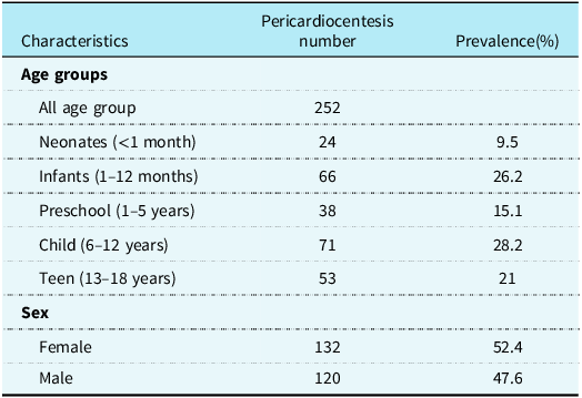
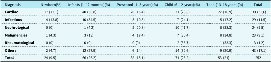
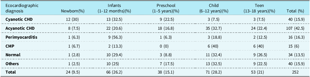
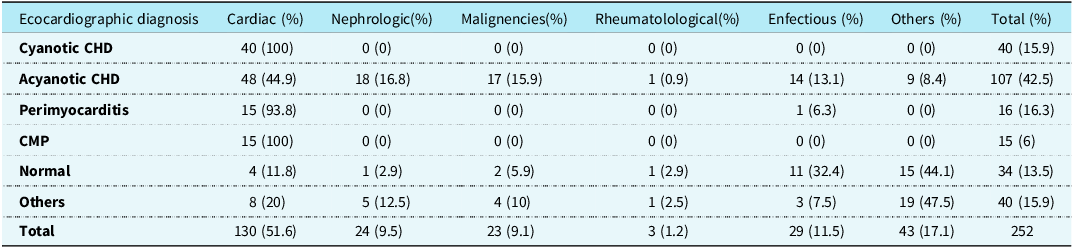
All data were analysed using IBM SPSS Statistics for Windows, Version 21.0 (IBM Corp., Armonk, NY, USA). Descriptive statistics were used to summarise patient demographics, clinical characteristics, and etiologic distributions. Categorical variables, including age groups, sex, underlying diagnoses, and clinical interventions, were expressed as frequencies and percentages and presented in tabular form (Tables 1–5).
Demographic characteristics of children who underwent pericardiocentesis

The age group distribution of the aetiology of pericardial tamponade

The age group distribution of echocardiographic diagnoses

The distribution of echocardiographic diagnosis with primary aetiologies

Distribution of echocardiographic diagnoses in patients with a history of cardiac catheterisation and/or surgery

Results
Over the 22-year study period, pericardiocentesis was performed on 252 paediatric patients who either presented to our emergency department or were referred for urgent pericardial drainage to our hospital. The age and sex distributions of the patients are summarised in Table 1.
The distribution of primary etiologic diagnosis according to patients’ age is presented in Table 2. Structural heart diseases (cyanotic, acyanotic heart diseases, and cardiomyopathies) were the highest in all age groups, with a prevalence of 51.6%. As expected, cardiac diseases were the most common during the neonatal period, and nephrological and infectious diseases were the second most common in older age groups after cardiac causes. Pericardiocentesis was most frequently performed in children aged 6–12 years (28.2%).
The distribution of patients’ echocardiographic diagnoses by age is presented in Table 3. Cardiomyopathies were more commonly identified in older children, whereas structural heart diseases, especially complex CHDs, were higher during the neonatal and infant periods.
Table 4 presents the distribution of primary etiologic diagnoses according to underlying heart diseases. Infectious aetiologies were the most common indication for pericardiocentesis after cardiac diseases (11.5%)
Table 5 classifies patients based on a history of catheterisation and/or cardiac surgery within the two months before presentation. It was found that cardiac tamponade occurred more frequently in patients with a history of cardiac surgery compared to those who had undergone catheterisation (12.3%).
Discussion
To the best of our knowledge, our study represents the most considerable single-centre experience of paediatric cardiac tamponade requiring percutaneous pericardiocentesis over 22 years. As our centre is the only referral hospital for paediatric cardiac intervention and surgery in the region, the number of patients was relatively high for a single centre. Our findings highlight the diverse etiologic spectrum, the age group distribution of aetiology, and the predominance of cardiac causes in children undergoing emergent intervention for cardiac tamponade.
Structural heart diseases, including cyanotic and acyanotic defects as well as cardiomyopathies, constituted the most common aetiology across all age groups, accounting for 51.6% of the cases in our study. Similar to our findings, Sasaki et al., in their 20-year multi-institutional analysis, reported that pericardial effusion and tamponade were more frequently observed in patients with structural heart disease and/or a history of cardiac surgery. Reference Sasaki, Prithvi, Matthew, Cole, Nao and Balagangadhar3
When classified by age distribution, the majority of pericardiocentesis procedures (49.2%) were performed in children aged 6–18 years. In the neonatal period, as expected, most cases were associated with cyanotic CHDs. Although pericarditis is an extremely rare and rapidly fatal disease in neonates, one of the successfully managed and published cases in our clinic was a 1-day-old newborn with haemorrhagic cardiac tamponade. Reference Başpinar, Kor and Bayraktaroğlu4 Figure 1 demonstrates marked pericardial effusion with right ventricular collapse, and Figure 2 shows a little contrast material given to the pericardial space.
Subcostal echocardiographic image of massive pericardial fluid. Note the compression of the right atrium ( arrow). ( RV;Right Ventricle, LV; Left Ventricle, RA;Right Atrium).

After of an introducer sheet into the pericardial space, small amounts of contrast material was injected. Chest radiograph showing large pericardial effusions and normal heart shifted toward the right side of the chest.

Patent ductus arteriosus stenting is a viable alternative procedure for managing patients with duct-dependent pulmonary circulation, offering several advantages, including shorter ICU and hospital stays, as well as a reduced need for mechanical ventilation. Reference Rimoni, Stern and Mainzer5 The procedure is technically challenging with a notable learning curve. As a referral centre, we perform numerous high-risk interventional procedures, including patent ductus arteriosus stenting. We have a considerable number of patients who underwent patent ductus arteriosus stenting; in fact, 31 of the patients who underwent pericardiocentesis had previously undergone patent ductus arteriosus stenting.
In developing countries, large pericardial effusions secondary to bacterial pericarditis still constitute the most common cause of cardiac tamponade. Reference Tutar, Yilmaz and Atalay6,Reference Akhtar, Malik, Anwar-ul-Haq and Atiq7 In our study, infectious causes were identified in 11% of the cases. This may be attributed to the fact that most of these studies are single-centre studies with limited patient diversity, and also due to the different study methodologies, which specifically exclude post-pericardiotomy patients. Reference Kühn, Peters, Marx and Breitbart8,Reference Başpinar, Karaaslan, Oran, Baysal and Yeniterzi9
As a referral centre, the large number of procedures performed may also be associated with some rare complications. In our data, a total of six patients developed traumatic pericardial tamponade. In two patients, iatrogenic haemorrhagic tamponade occurred due to ventricular perforation during pericardiocentesis, while in the remaining four, iatrogenic tamponade developed secondary to vascular and/or ventricular perforation during transcatheter interventions. One of these cases involved a one-year-old patient who experienced ventricular perforation during transcatheter patent ductus arteriosus closure; we successfully managed the case and reported it as the smallest case of transcatheter closure with an occluder in an iatrogenic perforation in a paediatric age group. Reference Temel, Selçuk and Başpınar10 The remaining iatrogenic cases occurred during transcatheter Atrial septal defect closure, balloon valvuloplasty for critical aortic stenosis, and patent ductus arteriosus stenting.
A total of 31 patients developed delayed tamponade within the first two months following cardiothoracic surgery. Recent studies have documented the prevalence of post-operative pericardial effusions ranging from 10–35% Reference Dalili, Zamani and Aarabi-Moghaddam11,Reference Cheung, Ho and Tang12 . However, only a small subset of these effusions is of a severity that requires pericardiocentesis and/or surgical intervention. The incidence of cardiac tamponade after cardiothoracic surgery ranges from 0.5 to 8.8%. Reference Price, Prout, Jaggar, Gibson and Pepper13,Reference Kuvin, Harati, Pandian, Bojar and Khabbaz14 In our study, 12.3% of the patients had a history of cardiothoracic surgery. This relatively high rate may be attributed to the fact that our institution serves as a referral centre for paediatric cardiac surgical cases.
A total of 30 patients in our cohort were syndromic, including 22 with Down syndrome. Except for two patients with Joubert syndrome and chronic kidney disease and a patient with congenital disorder of glycosylation Ia with recurrent pericardial effusion, all had undergone cardiac surgery and/or catheter-based interventions for CHD. Reference Işikay, Başpınar and Yılmaz15 We reported that our genetic anomalies are known to play a significant role in the development of CHDs. The comprehensive Baltimore–Washington Infant Study conducted in the mid-1980s also supported this association, demonstrating that approximately 25% of children with CHD had accompanying extracardiac malformations, and that a known genetic syndrome was identified in one-third of these cases. These findings highlight the pivotal role of genetic abnormalities in the etiopathogenesis of CHDs. Reference Greenwood, Rosenthal, Parisi, Fyler and Nadas16,Reference Ferencz, Rubin Judith and MCCARTER17
Although our study represents an extensive and comprehensive series, certain limitations should be acknowledged. The retrospective design carries the risk of data inconsistency or missing information. Furthermore, being conducted in a single centre may limit the generalizability of the findings to other healthcare systems or geographic populations.
Conclusion
Cardiac tamponade in children can occur due to various causes and poses a life-threatening condition if not treated promptly. The underlying aetiologies may vary depending on regional factors and the patient population of the medical centre; however, structural heart diseases have been reported as the leading cause in many studies; therefore, it is appropriate that patients with such conditions are prioritised for evaluation in this regard, particularly when presenting with relevant clinical symptoms.