INTRODUCTION
The infective cercaria of Schistosoma mansoni enters the mammalian host directly across the skin using secretions from its acetabular glands (Dorsey et al. Reference Dorsey, Cousin, Lewis and Stirewalt2002). Some days later it uses secretions from a tiny head gland, located within the muscular head capsule, to leave the skin via a blood or lymphatic vessel and begin its intravascular migration to the portal system (Crabtree and Wilson, Reference Crabtree and Wilson1985). The macroscopic adult worm resident in the bloodstream lacks any obvious physical barriers to protect its epithelial surfaces from immune attack. The tegument that covers the body and the gastrodermis lining the gut are both syncytia, bounded by membranes that come into contact with host plasma and cells. Nonetheless, adult worms can persist in the host for decades, free from any detectable damage, an ability that must depend on the molecular architecture of these surfaces. The blind-ending schistosome gut is the site for extracellular digestion of blood meals, with the residues expelled into the bloodstream. Finally schistosome eggs, deposited by females in the wall of the intestine (and accidently embolising in the liver), must first develop to maturity over approximately 5–7 days (Michaels and Prata, Reference Michaels and Prata1968). Only then do they begin secretion of proteins (Ashton et al. Reference Ashton, Harrop, Shah and Wilson2001b), presumably to aid escape, and pass through the gut tissues to the lumen to continue the life cycle.
A problem for researchers exploring the composition of these parasite-host interfaces has been how to identify the protein and glycan constituents. Only limited amounts of the larval secretions can be recovered for conventional biochemical anaylsis; nevertheless, several serine proteases, termed elastases, were cloned and characterized (Salter et al. Reference Salter, Choe, Albrecht, Franklin, Lim, Craik and McKerrow2002). Methods were developed in the early 1980s to isolate the tegument and enrich its membrane proteins but the identity of constituents remained elusive, much of our early knowledge being gleaned from the targets of monoclonal antibodies. Unfortunately, the supposed surface localization of many of these tegument proteins has subsequently proved erroneous, in part due to a tendency to fix and permeabilise worms before reacting with antibody. A number of gut proteases were identified by conventional biochemistry and cloning of their cDNAs (Caffrey et al. Reference Caffrey, McKerrow, Salter and Sajid2004). Circulating anodic and cathodic polysaccharide antigens were shown to comprise the glycocalyx lining the gut (Nash and Deelder, Reference Nash and Deelder1985) and subsequently developed as the basis for diagnostic tests (Deelder et al. Reference Deelder, Qian, Kremsner, Acosta, Rabello, Enyong, Simarro, van Etten, Krijger and Rotmans1994), at least as sensitive as the conventional faecal smear (Wilson et al. Reference Wilson, van Dam, Kariuki, Farah, Deelder and Coulson2006).
Approximately one decade ago the advent of proteomic techniques dramatically improved the possibilities for identification of parasite proteins (Ashton et al. Reference Ashton, Curwen and Wilson2001a). The marketing of immobilised pH gradient gels for first dimension separations made 2D electrophoresis highly reproducible, while methods for estimating spot density together with gel comparison software made quantitation of soluble proteins possible. The more intractable membrane proteins were tackled by liquid chromatographic separations of whole proteins or their constituent tryptic peptides (Wilson et al. Reference Wilson, Ashton, Braschi, Dillon, Berriman and Ivens2007). Latterly, quantitation has also been applied to the insoluble membrane protein preparations using the QconCAT technique (Castro-Borges et al. Reference Castro-Borges, Simpson, Dowle, Curwen, Thomas-Oates, Beynon and Wilson2011b). The development of MALDI ToF/ToF and electrospray mass spectrometers, powerful search engines such as MASCOT, and comprehensive sequence databases simplified the identification of proteins in a mixture. For schistosomes, the first ten years of cDNA sequencing produced about 10 000 sequences, but with much redundancy and <30 secreted or membrane proteins. Development of the ORESTES method that compensated for unequal message abundance (Dias Neto et al. Reference Dias Neto, Harrop, Correa-Oliveira, Wilson, Pena and Simpson1997), permitted the generation of >120 000 cDNA sequences across six life cycle stages by a Brasilian consortium (Verjovski-Almeida et al. Reference Verjovski-Almeida, DeMarco, Martins, Guimaraes, Ojopi, Paquola, Piazza, Nishiyama, Kitajima, Adamson, Ashton, Bonaldo, Coulson, Dillon, Farias, Gregorio, Ho, Leite, Malaquias, Marques, Miyasato, Nascimento, Ohlweiler, Reis, Ribeiro, Sa, Stukart, Soares, Gargioni, Kawano, Rodrigues, Madeira, Wilson, Menck, Setubal, Leite and Dias-Neto2003), giving the main impetus for schistosome proteomics. The authors claimed to sample ∼92% of the S. mansoni transcriptome, a level of coverage allowing representative proteomic profiles to be constructed that were not grossly biased towards abundant housekeeping proteins. Publication of the draft sequence of the S. mansoni genome followed in 2009 (Berriman et al. Reference Berriman, Haas, LoVerde, Wilson, Dillon, Cerqueira, Mashiyama, Al-Lazikani, Andrade, Ashton, Aslett, Bartholomeu, Blandin, Caffrey, Coghlan, Coulson, Day, Delcher, DeMarco, Djikeng, Eyre, Gamble, Ghedin, Gu, Hertz-Fowler, Hirai, Hirai, Houston, Ivens, Johnston, Lacerda, Macedo, McVeigh, Ning, Oliveira, Overington, Parkhill, Pertea, Pierce, Protasio, Quail, Rajandream, Rogers, Sajid, Salzberg, Stanke, Tivey, White, Williams, Wortman, Wu, Zamanian, Zerlotini, Fraser-Liggett, Barrell and El-Sayed2009), with ∼11 800 partial or complete coding sequences. This review details the progress made in characterizing the interface between S. mansoni and its mammalian host using proteomic techniques, since the first publication (Curwen et al. Reference Curwen, Ashton, Johnston and Wilson2004), and considers potential implications for diagnostics and vaccines.
THE SOLUBLE PROTEOME: WHAT DOES THE SCHISTOSOME CYTOSOL COMPRISE?
The first application of proteomics to schistosomes was a comparative anaylsis of the soluble proteins from different life cycle stages, using 2D electrophoresis for separation and quantitation and peptide mass fingerprinting for identification (Curwen et al. Reference Curwen, Ashton, Johnston and Wilson2004). Immunologists had devised a series of soluble antigen preparations derived from cercariae, lung schistosomula, adult worms and eggs (termed SCAP, SLAP, SWAP and SEA, respectively) for probing host humoral and cellular immune reactions to schistosome infection. The unstated assumption was that stage-specific aspects of the immune response could be characterized, an important consideration when vaccine candidates or pathogenic proteins were being sought. In fact, the study revealed that the four preparations had a remarkably similar composition, with more than 70% of their gel spots in common. Expressed in terms of total spot volume, i.e. protein content, the shared spots accounted for an even greater proportion than spot number alone indicates. Thus shared protein content ranged from 66% for SEA to 90·4% for SLAP, suggesting that very few proteins are specific to the lung schistosomulum, reinforcing previous ultrastrucural observations that revealed few unique features (Crabtree and Wilson, Reference Crabtree and Wilson1986). Put another way, approximately 71% of the protein content was shared between the four stages, on the basis of spot volume. Only the egg showed 9% of gel spots that were not shared with another stage. This may reflect both that the egg represents a completely different generation of the life cycle (ovum-miracidium-mother sporocyst), and that its role is to escape from the host, not to enter and persist in it.
Although the actual order of abundance differed between the stages, the common proteins comprised what might be called ‘housekeeping gene products’ involved in functional and structural processes to maintain parasite viability. Among them were glycolytic enzymes (aldolase, enolase, glyceraldehyde-3-phosphate dehydrogenase, phosphoglycerate kinase, triose phosphate isomerase), chaperones (14-3-3, cyclophilin, HSP70), cytoskeletal/motor proteins (actin, myosin light chain, dynein light chain, tropomyosin), calcium binding proteins (calreticulin, EF hand proteins Sm 20.8 and Sm 21.7, calponin) and anti-oxidants (glutathione-S-transferases 26 and 28, superoxide dismutase, thioredoxin). Collectively they represent a cross section of the cytosol of the ‘average’ schistosome cell and provide a useful benchmark against which to judge the enrichment of other preparations being analysed. Where we have encountered these proteins in later studies, we largely interpreted their presence as an indicator of cell damage leading to leakage of the cytosol. Sample preparation then becomes crucial to the generation of ‘sensible’ proteomic data, an important consideration given later claims for the composition of schistosome ‘secretomes’ (Cass et al. Reference Cass, Johnson, Califf, Xu, Hernandez, Stadecker, Yates and Williams2007; Liu et al. Reference Liu, Cui, Hu, Feng, Wang and Han2009). It should also be noted that, over the years, several of the ‘housekeeping’ proteins have been proposed as vaccine candidates in spite of their evident intracellular origin (Wilson and Coulson, Reference Wilson and Coulson2006).
WHAT DO INFECTIVE CERCARIAE AND MIGRATING SCHISTOSOMULA SECRETE?
The ideal schistosome vaccine would prevent parasite establishment by blocking the skin invasion process, always a tall order because the immune system would need to be on permanent ‘red alert’. Firstly, each incoming parasite does not provide much of a stimulus to the sensitized host and secondly, there is no time for an anamnestic response during the small window when it is susceptible to antibody-dependent cellular cytotoxicity (ADCC) in vitro (Wilson and Coulson, Reference Wilson and Coulson2006), features neglected by proponents of this effector mechanism. The cercaria uses the contents of its pre-and post-acetabular glands, comprising about 16% of body protein (Harrop and Wilson, Reference Harrop and Wilson1993), to penetrate into host skin. Irrespective of their status as vaccine candidates, characterization of these secretions in two proteomic studies has revealed hitherto unsuspected aspects of the infection process. Note that both investigated only the soluble fraction of the acetabular gland secretions. To date, no-one has characterized the pelletable and sticky components that probably comprise proteoglycans and/or glycoproteins, used by the cercaria to aid adhesion to the stratum corneum (Stirewalt, Reference Stirewalt1959).
The first proteomic analysis of material released when cercariae are transformed into schistosomula in vitro used skin lipid-induced secretion and shearing through a small bore syringe (needle) (Knudsen et al. Reference Knudsen, Medzihradszky, Lim, Hansell and McKerrow2005) as the stimuli. It highlighted three known elastase isoforms plus Sm16 ‘anti-inflammatory protein’ (SPO-1), in part due to the restricted scope provided by searching of the NCBInr database rather than the full transcriptome. However, the vast majority of proteins identified were the internal soluble ‘housekeeping proteins’ listed above, and also included several nuclear histones. To what extent this composition can be attributed to damage incurred during artificial transformation, e.g. by syringe-shearing, or is the result of holocrine secretion of the acetabular gland cell contents, is a matter of conjecture.
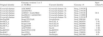
The second study of acetabular gland secretions (Curwen et al. Reference Curwen, Ashton, Sundaralingam and Wilson2006) used the method of tail detachment by vortexing of the suspension (Ramalho-Pinto et al. Reference Ramalho-Pinto, Gazzinelli, Howells, Mota-Santos, Figueiredo and Pellegrino1974), known to provide viable schistosomula for in vitro culture, and 70–87% maturation when these are delivered to the portal vasculature of naïve mice (as good as that for ex vivo schistosomula from the lungs, returned to mice; Harrop and Wilson, Reference Harrop and Wilson1993). Two-dimensional electrophoretic separation of the proteins released during the first three hours after transformation revealed a relatively simple pattern of 144 spots on the gel, versus 1314 spots in a comparable separation of soluble cercarial extract (SCAP). The 50 most abundant spots accounted for 79% of the protein content, with 54% of vesicular and 25% of cytosolic origin (Table 1). Collectively the elastase isoforms accounted for the largest proportion (34·4%) with a hitherto unsuspected invadolysin metalloprotease (12·8%) as the second most abundant protein. Also notable were three of the enigmatic SCP (sperm coat protein) domain-containing proteins now classified as SmVALs. The inference must be that these particular VALs exert their functions on the host during or shortly after entry to the skin. Similar venom allergen-like proteins have recently been reported in proteomic analyses of the secretory products of the nematodes Ancylostoma caninum (Mulvenna et al. Reference Mulvenna, Hamilton, Nagaraj, Smyth, Loukas and Gorman2009) and Heligmosomoides polygyrus (Hewitson et al. Reference Hewitson, Harcus, Murray, van Agtmaal, Filbey, Grainger, Bridgett, Blaxter, Ashton, Ashford, Curwen, Wilson, Dowle and Maizels2011). The most abundant cytosolic protein was a serine protease inhibitor (4·9%). The other principal proteins in descending order of concentration were cyclophillin, thioredoxin, glutathione-S-transferase 28, aldolase, triose phosphate isomerase and Sm14-fatty acid binding protein, all typical constituents of the soluble proteome (Curwen et al. Reference Curwen, Ashton, Johnston and Wilson2004); the holocrine secretion of the acetabular glands is the obvious explanation for their presence.
Major proteins in the cercarial acetabular gland secretions

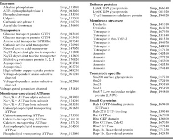
Membrane and membrane-associated tegumental proteins identified by proteomic analysis of the isolated tegument membranes

Data from Braschi et al., Reference Braschi, Borges and Wilson2006a, Reference Braschi, Curwen, Ashton, Verjovski-Almeida and Wilson2006b; Braschi & Wilson Reference Braschi and Wilson2006; Braschi and Wilson, unpublished.
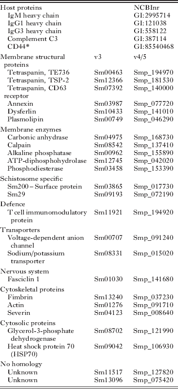
Proteins identified by external biotinylation of live worms
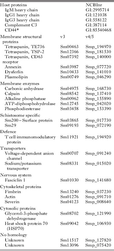
* CD44 was identified in an unfractionated sample not included in the publication
Data from Braschi and Wilson (2006), which used v3 of the genome assembly.
Annotations are updated to version 4/5 in the third column.
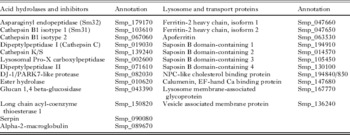
Proteins identified by analysis of worm vomitus

Data from Hall et al., Reference Hall, Braschi, Truscott, Mathieson, Cesari and Wilson2011
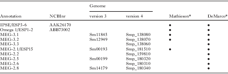
Secreted proteins identifed after in vitro culture of eggs
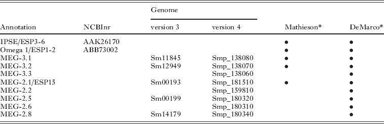
* Mathieson et al. 2010; DeMarco et al. 2010.
The secretions of the migrating schistosomula have proved more difficult to characterize, not least because of the small amounts of material released. However, leaving aside possible gut contributions, there are two sources for such material, the head gland, which persists up to the lung stage of migration, and the tegument, which possesses a unique inclusion, the homogeneous body (McLaren et al. Reference McLaren, Hockley, Goldring and Hammond1978). Biosynthetic labelling of cercariae in-snail prior to transformation, or of schistosomula in culture, using radiolabelled methionine established that schistosomula released a dominant protein of ∼ Mr 20 kDa into the external environment (Harrop and Wilson, Reference Harrop and Wilson1993). This protein appeared very relevant to protective immunity, in view of work on the radiation-attenuated cercarial vaccine that had revealed the lung-stage schistosomulum as the target of protective responses in immunised mice (reviewed by Coulson, Reference Coulson1997). However, its identity proved elusive in spite of attempts to clone it and only in 2010 was it identified by proteomics on 2D gel spots as a mixture of two proteins, MEG-3.1 and MEG-3.2, encoded by microexon genes (DeMarco et al. Reference DeMarco, Mathieson, Manuel, Dillon, Curwen, Ashton, Ivens, Berriman, Verjovski-Almeida and Wilson2010 – see below). Furthermore, gene expression was localized to the head gland of the cercaria/schistosomulum by whole mount in situ hybridisation (WISH), the only transcript so far identified from that tissue (DeMarco et al. Reference DeMarco, Mathieson, Manuel, Dillon, Curwen, Ashton, Ivens, Berriman, Verjovski-Almeida and Wilson2010).
It is worth recording, given the description of the adult tegument proteome below, that our attempts to characterize the surface of the lung schistosomulum have proved problematic. The surface does not detach easily, and even a small number of dead or damaged larvae in the medium thwarts the use of surface biotinylation or enzymatic shaving techniques. This is especially disappointing in view of the apparent role the tegument surface in priming the protective response in mice exposed to irradiated cercariae (Riengrojpitak et al. Reference Riengrojpitak, Anderson and Wilson1998). We have therefore had to rely on comparative transcriptome data (Dillon et al. Reference Dillon, Feltwell, Skelton, Ashton, Coulson, Quail, Nikolaidou-Katsaridou, Wilson and Ivens2006; Verjovski-Almeida et al. Reference Verjovski-Almeida, DeMarco, Martins, Guimaraes, Ojopi, Paquola, Piazza, Nishiyama, Kitajima, Adamson, Ashton, Bonaldo, Coulson, Dillon, Farias, Gregorio, Ho, Leite, Malaquias, Marques, Miyasato, Nascimento, Ohlweiler, Reis, Ribeiro, Sa, Stukart, Soares, Gargioni, Kawano, Rodrigues, Madeira, Wilson, Menck, Setubal, Leite and Dias-Neto2003) to infer the likely tegument surface composition of intravascular schistosomula (Castro-Borges et al. Reference Castro-Borges, Dowle, Curwen, Thomas-Oates and Wilson2011a).
WHAT DOES ANALYSIS OF THE TEGUMENT SURFACE REVEAL ABOUT LIFE IN THE BLOODSTREAM AND IMMUNE EVASION?
The external surface of the adult worm that forms its interface with the host bloodstream has attracted attention since the first reports that it was a naked syncytial layer of cytoplasm, termed a tegument, and not an inert cuticle (Morris and Threadgold, Reference Morris and Threadgold1968). A key observation, not apparent in the initial studies, was that when worms were given an additional fixation in uranyl acetate, the apical surface appeared multilaminate (Hockley and McLaren, Reference Hockley and McLaren1973). The multilayered organisation was interpreted as a normal plasma membrane overlain by a secreted bilayer, termed a membranocalyx (Wilson and Barnes, Reference Wilson and Barnes1974), on the basis of detailed ultrastructural analysis and by analogy with the glycocalyx of normal cells. This model for the molecular architecture of the tegument surface is adopted for what follows. The tegument cytoplasm contains two inclusions, multilaminate vesicles and discoid granules, which originate in the cell bodies beneath the musculature (see review by Skelly and Wilson, Reference Skelly and Wilson2006). The fate of the glycan-rich discoid granules appears to be formation of the tegument ground substance (there is no convincing evidence for the external secretion of their contents). The much scarcer multilaminate vesicles, on the other hand, fuse with the plasma membrane at the base of the tegumental pits to release their contents; these unfold and spread out laterally over the surface to form the membranocalyx (Wilson and Barnes, Reference Wilson and Barnes1974, 1977). As this is a continuous albeit slow process, it follows that the membranocalyx must turn over into the external medium. It was suggested that it forms a physical barrier to protect the underlying proteins of the plasma membrane from attack (Wilson and Barnes, Reference Wilson and Barnes1974). Its extrusion over the surface in the first 24 h after transformation to the schistosomulum coincides with the loss of susceptibility to ADCC in vitro (Dessein et al. Reference Dessein, Samuelson, Butterworth, Hogan, Sherry, Vadas and David1981).
Compositional analysis
The approach adopted in York to characterize tegument composition involved an initial freeze/thaw/vortex (f/t/v) step that had the advantage of detaching areas of the surface in large sheets without the complications of detergent extraction (Roberts et al. Reference Roberts, MacGregor, Vojvodic, Wells, Crabtree and Wilson1983). Using alkaline phosphatase as a marker it was possible to obtain a 130-fold enrichment of the surface relative to the worm body and very reproducible patterns of protein separation by electrophoresis. We had to wait 20 years for the advent of proteomics to make any progress in defining tegument surface composition. The first analysis using the f/t/v method to isolate the whole tegument, fractionated the proteins by 1D SDS PAGE, digested in-gel and separated the tryptic peptides by liquid chromatography (LC, van Balkom et al. Reference van Balkom, van Gestel, Brouwers, Krijgsveld, Tielens, Heck and van Hellemond2005). It revealed only 43 out of 1543 distinct proteins (<3%) were unique to the tegument, compared with the stripped bodies, unsurprising given that all tegument proteins are synthesised in the cell bodies that lie below the musculature. The authors drew attention to five previously identified tegument proteins only one of which, the glucose transporter SGTP-4, was a true membrane-spanning protein.
Our initial experience with the tegument recovered after f/t/v and highly enriched by density gradient centrifugation (gradient pellet, GP) was similar, with glycolytic enzymes and cytoskeletal proteins the predominant components identified (Braschi et al. Reference Braschi, Curwen, Ashton, Verjovski-Almeida and Wilson2006b). The GP was therefore subjected to a sequence of extractions with chaotropic agents of increasing potency (1, Tris buffer; 2, urea/thiourea; 3, urea/thiourea/CHAPS/sulfobetaine SB 3.10 (UTCS)) to produce three supernantants and a final insoluble pellet (FP). The first three soluble extracts were subjected to 2D electrophoretic separation before MALDI ToF tandem mass spectrometry. The FP was divided equally and processed by either 1-D electrophoretic separation followed by trypsinisation and fractionation by liquid chromatography (GeLC-MS/MS) or digestion of the sample in solution, followed by two different liquid chromatography steps (multidimensional LC, MDLC-MS/MS). In this procedure, the bulk of glycolytic enzymes and some cytoskeletal material was recovered in the Tris extract. Cytoskeletal proteins predominated in the urea/thiourea extract whilst the UTCS sample contained relatively little novel material. The majority of membrane proteins were detected in the FP. It is clear from these results and later compositional studies (Braschi et al. Reference Braschi, Borges and Wilson2006a and unpublished data) that the tegument surface, rather than being inert, is metabolically very active. Solute and ion transporters with associated ATPases are particularly prominent membrane proteins (Table 2). A number of hydrolytic enzymes has been identified, as well as proteins potentially concerned with immune defence. A series of proteins associated with maintenance of membrane structure (tetraspanins, annexins, ferlins) is evident, the diversity perhaps reflecting the morphological complexity of the highly pitted tegument surface. Surprisingly, no signalling receptors capable of binding external ligands have been found, in spite of reports in the literature that adult schistosomes can respond to host hormones and cytokines such as insulin, TGFβ and TNFα (LoVerde et al. Reference LoVerde, Osman and Hinck2007; Oliveira et al. Reference Oliveira, Carvalho, Venancio, Miyasato, Kawano, DeMarco and Verjovski-Almeida2009; You et al. Reference You, Zhang, Moertel, McManus and Gobert2009, but see review by Wilson, Reference Wilson2011). The only indication of potential signalling pathways has been provided by the presence of a number of small guanosine triphosphate-binding (G) proteins, presumably localized on the cytoplasmic face of the apical tegument membrane. This compositional analysis of the isolated tegument membranes needs to be treated cautiously as it tells us nothing about the precise membranes being analysed or their location in the tegument. The basal invaginations of the tegument are also removed by the f/t/v procedure, so additional evidence is needed to confirm the location of particular membrane proteins at the tegument surface.
Several trematode-specific proteins were identified by direct compositional analysis, known from previous molecular studies only by their molecular weights (Sm200, Sm29, Sm25, Sm13 and LMWP). Remarkably, in view of the apparent invulnerability of the adult worm to attack, one of the most surprising findings was the presence of host antibodies (IgM, IgG1 and IgG3 classes) and components of the complement cascade (C3 and C4) but not those of the membrane attack complex C5-C9. The inference must be that complement fixation is initiated but then inhibited at an early stage. In this context a mouse complement inhibitor (Crry) was also detected (Braschi et al. Reference Braschi, Borges and Wilson2006a) and there is a previous report, based on inhibition assays and immunocytochemistry, of decay accelerating factor on the tegument surface (Pearce et al. Reference Pearce, Hall and Sher1990), suggesting that the parasite may utilise host proteins to inhibit complement. Another surprising observation was the presence of host CD44 in the membrane fractions. Recent observations have confirmed that the entire tegument surface is covered in this protein (Castro-Borges et al. Reference Castro-Borges, Dowle, Curwen, Thomas-Oates and Wilson2011a). In the host, CD44 is found on many cell types, a range of isoforms being generated by alternative splicing. It is decorated by a variety of glycan structures that affect its functions in cell adhesion and migration. The ramifications of its presence on the worm surface are currently being investigated.
Biotinylation
Given the detection of antibodies and complement factors on the tegument surface by tandem MS analysis, it is apparent that the membranocalyx does not present an impenetrable barrier to immune attack since some schistosome proteins must be exposed as targets. We used the biotinylation of live worms in vitro with two membrane-impermeant sulfo-NHS-biotin reagents to label proteins with amine groups exposed either at lysine residues or at the N-terminus (Braschi and Wilson, Reference Braschi and Wilson2006). We then recovered the labelled proteins from worms by f/t/v, sequential differential extraction of the pellet with five different regimes, and streptavidin affinity beads. The reaction must be carried out in Hank's balanced salt solution to avoid quenching by free amino acids in the medium, and for only a brief period as worms are easily damaged in salt solution alone. The labelling of proteins in the soluble proteome (as defined earlier; Curwen et al. Reference Curwen, Ashton, Johnston and Wilson2004) was taken as evidence that such damage had occurred, and only experiments yielding a minimal number of biotinylated cytosolic or cytoskeletal proteins were analysed. With this level of stringency, we can be more confident that tagged proteins are at or close to the tegument surface.
Of the five sequential extraction regimes used on f/t/v-released material, the urea/thiourea, the urea/thiourea/CHAPS/SB3-10 and the 0·1% SDS recovered virtually all the biotinylated proteins, some being present in more than one extract. Two schistosome proteins of unknown function (Sm29 and Sm200) identified in previous studies as present at the tegument surface, act as a positive control for the technique (Table 3). The host proteins IgM, IgG1 and IgG3, plus complement C3 and CD44 were all detected. Three membrane structural tetraspanins and plasmolipin, which possess external loops, were found, along with one annexin. The presence of plasmolipin suggests the occurrence of lipid rafts in some regions of the surface. The labelling of the three phosphohydrolases and carbonic anhydrase strongly suggests that the ecto-domains and catalytic sites of these enzymes are external to the plasma membrane. Only two of the tegument transporters identified by compositional analysis were detected, perhaps indicating a lack of external loops. Two other proteins, originally not assigned a function, can now be given identities. The presence of a schistosome homologue of the host integrin, T cell immunomodulatory protein, on the external surface is intriguing. In the host it is a modulator of T cell function and has a protective effect in graft-v-host disease models. Does it modify the interaction of host leucocytes with the tegument surface? Fasciclin 1, well characterized in Drosophila (Elkins et al. Reference Elkins, Hortsch, Bieber, Snow and Goodman1990; Zinn et al. Reference Zinn, McAllister and Goodman1988) is a neural cell adhesion molecule, raising the possibility that it anchors the numerous ciliated sensory endings, exposed on the worm surface, to the tegument plasma membrane. Two proteins that by reference to other cells might be expected on the cytoplasmic face of the plasma membrane were labelled, dysferlin and the protease calpain, perhaps an indication of deeper penetration of the labelling reagent. The labelling of three constituents of the tegumental spines, actin, fimbrin and severin, confirms that the reagent did enter the cell but it should be noted that the plasma membrane adheres directly to the spines of the male dorsal tubercles so the penetration distance may only be nanometres. Two presumed cytosolic proteins, GAPDH and HSP70, were also labelled. Many of these findings of the membrane proteins were confirmed in a recent biotinylation study of the S. japonicum tegument using very similar methods (Mulvenna et al. Reference Mulvenna, Moertel, Jones, Nawaratna, Lovas, Gobert, Colgrave, Jones, Loukas and McManus2010), which added aquaporin, a glucose transporter and the heavy chain of an amino acid transporter, not found by Braschi and Wilson (Reference Braschi and Wilson2006). However, more of the major cytosolic components such as glutathione-S-transferases 26 and 28, enolase, the chaperone 14-3-3, and elongation factor 1α were also identified, with the implication of a deeper penetration of the reagent. In S. japonicum the biotin label was detected on the worm surface, but also in the cytoplasmic connections to the cell bodies and in discrete regions in sub-muscle cells that could be the cell bodies themselves (Mulvenna et al. Reference Mulvenna, Moertel, Jones, Nawaratna, Lovas, Gobert, Colgrave, Jones, Loukas and McManus2010).
Enzymatic shaving
The incubation of live worms with hydrolytic enzymes to release surface-exposed proteins has also been explored (Castro-Borges et al. Reference Castro-Borges, Dowle, Curwen, Thomas-Oates and Wilson2011a). Two enzymes, trypsin and phosphatidyl-inositol phospholipase C (PiPLC) were successful in producing highly enriched protein preparations for MS/MS analysis. The concentration of trypsin used was 100 times lower than that used to detach adherent mammalian cells from a culture dish (this is not the same as treating already fixed worms with trypsin; Perez-Sanchez et al. Reference Perez-Sanchez, Valero, Ramajo-Hernandez, Siles-Lucas, Ramajo-Martin and Oleaga2008). Another complication arises with these live worm incubations that is not inherent in the tegument compositional analysis. Although the incubation period is quite brief, enough worms vomit into the medium to contaminate it with gut-derived proteins such as cathepsin B1, asparaginyl endopeptidase, and host haemoglobin from ingested erythrocytes. The host proteins released by trypsin incubation were complement C3 and C4, and CD44. Three schistosome annexins were found, including Smp 077720, as well as Sm200 and calpain, more evidence that this last is external to the plasma membrane. A previously unreported proteoglycan was identified and two BAR (Bin–Amphiphysin–Rvs) domain-containing endophilins, which in mammalian cells are involved in vesicle-plasma membrane fusions. The detection of endophilins suggests that the trypsin is able to access the compartment from which the membranocalyx is exported onto the tegument surface; the schistosome-specific Sm25 may be associated with the same compartment (Ali et al. Reference Ali, Jeffs, Meadows, Hollyer, Owen, Abath, Allen, Hackett, Smithers and Simpson1991; Abath et al. Reference Abath, Hirst, Hagan and Simpson1999). The other protein released was SmKK7, originally described as a putative potassium channel blocker in the secretions of cercariae (Curwen et al. Reference Curwen, Ashton, Sundaralingam and Wilson2006), but now known to be a marker for the peripheral nervous system (Manuel, Reference Manuel2010).
PiPLC treatment of live worms to release any accessible GPI-anchored proteins produced a rather different subset of identities. The iTRAQ labelling method was used to distinguish between those proteins originating from the gut or from tissue damage versus those resulting from enzyme activity, producing a very select group of seven parasite and two host proteins, CD48 and CD90. These last two, both GPI-anchored, together with CD44 are a testament to the acquisitive properties of the tegument surface for host proteins. The parasite proteins released were the enzymes carbonic anhydrase, alkaline phosphatase and ADP-ribosyl cyclase, the two trematode-specific proteins Sm200 and Sm29, and two homologues of the host complement inhibitor CD59 (Smp_019350, Smp_105220). In humans, CD59 protects self-cells against complement fixation by blocking formation of the C5-C9 membrane attack complex (Huang et al. Reference Huang, Qiao, Abagyan, Hazard and Tomlinson2006) and a similar role is attractive in schistosomes as part of the immune evasion strategy. However, experimental proof is still lacking. All we can conclude is that while C3 and C4 are present at the surface, there is no evidence for factors C5-C9 that comprise the membrane attack complex.
It is worth noting that the incubation of viable adult worms +/− PiPLC also represents a short-term culture experiment after which any proteins truly secreted from the tegument might be detected, provided they can be distinguished from those leaking due to damage. In this category we can include a tetraspanin (TSP-2), already known to be associated with the tegument surface (Tran et al. Reference Tran, Pearson, Bethony, Smyth, Jones, Duke, Don, McManus, Correa-Oliveira and Loukas2006), an annexin (Smp_074140) also released by trypsin treatment and an 8 kDa low molecular weight protein (LMWP), that lacks a membrane anchor. Among the enzymes with ecto-domains, we have suggested that carbonic anhydrase plays an important role in acid base balance (Castro-Borges et al. Reference Castro-Borges, Dowle, Curwen, Thomas-Oates and Wilson2011a), pertinent in the context of lactic acid export via tegument surface aquaporin (Faghiri et al. Reference Faghiri, Camargo, Huggel, Forster, Ndegwa, Verrey and Skelly2010). A role in calcium mobilization has been proposed for ADP-ribosyl cyclase but alternatively it could function in immune evasion by regulating ecto-NAD+ levels, thereby reducing substrate availability for CD38- and CD157-mediated effector functions of lymphocytes (Goodrich et al. Reference Goodrich, Muller-Steffner, Osman, Moutin, Kusser, Roberts, Woodland, Randall, Kellenberger, LoVerde, Schuber and Lund2005).
Quantitation
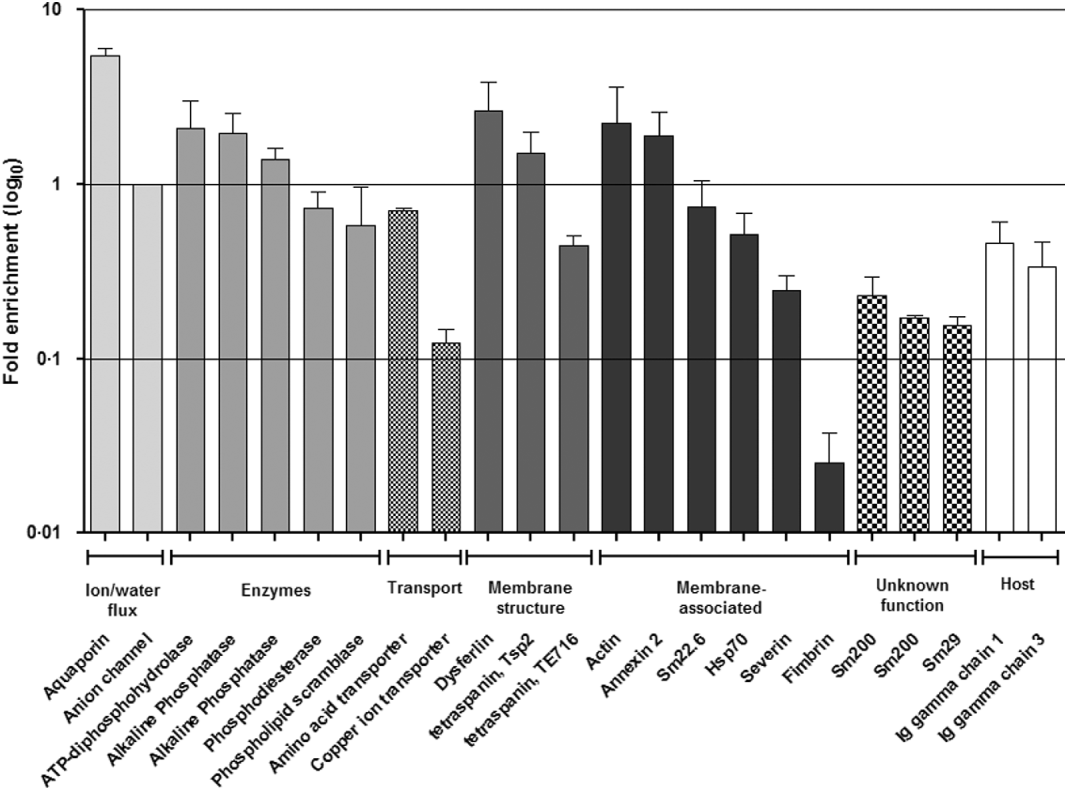
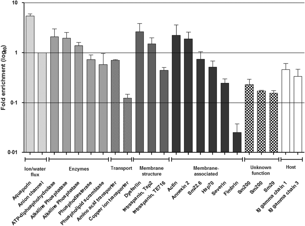
The abundance of the major constituents of the tegument surface has been estimated using the QconCAT technique (Castro-Borges et al. Reference Castro-Borges, Simpson, Dowle, Curwen, Thomas-Oates, Beynon and Wilson2011b). An artificial protein was produced that is a concatamer of Quantitation (Q) peptides (hence QconCAT; Rivers et al. Reference Rivers, Simpson, Robertson, Gaskell and Beynon2007), each one having a different mass but otherwise identical physiochemical properties to its target peptide. These were selected from proteins we had already found in the tegument, thus facilitating their simultaneous quantitative estimation. A gene encoding 33 signature peptides was designed, and the concatamer was expressed heterologously in Escherichia coli grown in the presence of [13C6] lysine and [13C6] arginine (Rivers et al. Reference Rivers, Simpson, Robertson, Gaskell and Beynon2007) to label it with stable isotopes. The labelled QconCAT protein was then purified, quantified and a known amount added to two tegument preparations. Tryptic digestion of the QconCAT-sample mixture released each of the QconCAT peptides in a strict 1:1 stoichiometry and subsequent MS analysis allowed the quantification of each peptide present in the sample. Addition of the concatamer to the gradient pellet preparation of isolated tegument surface (Roberts et al. Reference Roberts, MacGregor, Vojvodic, Wells, Crabtree and Wilson1983) revealed aquaporin to be relatively the most abundant, followed by dysferlin, ATP-diphosphohydrolase, alkaline phosphatase, actin, annexin Smp_077720 and tetraspanin TSP-2 in descending order (Fig. 1). There is a question mark over the dysferlin that will only be resolved by localization studies, as in higher animals it is associated with the plasma membranes of muscle cells. The amounts of IgG1 and IgG3 detected were small both in molar (one third to one half that of the anion channel reference protein) and absolute terms (2·3 and 0·8%, respectively of the total GP). The number of molecules of C3 detected was only one tenth that of the anion channel, i.e. less than a third that of the two IgGs, amounting to 1% of the GP mass. This means that the bound immunoglobulins and complement factors are present at the tegument surface in small amounts, relative to their potential targets. The paucity of complement C3 is further evidence that no amplification of the complement cascade occurs.
Relative abundance of 21 S. mansoni tegument proteins in the purified gradient pellet using the QconCAT approach. The mean fold enrichment±S.D is expressed on a log scale relative to the anion channel as unity. The height of the bars is proportional to the number of molecules of each of the proteins, which are grouped according to their molecular function. Data reproduced from Castro-Borges et al. (Reference Castro-Borges, Simpson, Dowle, Curwen, Thomas-Oates, Beynon and Wilson2011b).
When the concatamer was added to the preparation released by PiPLC treatment of live worms, a very different result emerged (Castro-Borges et al. Reference Castro-Borges, Simpson, Dowle, Curwen, Thomas-Oates, Beynon and Wilson2011b). The dominant protein was Sm200 (87%) that represented only 2–3% of the GP in spite of its mass. Alkaline phosphatase (6·8%) and Sm29 (0·44%) were the other two GPI-anchored proteins estimated. It was concluded, on the basis of protein ratios, that Sm200 is present in a different ‘surface compartment’ from Sm29 and alkaline phosphatase. It may simply be that Sm200 is easily detached from the surface due to its size and GPI-anchor, or conceivably that it uniquely resides in the membranocalyx, which separates from the plasma membrane during GP preparation.
Cationised ferritin-stimulated sloughing
Another angle on tegument surface organisation may be provided by the observation in early electron microscope studies that cationised ferritin (CF) will bind to live worms, suggesting that they carry a negative charge (Wilson and Barnes, Reference Wilson and Barnes1977). Furthermore, after a 30 minute pulse of worms with CF, chase incubation in Eagle's medium alone without supplements for four hours resulted in sloughing of the tag attached to sheets of membranocalyx. The proteomic observations need to be treated with caution due to lack of replication but a single MS/MS analysis of the pelleted material for known tegument membrane components identified six proteins: Sm200, Sm29, Sm13, Annexin Smp_077720, Tetraspanin-TSP2, and LMWP. The inference must be that these six are the best candidates for location in the membranocalyx. It is noteworthy that immunoglobulins, complement factors and CD44 were not identified in the pellet, with the implication that these host proteins are associated with the plasma membrane constituents, rather than the membranocalyx. The sloughing also resulted in the release of signature cytosolic proteins into the culture supernatant, including glycolytic enzymes (enolase, aldolase), chaperones (HSP70, cyclophilin) and defence proteins (glutathione-S-transferase, superoxide dismutase), implying that a degree of tegument damage is inflicted by the procedure. The enzyme calpain was also detected in the supernatant but not the pellet, along with a schistosome serpin, Smp_090080, first cloned twenty years ago (GenBank: AAA29938.1). This result suggests that both are in a soluble rather than membrane compartment of the tegument.
A model for the molecular architecture of the tegument surface
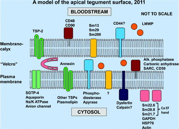
We first attempted to put tegument proteins into a molecular model of the tegument surface in 2006, based on the results of the first proteomic analyses (Braschi et al. Reference Braschi, Curwen, Ashton, Verjovski-Almeida and Wilson2006b; Braschi and Wilson, Reference Braschi and Wilson2006). This is very much ‘work in progress’ but we can expand on the earlier observations by adding newly discovered proteins, and also tentatively assign some to the membranocalyx (Fig. 2). It should be noted that the size and shape of the various components, has not been taken into account relative to the thickness of the two lipid bilayers. The most plausible membranocalyx component is Sm200, found in both membrane and cytosolic fractions. We still know little about the lipids that comprise the membranocalyx and give it such tremendous avidity for host proteins. Its permeability to the ions and solutes that must reach the transporters and enzymes of the plasma membrane suggests a relatively fluid organisation, rather than the presence of organised pores, particularly as the enzyme PiPLC (MW, 30 kDa) can apparently access the lipid anchors of proteins inserted in the outer leaflet of the plasma membrane (Castro-Borges et al. Reference Castro-Borges, Dowle, Curwen, Thomas-Oates and Wilson2011a). This fluidity, compared to the underlying plasma membrane, could have many sources. These include shorter chain fatty acids in phospholipids, a preponderance of glycoinositol phospholipids, no cholesterol, fewer membrane structural tetraspanins so an absence of the tetraspanin web, and almost certainly no linkage to the actin cytoskeleton. The way in which the membranocalyx can adhere to the plasma membrane yet also slide laterally over it has lead to the suggestion that there must be some kind of adhesion mechanism, a molecular Velcro, keeping the two layers together. The presence of annexin Smp_077720 external to, but associated with the plasma membrane, coupled with the lipid binding properties of annexins, provides one plausible candidate for the role. This hypothesis could be tested using RNA interference to knock down gene expression and determine the effect of tegument surface structure.
Diagram of the putative location of proteins in the membranocalyx and apical plasma membrane of the tegument surface. Note that the proteins and lipid bilayers are not depicted to scale. Host CD44 may be anchored in the plasma membrane, but with its N-terminus protruding above the membranocalyx surface. Data suggest that calpain may be outside the plasma membrane. The location of immunoglobulins and complement factors C3 and C4 are not shown.

In the current model, host CD44 is placed in the membranocalyx because it is readily detected by antibodies on the surface of live worms (Castro-Borges et al. Reference Castro-Borges, Dowle, Curwen, Thomas-Oates and Wilson2011a). However, its apparent absence from the CF pellet of sloughed membranocalyx suggests that it may be anchored in the plasma membrane. This is actually a serious issue because the first estimates of membranocalyx turnover, based on the CF sloughing, gave a t½ of 2–3 h (Wilson and Barnes, Reference Wilson and Barnes1977). This was later revised upwards to 5·4 days on the basis of a solid phase radio-immune assay using antibodies to erythrocyte markers on the worm surface (Saunders et al. Reference Saunders, Wilson and Coulson1987), on the assumption that these were glycolipids inserted into the membranocalyx (Goldring et al. Reference Goldring, Clegg, Smithers and Terry1976, Reference Goldring, Kusel and Smithers1977). If, as now seems likely, the antibodies were directed against CD44 and this host protein is anchored in the plasma membrane, then the turnover values obtained were not for the half-life of the membranocalyx, but for the slow displacement of host CD44.
BLOOD FEEDING: WHAT DOES WORM VOMITUS TELL US ABOUT GUT FUNCTION?
The second major interface with the host, the alimentary tract, is more difficult to investigate than the tegument because of its inaccessibility. At the cercaria/skin schistosomulum stage, a rudimentary gut is present, and actively ingesting material (Thornhill et al. Reference Thornhill, McVeigh, Jurberg and Kusel2010), but erythrocyte ingestion does not begin until the parasite has reached the hepatic portal distributaries of the liver (Clegg, Reference Clegg1965). The gut is blind-ending and in the adult runs the whole length of the body, partly as single and partly as bifurcated caeca. It comprises a single epithelial layer invested with tiny muscle fibres that provide the motive force for peristalsis. The absorptive surface is extended by lamellar plates, rather than more conventional microvilli (Hall et al. Reference Hall, Braschi, Truscott, Mathieson, Cesari and Wilson2011). Immediately anterior to the gut, and ventral to the oesophagus lies an oesophageal gland comprising 20–30 cells, really modified tegument cell bodies, that secretes material into the lumen.
Analysis of gene expression in the gut epithelium by RT-PCR has been performed using laser capture microdissection to recover tissue from sections (Nawaratna et al. Reference Nawaratna, McManus, Moertel, Gobert and Jones2011). This approach identified transcript for several transporters (Ca++ATPase, phospholipid transporter, amino acid transporter, multi-drug resistance protein), a small number of lysosomal proteins (LAMP, NPC-2, acid lipase and phospholipase A2) and Cathepsins L, B and D2. However, collection of regurgitated vomitus has also provided valuable insights into gut function (Delcroix et al. Reference Delcroix, Medzihradsky, Caffrey, Fetter and McKerrow2007; Hall et al. Reference Hall, Braschi, Truscott, Mathieson, Cesari and Wilson2011). Fortuitously for tegument studies, as it turns out, schistosomes in vitro are very reluctant to open their mouths. This is in contrast to the liver fluke Fasciola hepatica that regurgitates quantities of vomitus from its proportionally larger gut continuously, playing havoc with tegument analysis (Wilson et al. Reference Wilson, Wright, de Castro Borges, Parker-Manuel, Dowle, Ashton, Young, Gasser and Spithill2011). Giving schistosomes cold shocks or even putting them in distilled water results in preparations highly contaminated with cytosolic and cytoskeletal proteins from the tegument and other tissues (Delcroix et al. Reference Delcroix, Medzihradsky, Caffrey, Fetter and McKerrow2007; Planchart et al. Reference Planchart, Incani and Cesari2007). The cleanest vomitus preparations have been obtained by the culture of ex vivo day 28 juvenile worms in RPMI-1640 medium for 4 days without protein supplements (Hall et al. Reference Hall, Braschi, Truscott, Mathieson, Cesari and Wilson2011). Proteomic analysis of the collected vomit confirmed the earlier biochemical studies (Caffrey et al. Reference Caffrey, McKerrow, Salter and Sajid2004; Delcroix et al. Reference Delcroix, Sajid, Caffrey, Lim, Dvorak, Hsieh, Bahgat, Dissous and McKerrow2006) that described the presence of a series of cathepsins and the pro-enzyme convertase, asparaginyl endopeptidase. Additional hydrolytic enzymes, including several proteases, largely of lysosomal origin, were also identified (Table 4 and Hall et al. Reference Hall, Braschi, Truscott, Mathieson, Cesari and Wilson2011). In the context of the hydrolytic environment, two protease inhibitors, a serpin and alpha-2-macroglobulin were identified, potentially performing a self-protective function to prevent auto-digestion. A series of carrier or binding proteins was also present, likely originating from lysosomes. These included saposins, NPC-2 (Niemann-Pick disease, type C), ferritin isoforms and calumenin, capable of binding lipids, cholesterol, ferric iron and calcium, respectively. The lysosomal markers, lysosome membrane-associated glycoprotein (LAMP) and vesicle associated membrane protein (VAMP), were also detected. In view of the known acid pH of the gut and the presence in vomitus of so many lysosomal proteins it was suggested that digestion of the haemoglobin and plasma proteins that comprise the schistosome diet was effected by secretion of lysosomes from the gut epithelium into the lumen. The long chain acyl-coenzyme thioesterase 1 identified, is normally a constituent of peroxisomes and may indicate that these organelles also make a contribution to luminal digestion.
As a corollary to the above, it was suggested that the function of the carrier proteins identified was to scavenge lipids and inorganic ions from the gut lumen by endocytosis, potentially receptor mediated. All the necessary components for receptor-mediated endocytosis have been identified in the S. mansoni transcriptome or genome, but none have yet been localised to any tissue (Wilson, Reference Wilson2011). Feeding experiments with starved adult worms, using fluorescent space-filling dextran as the probe, have revealed for the first time the occurrence of endocytosis at the luminal surface of the gut epithelium (Hall et al. Reference Hall, Braschi, Truscott, Mathieson, Cesari and Wilson2011). Similar experiments with labelled erythrocytes have also shown their uncoating as they pass through the oesophagus, with transfer of the lipophilic label PKH2 to the oesophageal lining (Hall et al. Reference Hall, Braschi, Truscott, Mathieson, Cesari and Wilson2011). This observation implicates the one known product of the oesophageal gland, the microexon-encoded protein MEG-4.1 (DeMarco et al. Reference DeMarco, Mathieson, Manuel, Dillon, Curwen, Ashton, Ivens, Berriman, Verjovski-Almeida and Wilson2010) originally described as Ag 10.3 (Davis et al. Reference Davis, Davis, Carroll, Rajkovic and Rottman1988), in the uncoating process.
ARE MICRO-EXON GENE PRODUCTS THE SCHISTOSOME'S ANSWER TO LIFE IN THE BLOODSTREAM?
The description of micro exon genes (MEGs), briefly in the S. mansoni genome paper (Berriman et al. Reference Berriman, Haas, LoVerde, Wilson, Dillon, Cerqueira, Mashiyama, Al-Lazikani, Andrade, Ashton, Aslett, Bartholomeu, Blandin, Caffrey, Coghlan, Coulson, Day, Delcher, DeMarco, Djikeng, Eyre, Gamble, Ghedin, Gu, Hertz-Fowler, Hirai, Hirai, Houston, Ivens, Johnston, Lacerda, Macedo, McVeigh, Ning, Oliveira, Overington, Parkhill, Pertea, Pierce, Protasio, Quail, Rajandream, Rogers, Sajid, Salzberg, Stanke, Tivey, White, Williams, Wortman, Wu, Zamanian, Zerlotini, Fraser-Liggett, Barrell and El-Sayed2009) and subsequently as a fuller exposition (DeMarco et al. Reference DeMarco, Mathieson, Manuel, Dillon, Curwen, Ashton, Ivens, Berriman, Verjovski-Almeida and Wilson2010) has added another dimension to our view of the parasite-host interface. MEGs appear to be a unique feature of the schistosome genome. At least 75% of the protein coding region is composed of microexons ranging in length from 6 to 36 base pairs, 92% of which are symmetrical in multiples of three (the commonest frequency being 21). This means that individual microexons can be spliced out without altering the reading frame, potentially forming variant transcripts by exon skipping. Other than a shared gene structure, the 18 MEG families (with up to 23 members) so far identified show no similarity with each other or with annotated genes from outside the Schistosoma genus. They also encode no identifiable motifs or functional domains, apart from a signal peptide, suggesting that they are secreted. Two have already been mentioned in this review (MEG-3; MEG-4.1), but it was research on the secretome of the schistosome egg that first brought them to prominence (Mathieson and Wilson, Reference Mathieson and Wilson2010). In addition, microarray analysis of changes in gene expression associated with infection of, and establishment in the mammalian host has shown that transcripts representing the vast majority of MEG families are highly enriched in the intramolluscan germ balls, cercariae, or day 3 schistosomula (Parker-Manuel et al. Reference Parker-Manuel, Ivens, Dillon and Wilson2011). Indeed, in the schistosomulum, MEGs 4.1, 8, 12, 14, 15, 16, and 17 showed the highest fold increases in expression, indicating the probable importance of the proteins they encode in parasite establishment and subsequent persistence in the mammalian host.
From studies on immune-compromised mice it is apparent that schistosome eggs secrete proteins (Ashton et al. Reference Ashton, Harrop, Shah and Wilson2001b), and recruit the host immune response to aid their passage through the tissues (Doenhoff et al. Reference Doenhoff, Hassounah, Murare, Bain and Lucas1986). The egg is laid undeveloped in an intestinal blood vessel and the miracidium differentiates from the ovum over 5–7 days. A syncytial sub-shell envelope arises but only when the miracidium is mature does this layer begin to release secretions that are crucial for escape (Ashton et al. Reference Ashton, Harrop, Shah and Wilson2001b), as opposed to those proteins that mediate granuloma formation and could originate from any compartment. The first published account of the egg secretome detailed the presence of a large number of cytosolic and cytoskeletal proteins (and even nuclear histones) released from eggs in culture (Cass et al. Reference Cass, Johnson, Califf, Xu, Hernandez, Stadecker, Yates and Williams2007). As many of these lacked a conventional leader sequence, the authors resorted to the use of SecretomeP to justify their presence in the secretions, released via a proposed non-classical secretion pathway. This programme was developed to predict the small number of bacterial and mammalian proteins that do exit cells via a novel route (Bendtsen et al. Reference Bendtsen, Jensen, Blom, Von Heijne and Brunak2004), and its use to justify the presence of cytosolic proteins in schistosome secretions has already been criticised (DeMarco and Verjovski-Almeida, Reference DeMarco and Verjovski-Almeida2009). The authors do not entertain the alternative possibility that a proportion of damaged or dead eggs in their cultures might be leaking the internal proteins (Cass et al. Reference Cass, Johnson, Califf, Xu, Hernandez, Stadecker, Yates and Williams2007). Mathieson and Wilson reported a much smaller number of proteins in the secretions of eggs, in which post-culture viability was >96%, assessed by observation of muscular and flame cell activity in unhatched miracidia (Mathieson and Wilson, Reference Mathieson and Wilson2010). These proteins were dominated by the now well characterized IPSE and Omega-1 (Fitzsimmons et al. Reference Fitzsimmons, Schramm, Jones, Chalmers, Hoffmann, Grevelding, Wuhrer, Hokke, Haas, Doenhoff and Dunne2005; Schramm et al. Reference Schramm, Gronow, Knobloch, Wippersteg, Grevelding, Galle, Fuller, Stanley, Chiodini, Haas and Doenhoff2006). On 2D gel separations IPSE alone accounted for >83% of the released material, while a few minor spots could also be discerned (Mathieson and Wilson, Reference Mathieson and Wilson2010). Using a lectin column to deplete the IPSE and Omega-1 proteins (Schramm et al. Reference Schramm, Gronow, Knobloch, Wippersteg, Grevelding, Galle, Fuller, Stanley, Chiodini, Haas and Doenhoff2006), the background spots were greatly enriched and could be identified by tandem mass spectrometry (DeMarco et al. Reference DeMarco, Mathieson, Manuel, Dillon, Curwen, Ashton, Ivens, Berriman, Verjovski-Almeida and Wilson2010; Mathieson and Wilson, Reference Mathieson and Wilson2010). They proved to be predominantly protein products of the MEG-2 and MEG-3 gene families, each represented by one to several spots on the 2D gel (Table 5). A lack of evidence for post-translational modifications strongly suggests that these are variant protein versions of the variant transcripts found by RT-PCR (DeMarco et al. Reference DeMarco, Mathieson, Manuel, Dillon, Curwen, Ashton, Ivens, Berriman, Verjovski-Almeida and Wilson2010). At least 13 spots were encoded by MEG-3 variants and 10 spots by MEG-2 variants.
The production of variant proteins by other blood dwelling parasites is a well-established phenomenon. The protective variant surface glycoprotein (VSG) coat of Trypanosoma brucei, encoded by >1000 VSG genes that are transcribed one at a time at telomeric expression sites, has been thoroughly documented (Taylor and Rudenko, Reference Taylor and Rudenko2006). An infected host mounts an effective antibody response against the predominant VSG variants, leading to antibody-mediated lysis. However, trypanosomes continually appear that have switched to antigenically different VSGs, leading to a chronic infection that can last for years. Similarly, Plasmodium falciparum possesses a family of clonally variant surface antigens, collectively termed PfEMP1 and expressed on the parasitised erythrocyte surface, encoded by about 60 var genes per haploid genome (Kyes et al. Reference Kyes, Kraemer and Smith2007). Switching of var gene expression allows the parasite to modify the antigenic and functional properties of infected erythrocytes, thereby evading immunity and affecting infection outcome.
The demonstration of the variant proteins in schistosomes raises many questions. The secretion of variant versions of a protein, not all of which can be functional, comes at a metabolic cost to the parasite, and leads to the obvious conclusion that they are involved in immune evasion (DeMarco et al. Reference DeMarco, Mathieson, Manuel, Dillon, Curwen, Ashton, Ivens, Berriman, Verjovski-Almeida and Wilson2010). If so, then schistosomes are unlike populations of the protozoan blood parasites Trypanosoma and Plasmodium, which produce their variants sequentially by switching gene expression, to confuse the immune system. Schistosomes generate their variants simultaneously, so any mechanism of evasion must differ from that of the protozoans. Potentially, the variants could interfere with immune priming of T cells by accessory cells if they increased the range of peptides presented by MHCII molecules, at the same time reducing the frequency of each. This is a question for the future and the most urgent task is to discover the function of one or more of these proteins at the parasite-host interface. An inference must be that if, in the course of schistosome evolution, breaking down exons into small coding cassettes confers a selective advantage then the functions of the encoded proteins must be important for parasite survival. That ties in neatly with their apparent secreted status.
CONCLUSIONS: DOES ANALYSIS OF THE SCHISTOSOME-MAMMALIAN HOST INTERFACE HOLD ANY PROSPECTS FOR DIAGNOSTICS OR VACCINES?
The conclusions drawn from this review need to be prefaced by a major caveat. The identification of schistosome proteins by tandem mass spectrometry is now so easy that researchers need to be severely critical in interpreting their accumulated datasets. Schistosomes obey the normal rules of cell biology, they are not leaky sieves oozing cytosolic proteins across their surface membranes. The first response to finding such proteins in parasite secretions should be to see them as evidence for damage, unless circumstances (such as holocrine secretion) indicate otherwise. The danger is in confusing the ‘necrotome’ with the ‘secretome’. That said, the characterization of proteins expressed on, or secreted from schistosome parasites in the mammalian host should provide opportunities to develop new diagnostics and vaccine candidates. The information obtained is most immediately applicable to the search for diagnostics; its translation to the vaccine sphere requires greater scrutiny.
Diagnostics
The use of immunological tests to detect schistosome antigens in the host circulation is well established, and preferable to detection of anti-schistosome antibodies; these only indicate past exposure, not concurrent infection. The current test detects the gut proteoglycan circulating cathodic antigen (CAA). However, its sensitivity and that of the faecal smear technique, are inadequate to identify patients with low worm burdens. As there is no ethical way to determine actual worm burden in humans for correlation with the surrogate measures, the precise sensitivity of current diagnostics has remained vague. A correlative study in baboons, a realistic permissive host, where worm burden can be measured by portal perfusion suggests a detection limit of 27 worms for CAA and 40 worms for the smear technique (Wilson et al. Reference Wilson, van Dam, Kariuki, Farah, Deelder and Coulson2006). Clearly there is scope for improvement. Characterization of the vomitus highlights several proteins, released into the bloodstream that could act as indicators of infection. As such they might not suffer disadvantages inherent in the use of glycans as diagnostic molecules, namely the ubiquity of glycan epitopes across species, as well as the greater promiscuity and lower avidity of anti-glycan antibodies for their targets.
One important parameter is the rate of antigen clearance from the bloodstream; in this context it is possible that the two protease inhibitors, serpin and α2 macroglobulin, might prove ideal if they are resistant to the action of blood proteases. One recently reported approach (Sulbaran et al. Reference Sulbaran, Ballen, Bermudez, Lorenzo, Noya and Cesari2010), is to capture the gut hydrolases using antibodies and measure their enzymatic activity as an amplification step to increase sensitivity. At least two tegument surface proteins appear to have diagnostic potential. Sm200 seems very loosely attached and has been detected in the ‘lipid’ fraction of host serum using specific antibodies (Sprong et al. Reference Sprong, Suchanek, van Dijk, van Remoortere, Klumperman, Avram, van der Linden, Leusen, van Hellemond and Thiele2006). The other is LMWP, apparently a true tegument secretion; its small size (MW, 8·8 kDa) could mean that it passes into urine through the glomerular filters. (Urine would be a much simpler body fluid on which to base a diagnostic test for S. mansoni.) Into this category also come some MEG-2 and MEG-3 family proteins secreted by eggs, which may also be small enough to pass into urine. Eggs have another advantage as a source of diagnostic proteins, since they are produced at the rate of 300 per female per day. When mature, they secrete their ‘escape proteins’ for days to weeks so that a single worm pair will be responsible for the secretions of some thousands of viable tissue eggs at any one time.
For vaccines, is there a chink in the armour?
An inherent problem for schistosome vaccine development has been on what to base the strategy. In spite of much effort, research on human response has produced few concrete leads, and where protective immunity has been implicated, it appears to be related to IgE production (Dunne et al. Reference Dunne, Butterworth, Fulford, Kariuki, Langley, Ouma, Capron, Pierce and Sturrock1992). Protection mediated by IgE is unlikely to form the basis of an effective vaccine because of associated regulatory issues. We therefore have to turn to animal models for a lead and we have argued that only two are sufficiently robust to provide a suitable paradigm (Wilson and Coulson, Reference Wilson and Coulson2009). The first is the radiation-attenuated (RA) cercarial vaccine, applied to rodents and primates, for which there is a wealth of literature (Coulson, Reference Coulson1997; Hewitson et al. Reference Hewitson, Hamblin and Mountford2005). Latterly we have added the self-cure phenomenon in the rhesus macaque (Wilson et al. Reference Wilson, Langermans, van Dam, Vervenne, Hall, Borges, Dillon, Thomas and Coulson2008; Wilson and Coulson, Reference Wilson and Coulson2009). In neither of these models is there credible in vivo evidence for the operation of ADCC as a mechanism for parasite elimination. In mice, the target of the RA vaccine is the lung-stage schistosomulum, and the mechanism appears to be the blocking of intravascular migration through the pulmonary capillaries by the inflammation induced. In the rhesus macaque, mature, egg-laying worms are eliminated by a protracted process that first involves cessation of blood feeding, then egg laying and finally death by starvation after internal non-essential tissues have been consumed (Wilson et al. Reference Wilson, Langermans, van Dam, Vervenne, Hall, Borges, Dillon, Thomas and Coulson2008). In both these models, it is the proteins exposed on the surface or released into the environment that must mediate the protection.
There do not appear to have been any reports of success in protection experiments with the cercarial secretions, including the elastases, in spite of the fact that their rich assemblage of glycan residues (Jang-Lee et al. Reference Jang-Lee, Curwen, Ashton, Tissot, Mathieson, Panico, Dell, Wilson and Haslam2007) are highly immunogenic in baboons and mice (Kariuki et al. Reference Kariuki, Farah, Wilson and Coulson2008). Indeed, this property has lead to the ‘smokescreen hypothesis’, in which the strong immunogenicity of cercarial secretions disguises more vulnerable peptide epitopes from immune attack (Eberl et al. Reference Eberl, Langermans, Vervenne, Nyame, Cummings, Thomas, Coulson and Wilson2001). A variant on this idea is the ‘matador's cloak’, where glycans released from the acetabular glands into the skin decoy leukocytes away from the parasite during the brief period when it is acquiring its immune disguise (Jang-Lee et al. Reference Jang-Lee, Curwen, Ashton, Tissot, Mathieson, Panico, Dell, Wilson and Haslam2007).
The proteomic analyses of the tegument described in this review have been paralleled by a number of studies on the highlighted proteins. These include alkaline phosphatase, aquaporin, annexin 2 and phosphodiesterase but there are no reports of their vaccine potential (Rofatto et al. Reference Rofatto, Tararam, Borges, Wilson, Leite and Farias2009; Faghiri et al. Reference Faghiri, Camargo, Huggel, Forster, Ndegwa, Verrey and Skelly2010; Tararam et al. Reference Tararam, Farias, Wilson and Leite2010; Araujo-Montoya et al. Reference Araujo-Montoya, Rofatto, Tararam, Farias, Oliveira, Verjovski-Almeida, Wilson and Leite2011; Bhardwaj et al. Reference Bhardwaj, Krautz-Peterson, Da'dara, Tzipori and Skelly2011; Bhardwaj and Skelly, Reference Bhardwaj and Skelly2011). Other tegument surface proteins (TSP-2, Sm29, Calpain) have been targeted with more success (Tran et al. Reference Tran, Pearson, Bethony, Smyth, Jones, Duke, Don, McManus, Correa-Oliveira and Loukas2006; Cardoso et al. Reference Cardoso, Macedo, Gava, Kitten, Mati, de Melo, Caliari, Almeida, Venancio, Verjovski-Almeida and Oliveira2008; Ahmad et al. Reference Ahmad, Zhang, Torben, Damian, Wolf, White, Chavez-Suarez, Kennedy and Siddiqui2009). Unfortunately, there has been no spectacular breakthrough with consistent protection above 50% achieved. Note that the irradiated vaccine in baboons has elicited up to 86% protection (Yole et al. Reference Yole, Pemberton, Reid and Wilson1996), and in mice virtually sterile immunity (Anderson et al. Reference Anderson, Shires, Wilson and Mountford1998; Wynn et al. Reference Wynn, Reynolds, James, Cheever, Caspar, Hieny, Jankovic, Strand and Sher1996). There are other surface proteins that have yet to feature in protection experiments, including Sm200, Sm13, carbonic anhydrase, LMWP, ADP-ribosyl cyclase, and the CD59 isoforms. In the case of the RA vaccine model, those proteins that can be detached from the surface by accessory cells are attractive candidates. For the rhesus macaque model, the enzymes with ecto-domains, or transporters in the plasma membrane that could be blocked by antibodies are the most plausible targets.
It seems entirely possible with both models that multiple proteins need to be targeted to block migration in the lungs or disable adult worms in the portal system. That presents a dilemma, as antigens are almost always tested singly, eliminating the possibility of synergy. In the context of the RA vaccine, we used to refer to the major secreted protein of the lung schistosomulum as the ‘holy grail’ of schistosome vaccinology. The euphoria following its identification as a mixture of MEG-3 family variants (DeMarco et al. Reference DeMarco, Mathieson, Manuel, Dillon, Curwen, Ashton, Ivens, Berriman, Verjovski-Almeida and Wilson2010) was short lived as close behind came the unwelcome news that it was also secreted by mature eggs (Mathieson and Wilson, Reference Mathieson and Wilson2010). The common feature is that both stages secrete MEG-3 proteins when they are resident in the blood vessels, strongly suggesting that they are released to modulate host vascular physiology. Unfortunately for vaccine development, when highly immunogenic live eggs are administered to the peritoneal cavity of mice they fail completely to protect the recipients against a cercarial challenge (Kariuki et al. Reference Kariuki, Farah, Wilson and Coulson2008). Is this a case of micro-exon controlled protein variation in action to protect a vital biological function of the schistosomulum and egg? Do schistosomes have all the answers?